|
|
|
|
 Description
|
|
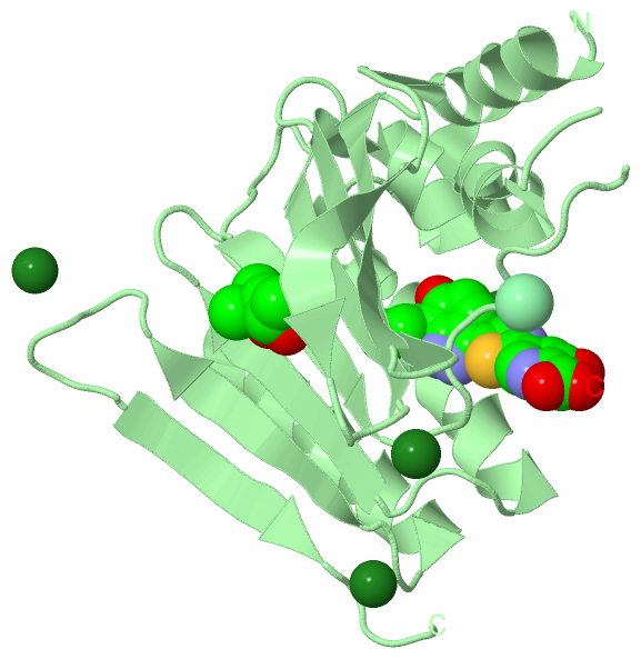
Compounds
|
||||||||||||||||||||||||||||||||||||||||||||||||||||
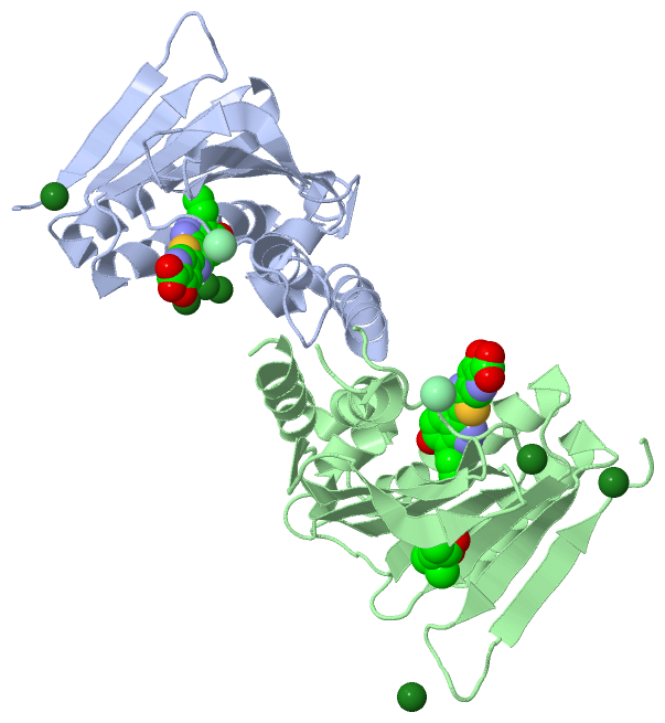
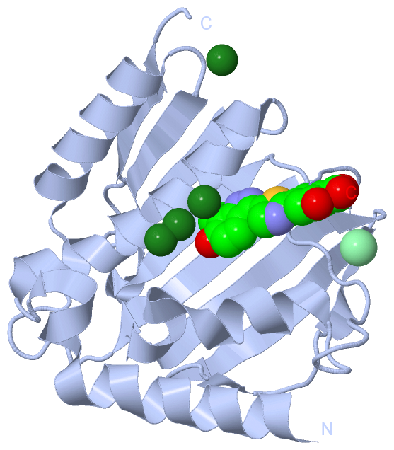
Chains, Units
Summary Information (see also Sequences/Alignments below) |
Ligands, Modified Residues, Ions (4, 13)
Asymmetric Unit (4, 13)
|
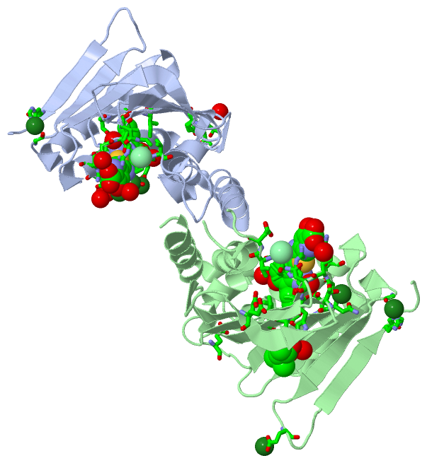
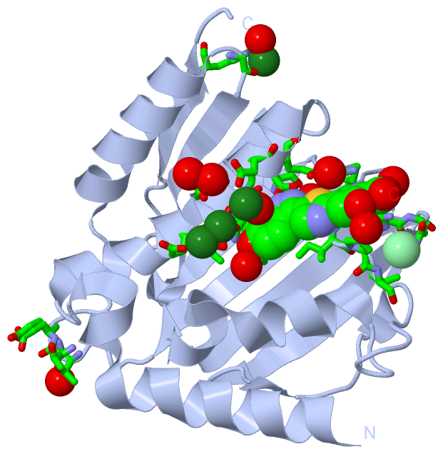
Sites (13, 13)
Asymmetric Unit (13, 13)
|
SS Bonds (0, 0)| (no "SS Bond" information available for 5D7R) |
Cis Peptide Bonds (0, 0)| (no "Cis Peptide Bond" information available for 5D7R) |
SAPs(SNPs)/Variants (0, 0)| (no "SAP(SNP)/Variant" information available for 5D7R) |
PROSITE Motifs (0, 0)| (no "PROSITE Motif" information available for 5D7R) |
Exons (0, 0)| (no "Exon" information available for 5D7R) |
Sequences/Alignments
Asymmetric Unit
Chain A from PDB Type:PROTEIN Length:193
SCOP domains ------------------------------------------------------------------------------------------------------------------------------------------------------------------------------------------------- SCOP domains
CATH domains ------------------------------------------------------------------------------------------------------------------------------------------------------------------------------------------------- CATH domains
Pfam domains ------------------------------------------------------------------------------------------------------------------------------------------------------------------------------------------------- Pfam domains
SAPs(SNPs) ------------------------------------------------------------------------------------------------------------------------------------------------------------------------------------------------- SAPs(SNPs)
PROSITE ------------------------------------------------------------------------------------------------------------------------------------------------------------------------------------------------- PROSITE
Transcript ------------------------------------------------------------------------------------------------------------------------------------------------------------------------------------------------- Transcript
5d7r A 15 GAGQIQVLEGLEAVRKRPGMYIGSTSERGLHHLVWEIVDNSIDEALAGYANQIEVVIEKDNWIKVTDNGRGIPVDIQEKMGRPAVEVILTSSVVNALSQDLEVYVHRNETIYHQAYKKGVPQFDLKEVGTTDKTGTVIRFKADGEIFTETTVYNYETLQQRIRELAFLNKGIQITLRDERDEENVREDSYHYE 230
24 34 44 54 64 74 84 94 104| 137 147 157 167 177 187 197 207 217 227
104|
128
Chain B from PDB Type:PROTEIN Length:187
SCOP domains ------------------------------------------------------------------------------------------------------------------------------------------------------------------------------------------- SCOP domains
CATH domains ------------------------------------------------------------------------------------------------------------------------------------------------------------------------------------------- CATH domains
Pfam domains ------------------------------------------------------------------------------------------------------------------------------------------------------------------------------------------- Pfam domains
SAPs(SNPs) ------------------------------------------------------------------------------------------------------------------------------------------------------------------------------------------- SAPs(SNPs)
PROSITE ------------------------------------------------------------------------------------------------------------------------------------------------------------------------------------------- PROSITE
Transcript ------------------------------------------------------------------------------------------------------------------------------------------------------------------------------------------- Transcript
5d7r B 19 IQVLEGLEAVRKRPGMYIGSTSERGLHHLVWEIVDNSIDEALAGYANQIEVVIEKDNWIKVTDNGRGIPVDIQGRPAVEVILTSSVVNALSQDLEVYVHRNETIYHQAYKKGVPQFDLKEVGTTDKTGTVIRFKADGEIFTETTVYNYETLQQRIRELAFLNKGIQITLRDERDEENVREDSYHYEG 231
28 38 48 58 68 78 88 || 101 || 134 144 154 164 174 184 194 204 214 224
91| 104|
95 128
|
||||||||||||||||||||
SCOP Domains (0, 0)| (no "SCOP Domain" information available for 5D7R) |
CATH Domains (0, 0)| (no "CATH Domain" information available for 5D7R) |
Pfam Domains (0, 0)| (no "Pfam Domain" information available for 5D7R) |
Gene Ontology (13, 13)|
Asymmetric Unit(hide GO term definitions) |
Interactive Views
|
||||||||||||||||||||||||||||||||||||||||||||||||||||||||||||||||||||||||||||||||||||||||||||||||||||||||||||||||||||||||||||||||||||||||||||||||||||||||||||||||||||||||||||||||||||||||||||||||||||||||||||||||||||||||||||||||||||||||||||||||||||||
Still Images
|
||||||||||||||||
Databases
|
||||||||||||||||||||||||||||||||||||||||||||||||||||||||||||||||||||||||||||||||||||||||||||||||||||||||||||||||||||||||||||||||||||||||||||||||||||||||||||||||
Analysis Tools
|
|||||||||||||||||||||||||||||||||||||||||||||||||||||||||||||
Entries Sharing at Least One Protein Chain (UniProt ID)
Related Entries Specified in the PDB File
|
|