Show PDB file:   
         Plain Text   HTML   (compressed file size)
QuickSearch:   
by PDB,NDB,UniProt,PROSITE Code or Search Term(s)  
(-)Asym./Biol. Unit
(-)Asym./Biol. Unit - sites
collapse expand < >
Image Asym./Biol. Unit
Asym./Biol. Unit  (Jmol Viewer)
Image Asym./Biol. Unit - sites
Asym./Biol. Unit - sites  (Jmol Viewer)

(-) Description

Title :  CRYSTAL STRUCTURE OF THE CTD OF DROSOPHILA OSKAR PROTEIN
 
Authors :  N. Yang, M. Hu, Z. Yu, M. Wang, R. Lehmann, R. M. Xu
Date :  03 Jul 15  (Deposition) - 02 Sep 15  (Release) - 30 Sep 15  (Revision)
Method :  X-RAY DIFFRACTION
Resolution :  2.10
Chains :  Asym./Biol. Unit :  A
Keywords :  3'-Utr, Dimerization, Rna Binding Protein (Keyword Search: [Gene Ontology, PubMed, Web (Google))
 
Reference :  N. Yang, Z. Yu, M. Hu, M. Wang, R. Lehmann, R. M. Xu
Structure Of Drosophila Oskar Reveals A Novel Rna Binding Protein
Proc. Natl. Acad. Sci. Usa V. 112 11541 2015
PubMed-ID: 26324911  |  Reference-DOI: 10.1073/PNAS.1515568112

(-) Compounds

Molecule 1 - MATERNAL EFFECT PROTEIN OSKAR
    ChainsA
    EngineeredYES
    Expression SystemESCHERICHIA COLI
    Expression System PlasmidPET28A-SUMO
    Expression System StrainBL21(DE3) CODON PLUS RIL
    Expression System Taxid562
    Expression System Vector TypePLASMID
    FragmentUNP RESIDUES 393-606
    GeneOSK, CG10901
    Organism CommonFRUIT FLY
    Organism ScientificDROSOPHILA MELANOGASTER
    Organism Taxid7227

 Structural Features

(-) Chains, Units

  1
Asymmetric/Biological Unit A

Summary Information (see also Sequences/Alignments below)

(-) Ligands, Modified Residues, Ions  (2, 13)

Asymmetric/Biological Unit (2, 13)
No.NameCountTypeFull Name
1MSE6Mod. Amino AcidSELENOMETHIONINE
2SO47Ligand/IonSULFATE ION

(-) Sites  (7, 7)

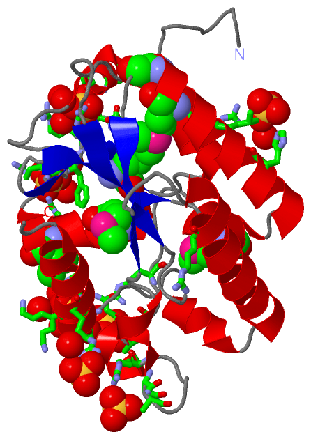
Asymmetric Unit (7, 7)
No.NameEvidenceResiduesDescription
1AC1SOFTWAREARG A:436 , ARG A:442 , PHE A:443 , ARG A:544 , HOH A:842 , HOH A:863binding site for residue SO4 A 701
2AC2SOFTWAREASN A:406 , SER A:408 , ARG A:444 , ARG A:445 , HIS A:446 , GLU A:447 , HOH A:805 , HOH A:814 , HOH A:816binding site for residue SO4 A 702
3AC3SOFTWAREGLN A:572 , ALA A:573 , SER A:574 , ARG A:576 , HOH A:835 , HOH A:837 , HOH A:866binding site for residue SO4 A 703
4AC4SOFTWAREGLN A:572 , ASN A:589 , LYS A:590 , ARG A:593 , HOH A:831 , HOH A:835 , HOH A:866 , HOH A:919 , HOH A:934binding site for residue SO4 A 704
5AC5SOFTWAREARG A:465 , HIS A:499 , LEU A:503 , HOH A:827binding site for residue SO4 A 705
6AC6SOFTWAREARG A:595 , HOH A:958binding site for residue SO4 A 706
7AC7SOFTWAREASN A:561 , GLU A:562 , ARG A:567binding site for residue SO4 A 707

(-) SS Bonds  (0, 0)

(no "SS Bond" information available for 5CD9)

(-) Cis Peptide Bonds  (0, 0)

(no "Cis Peptide Bond" information available for 5CD9)

 Sequence-Structure Mapping

(-) SAPs(SNPs)/Variants  (0, 0)

(no "SAP(SNP)/Variant" information available for 5CD9)

(-) PROSITE Motifs  (0, 0)

(no "PROSITE Motif" information available for 5CD9)

(-) Exons   (0, 0)

(no "Exon" information available for 5CD9)

(-) Sequences/Alignments

Asymmetric/Biological Unit
   Reformat: Number of residues per line =  ('0' or empty: single-line sequence representation)
  Number of residues per labelling interval =   
  UniProt sequence: complete  aligned part    
   Show mapping: SCOP domains CATH domains Pfam domains Secondary structure (by author)
SAPs(SNPs) PROSITE motifs Exons
(details for a mapped element are shown in a popup box when the mouse pointer rests over it)
Chain A from PDB  Type:PROTEIN  Length:209
                                                                                                                                                                                                                                                 
               SCOP domains ----------------------------------------------------------------------------------------------------------------------------------------------------------------------------------------------------------------- SCOP domains
               CATH domains ----------------------------------------------------------------------------------------------------------------------------------------------------------------------------------------------------------------- CATH domains
               Pfam domains ----------------------------------------------------------------------------------------------------------------------------------------------------------------------------------------------------------------- Pfam domains
         Sec.struct. author ........hhhhhh.......ee..eeeeehhhhhhhhhhhh...........ee.eee...hhhhhhhhhhh.......eeeee.hhhhhhh..hhhhhhhhhhhhhhhhhhh..eeeee....hhhhh.hhhhhhhhhhhhhhhhhhh....ee..hhhhh......hhhhh.................hhhhhhhhhhhhhhh... Sec.struct. author
                 SAPs(SNPs) ----------------------------------------------------------------------------------------------------------------------------------------------------------------------------------------------------------------- SAPs(SNPs)
                    PROSITE ----------------------------------------------------------------------------------------------------------------------------------------------------------------------------------------------------------------- PROSITE
                 Transcript ----------------------------------------------------------------------------------------------------------------------------------------------------------------------------------------------------------------- Transcript
                 5cd9 A 398 TILTSGTYNDSLLTINSDYDAYLLDFPLmGDDFmLYLARmELKCRFRRHERVLQSGLCVSGLTINGARNRLKRVQLPEGTQIIVNIGSVDImRGKPLVQIEHDFRLLIKEmHNmRLVPILTNLAPLGNYCHDKVLCDKIYRFNKFIRSECCHLKVIDIHSCLINERGVVRFDCFQASPRQVTGSKEPYLFWNKIGRQRVLQVIETSLEY 606
                                   407       417       427   |   437       447       457       467       477       487 |     497       507|  |   517       527       537       547       557       567       577       587       597         
                                                      426-MSE|     |                                                 489-MSE            508-MSE                                                                                              
                                                           431-MSE |                                                                       511-MSE                                                                                           
                                                                 437-MSE                                                                                                                                                                     

   Legend:   → Mismatch (orange background)
  - → Gap (green background, '-', border residues have a numbering label)
    → Modified Residue (blue background, lower-case, 'x' indicates undefined single-letter code, labelled with number + name)
  x → Chemical Group (purple background, 'x', labelled with number + name, e.g. ACE or NH2)
  extra numbering lines below/above indicate numbering irregularities and modified residue names etc., number ends below/above '|'

 Classification and Annotation

(-) SCOP Domains  (0, 0)

(no "SCOP Domain" information available for 5CD9)

(-) CATH Domains  (0, 0)

(no "CATH Domain" information available for 5CD9)

(-) Pfam Domains  (0, 0)

(no "Pfam Domain" information available for 5CD9)

(-) Gene Ontology  (26, 26)

Asymmetric/Biological Unit(hide GO term definitions)

 Visualization

(-) Interactive Views

Asymmetric/Biological Unit
  Complete Structure
    Jena3D(integrated viewing of ligand, site, SAP, PROSITE, SCOP information)
    WebMol | AstexViewer[tm]@PDBe
(Java Applets, require no local installation except for Java; loading may be slow)
    STRAP
(Java WebStart application, automatic local installation, requires Java; full application with system access!)
    RasMol
(require local installation)
    Molscript (VRML)
(requires installation of a VRML viewer; select preferred view via VRML and generate a mono or stereo PDF format file)
 
  Ligands, Modified Residues, Ions
    MSE  [ RasMol | Jena3D ]  +environment [ RasMol | Jena3D ]
    SO4  [ RasMol | Jena3D ]  +environment [ RasMol | Jena3D ]
 
  Sites
    AC1  [ RasMol ]  +environment [ RasMol ]
    AC2  [ RasMol ]  +environment [ RasMol ]
    AC3  [ RasMol ]  +environment [ RasMol ]
    AC4  [ RasMol ]  +environment [ RasMol ]
    AC5  [ RasMol ]  +environment [ RasMol ]
    AC6  [ RasMol ]  +environment [ RasMol ]
    AC7  [ RasMol ]  +environment [ RasMol ]
 
  Cis Peptide Bonds
(no "Cis Peptide Bonds" information available for 5cd9)
 

(-) Still Images

Jmol
  protein: cartoon or spacefill or dots and stick; nucleic acid: cartoon and stick; ligands: spacefill; active site: stick
Molscript
  protein, nucleic acid: cartoon; ligands: spacefill; active site: ball and stick

 Databases and Analysis Tools

(-) Databases

Access by PDB/NDB ID
  5cd9
    Family and Domain InformationProDom | SYSTERS
    General Structural InformationGlycoscienceDB | MMDB | NDB | OCA | PDB | PDBe | PDBj | PDBsum | PDBWiki | PQS | PROTEOPEDIA
    Orientation in MembranesOPM
    Protein SurfaceSURFACE
    Secondary StructureDSSP (structure derived) | HSSP (homology derived)
    Structural GenomicsGeneCensus
    Structural NeighboursCE | VAST
    Structure ClassificationCATH | Dali | SCOP
    Validation and Original DataBMRB Data View | BMRB Restraints Grid | EDS | PROCHECK | RECOORD | WHAT_CHECK
 
Access by UniProt ID/Accession number
  OSKA_DROME | P25158
    Comparative Protein Structure ModelsModBase
    Genomic InformationEnsembl
    Protein-protein InteractionDIP
    Sequence, Family and Domain InformationInterPro | Pfam | SMART | UniProtKB/SwissProt
 
Access by Enzyme Classificator   (EC Number)
  (no 'Enzyme Classificator' available)
    General Enzyme InformationBRENDA | EC-PDB | Enzyme | IntEnz
    PathwayKEGG | MetaCyc
 
Access by Disease Identifier   (MIM ID)
  (no 'MIM ID' available)
    Disease InformationOMIM
 
Access by GenAge ID
  (no 'GenAge ID' available)
    Age Related InformationGenAge

(-) Analysis Tools

Access by PDB/NDB ID
    Domain InformationXDom
    Interatomic Contacts of Structural UnitsCSU
    Ligand-protein ContactsLPC
    Protein CavitiescastP
    Sequence and Secondary StructurePDBCartoon
    Structure AlignmentSTRAP(Java WebStart application, automatic local installation, requires Java; full application with system access!)
    Structure and Sequence BrowserSTING
 
Access by UniProt ID/Accession number
  OSKA_DROME | P25158
    Protein Disorder PredictionDisEMBL | FoldIndex | GLOBPLOT (for more information see DisProt)

 Related Entries

(-) Entries Sharing at Least One Protein Chain (UniProt ID)

UniProtKB/Swiss-Prot
        OSKA_DROME | P251585a48 5a49 5a4a 5cd7 5cd8 5nt7

(-) Related Entries Specified in the PDB File

5cd7 5cd8