Show PDB file:   
         Plain Text   HTML   (compressed file size)
QuickSearch:   
by PDB,NDB,UniProt,PROSITE Code or Search Term(s)  
(-)Asymmetric Unit
(-)Asym. Unit - sites
(-)Biological Unit 1
(-)Biol. Unit 1 - sites
collapse expand < >
Image Asymmetric Unit
Asymmetric Unit  (Jmol Viewer)
Image Asym. Unit - sites
Asym. Unit - sites  (Jmol Viewer)
Image Biological Unit 1
Biological Unit 1  (Jmol Viewer)
Image Biol. Unit 1 - sites
Biol. Unit 1 - sites  (Jmol Viewer)

(-) Description

Title :  THE STRUCTURE OF HUMAN MAT2A IN COMPLEX WITH S-ADENOSYLETHIONINE AND PPNP.
 
Authors :  B. Murray, S. V. Antonyuk, A. Marina, S. C. Lu, J. M. Mato, S. S. Hasnain, A
Date :  30 Apr 15  (Deposition) - 17 Feb 16  (Release) - 09 Mar 16  (Revision)
Method :  X-RAY DIFFRACTION
Resolution :  1.83
Chains :  Asym. Unit :  A
Biol. Unit 1:  A  (4x)
Keywords :  Transferase, Methionine Adenosyltransferase, Cell Growth, Liver Cancer, Methylation (Keyword Search: [Gene Ontology, PubMed, Web (Google))
 
Reference :  B. Murray, S. V. Antonyuk, A. Marina, S. C. Lu, J. M. Mato, S. S. Hasnain, A. L. Rojas
Crystallography Captures Catalytic Steps In Human Methionine Adenosyltransferase Enzymes.
Proc. Natl. Acad. Sci. Usa V. 113 2104 2016
PubMed-ID: 26858410  |  Reference-DOI: 10.1073/PNAS.1510959113

(-) Compounds

Molecule 1 - S-ADENOSYLMETHIONINE SYNTHASE ISOFORM TYPE-2
    ChainsA
    EC Number2.5.1.6
    EngineeredYES
    Expression SystemESCHERICHIA COLI
    Expression System PlasmidPNIC28-BSA4
    Expression System StrainBL21
    Expression System Taxid511693
    Organism CommonHUMAN
    Organism ScientificHOMO SAPIENS
    Organism Taxid9606
    SynonymADOMET SYNTHASE 2, METHIONINE ADENOSYLTRANSFERASE 2, MAT 2, METHIONINE ADENOSYLTRANSFERASE II, MAT-II, METHIONINE ADENOSYLTRANSFERASE 2A

 Structural Features

(-) Chains, Units

  1
Asymmetric Unit A
Biological Unit 1 (4x)A

Summary Information (see also Sequences/Alignments below)

(-) Ligands, Modified Residues, Ions  (6, 8)

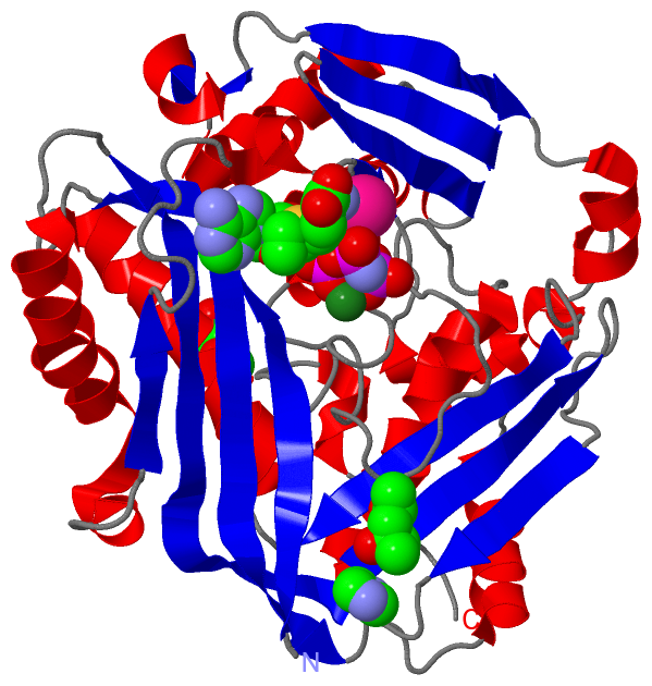
Asymmetric Unit (6, 8)
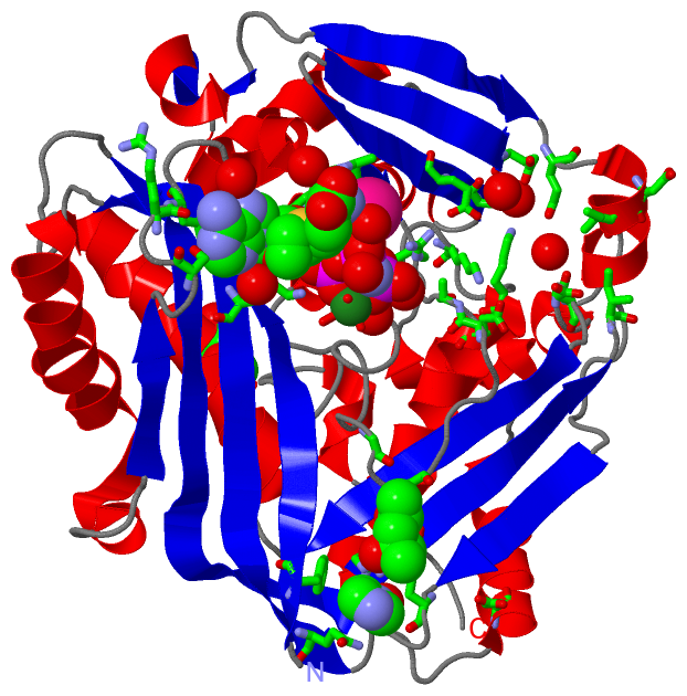
No.NameCountTypeFull Name
1IMD1Ligand/IonIMIDAZOLE
2K1Ligand/IonPOTASSIUM ION
3MG2Ligand/IonMAGNESIUM ION
4MPD2Ligand/Ion(4S)-2-METHYL-2,4-PENTANEDIOL
5PPK1Ligand/Ion(DIPHOSPHONO)AMINOPHOSPHONIC ACID
6S7M1Ligand/Ion[(3S)-3-AMINO-3-CARBOXYPROPYL]{[(2S,3S,4R,5R)-5-(6-AMINO-9H-PURIN-9-YL)-3,4-DIHYDROXYTETRAHYDROFURAN-2-YL]METHYL}ETHYLSULFONIUM
Biological Unit 1 (4, 20)
No.NameCountTypeFull Name
1IMD4Ligand/IonIMIDAZOLE
2K-1Ligand/IonPOTASSIUM ION
3MG-1Ligand/IonMAGNESIUM ION
4MPD8Ligand/Ion(4S)-2-METHYL-2,4-PENTANEDIOL
5PPK4Ligand/Ion(DIPHOSPHONO)AMINOPHOSPHONIC ACID
6S7M4Ligand/Ion[(3S)-3-AMINO-3-CARBOXYPROPYL]{[(2S,3S,4R,5R)-5-(6-AMINO-9H-PURIN-9-YL)-3,4-DIHYDROXYTETRAHYDROFURAN-2-YL]METHYL}ETHYLSULFONIUM

(-) Sites  (7, 7)

Asymmetric Unit (7, 7)
No.NameEvidenceResiduesDescription
1AC1SOFTWAREGLY A:273 , GLY A:275 , ARG A:313 , TYR A:335BINDING SITE FOR RESIDUE MPD A 400
2AC2SOFTWAREHIS A:29 , ASP A:31 , ASP A:134 , LYS A:181 , ARG A:264 , LYS A:265 , GLY A:280 , ALA A:281 , LYS A:285 , ASP A:291 , S7M A:404 , K A:405 , MG A:406 , MG A:407 , HOH A:2008 , HOH A:2015 , HOH A:2208 , HOH A:2209 , HOH A:2273 , HOH A:2274 , HOH A:2275BINDING SITE FOR RESIDUE PPK A 402
3AC3SOFTWAREPHE A:18 , GLN A:190 , GLU A:342 , HOH A:2154BINDING SITE FOR RESIDUE IMD A 403
4AC4SOFTWAREHIS A:29 , PRO A:30 , ALA A:55 , GLU A:70 , GLN A:113 , ASP A:116 , ILE A:117 , GLY A:133 , ASP A:134 , ASP A:179 , LYS A:181 , SER A:247 , ARG A:249 , PHE A:250 , ASP A:258 , LYS A:289 , ILE A:322 , PPK A:402 , HOH A:2040 , HOH A:2106 , HOH A:2204 , HOH A:2206 , HOH A:2274 , HOH A:2277BINDING SITE FOR RESIDUE S7M A 404
5AC5SOFTWAREGLU A:57 , ASP A:258 , ALA A:259 , PPK A:402 , MG A:406 , HOH A:2014 , HOH A:2035BINDING SITE FOR RESIDUE K A 405
6AC6SOFTWAREASP A:31 , LYS A:265 , PPK A:402 , K A:405 , HOH A:2014 , HOH A:2015BINDING SITE FOR RESIDUE MG A 406
7AC7SOFTWAREASP A:291 , PPK A:402 , HOH A:2006 , HOH A:2008 , HOH A:2273 , HOH A:2276BINDING SITE FOR RESIDUE MG A 407

(-) SS Bonds  (0, 0)

(no "SS Bond" information available for 5A1G)

(-) Cis Peptide Bonds  (0, 0)

(no "Cis Peptide Bond" information available for 5A1G)

 Sequence-Structure Mapping

(-) SAPs(SNPs)/Variants  (0, 0)

(no "SAP(SNP)/Variant" information available for 5A1G)

(-) PROSITE Motifs  (0, 0)

(no "PROSITE Motif" information available for 5A1G)

(-) Exons   (0, 0)

(no "Exon" information available for 5A1G)

(-) Sequences/Alignments

Asymmetric Unit
   Reformat: Number of residues per line =  ('0' or empty: single-line sequence representation)
  Number of residues per labelling interval =   
  UniProt sequence: complete  aligned part    
   Show mapping: SCOP domains CATH domains Pfam domains Secondary structure (by author)
SAPs(SNPs) PROSITE motifs Exons
(details for a mapped element are shown in a popup box when the mouse pointer rests over it)
Chain A from PDB  Type:PROTEIN  Length:380
                                                                                                                                                                                                                                                                                                                                                                                                                            
               SCOP domains -------------------------------------------------------------------------------------------------------------------------------------------------------------------------------------------------------------------------------------------------------------------------------------------------------------------------------------------------------------------------------------------- SCOP domains
               CATH domains -------------------------------------------------------------------------------------------------------------------------------------------------------------------------------------------------------------------------------------------------------------------------------------------------------------------------------------------------------------------------------------------- CATH domains
               Pfam domains -------------------------------------------------------------------------------------------------------------------------------------------------------------------------------------------------------------------------------------------------------------------------------------------------------------------------------------------------------------------------------------------- Pfam domains
         Sec.struct. author .eeeeeeeee...hhhhhhhhhhhhhhhhhhhhh...eeeeeeeee..eeeeeeeee.....hhhhhhhhhhhhhh.eehhhhee....eeeeeee..hhhhhhhhh...hhhhh.....eeeeeeee.......hhhhhhhhhhhhhhhhhhhh.....eeeeeeeeeeeeeeee..eeeeeeeeeeeeeeee....hhhhhhhhhhhhhhhhhhhhhhh....eeee........hhhhh..ee...........................hhhhhhhhhhhhhhhhhhhh....eeeeeeee........eeeee.......hhhhhhhhhhhhh..hhhhhhhhh......hhhhh.........hhhhh...... Sec.struct. author
                 SAPs(SNPs) -------------------------------------------------------------------------------------------------------------------------------------------------------------------------------------------------------------------------------------------------------------------------------------------------------------------------------------------------------------------------------------------- SAPs(SNPs)
                    PROSITE -------------------------------------------------------------------------------------------------------------------------------------------------------------------------------------------------------------------------------------------------------------------------------------------------------------------------------------------------------------------------------------------- PROSITE
                 Transcript -------------------------------------------------------------------------------------------------------------------------------------------------------------------------------------------------------------------------------------------------------------------------------------------------------------------------------------------------------------------------------------------- Transcript
                 5a1g A  16 GTFLFTSESVGEGHPDKICDQISDAVLDAHLQQDPDAKVACETVAKTGMILLAGEITSRAAVDYQKVVREAVKHIGYDDSSKGFDYKTCNVLVALEQQSPDIAQGVHLDRNEEDIGAGDQGLMFGYATDETEECMPLTIVLAHKLNAKLAELRRNGTLPWLRPDSKTQVTVQYMQDRGAVLPIRVHTIVISVQHDEEVCLDEMRDALKEKVIKAVVPAKYLDEDTIYHLQPSGRFVIGGPQGDAGLTGRKIIVDTYGGWGAHGGGAFSGKDYTKVDRSAAYAARWVAKSLVKGGLCRRVLVQVSYAIGVSHPLSISIFHYGTSQKSERELLEIVKKNFDLRPGVIVRDLDLKKPIYQRTAAYGHFGRDSFPWEVPKKLKY 395
                                    25        35        45        55        65        75        85        95       105       115       125       135       145       155       165       175       185       195       205       215       225       235       245       255       265       275       285       295       305       315       325       335       345       355       365       375       385       395

   Legend:   → Mismatch (orange background)
  - → Gap (green background, '-', border residues have a numbering label)
    → Modified Residue (blue background, lower-case, 'x' indicates undefined single-letter code, labelled with number + name)
  x → Chemical Group (purple background, 'x', labelled with number + name, e.g. ACE or NH2)
  extra numbering lines below/above indicate numbering irregularities and modified residue names etc., number ends below/above '|'

 Classification and Annotation

(-) SCOP Domains  (0, 0)

(no "SCOP Domain" information available for 5A1G)

(-) CATH Domains  (0, 0)

(no "CATH Domain" information available for 5A1G)

(-) Pfam Domains  (0, 0)

(no "Pfam Domain" information available for 5A1G)

(-) Gene Ontology  (11, 11)

Asymmetric Unit(hide GO term definitions)

 Visualization

(-) Interactive Views

Asymmetric Unit
  Complete Structure
    Jena3D(integrated viewing of ligand, site, SAP, PROSITE, SCOP information)
    WebMol | AstexViewer[tm]@PDBe
(Java Applets, require no local installation except for Java; loading may be slow)
    STRAP
(Java WebStart application, automatic local installation, requires Java; full application with system access!)
    RasMol
(require local installation)
    Molscript (VRML)
(requires installation of a VRML viewer; select preferred view via VRML and generate a mono or stereo PDF format file)
 
  Ligands, Modified Residues, Ions
    IMD  [ RasMol | Jena3D ]  +environment [ RasMol | Jena3D ]
    K  [ RasMol | Jena3D ]  +environment [ RasMol | Jena3D ]
    MG  [ RasMol | Jena3D ]  +environment [ RasMol | Jena3D ]
    MPD  [ RasMol | Jena3D ]  +environment [ RasMol | Jena3D ]
    PPK  [ RasMol | Jena3D ]  +environment [ RasMol | Jena3D ]
    S7M  [ RasMol | Jena3D ]  +environment [ RasMol | Jena3D ]
 
  Sites
    AC1  [ RasMol ]  +environment [ RasMol ]
    AC2  [ RasMol ]  +environment [ RasMol ]
    AC3  [ RasMol ]  +environment [ RasMol ]
    AC4  [ RasMol ]  +environment [ RasMol ]
    AC5  [ RasMol ]  +environment [ RasMol ]
    AC6  [ RasMol ]  +environment [ RasMol ]
    AC7  [ RasMol ]  +environment [ RasMol ]
 
  Cis Peptide Bonds
(no "Cis Peptide Bonds" information available for 5a1g)
 
Biological Unit
  Complete Structure
    Biological Unit 1  [ Jena3D ]

(-) Still Images

Jmol
  protein: cartoon or spacefill or dots and stick; nucleic acid: cartoon and stick; ligands: spacefill; active site: stick
Molscript
  protein, nucleic acid: cartoon; ligands: spacefill; active site: ball and stick

 Databases and Analysis Tools

(-) Databases

Access by PDB/NDB ID
  5a1g
    Family and Domain InformationProDom | SYSTERS
    General Structural InformationGlycoscienceDB | MMDB | NDB | OCA | PDB | PDBe | PDBj | PDBsum | PDBWiki | PQS | PROTEOPEDIA
    Orientation in MembranesOPM
    Protein SurfaceSURFACE
    Secondary StructureDSSP (structure derived) | HSSP (homology derived)
    Structural GenomicsGeneCensus
    Structural NeighboursCE | VAST
    Structure ClassificationCATH | Dali | SCOP
    Validation and Original DataBMRB Data View | BMRB Restraints Grid | EDS | PROCHECK | RECOORD | WHAT_CHECK
 
Access by UniProt ID/Accession number
  METK2_HUMAN | P31153
    Comparative Protein Structure ModelsModBase
    Genomic InformationEnsembl
    Protein-protein InteractionDIP
    Sequence, Family and Domain InformationInterPro | Pfam | SMART | UniProtKB/SwissProt
 
Access by Enzyme Classificator   (EC Number)
  2.5.1.6
    General Enzyme InformationBRENDA | EC-PDB | Enzyme | IntEnz
    PathwayKEGG | MetaCyc
 
Access by Disease Identifier   (MIM ID)
  (no 'MIM ID' available)
    Disease InformationOMIM
 
Access by GenAge ID
  (no 'GenAge ID' available)
    Age Related InformationGenAge

(-) Analysis Tools

Access by PDB/NDB ID
    Domain InformationXDom
    Interatomic Contacts of Structural UnitsCSU
    Ligand-protein ContactsLPC
    Protein CavitiescastP
    Sequence and Secondary StructurePDBCartoon
    Structure AlignmentSTRAP(Java WebStart application, automatic local installation, requires Java; full application with system access!)
    Structure and Sequence BrowserSTING
 
Access by UniProt ID/Accession number
  METK2_HUMAN | P31153
    Protein Disorder PredictionDisEMBL | FoldIndex | GLOBPLOT (for more information see DisProt)

 Related Entries

(-) Entries Sharing at Least One Protein Chain (UniProt ID)

UniProtKB/Swiss-Prot
        METK2_HUMAN | P311532p02 4ktt 4ktv 4ndn 5a19 5a1i 5ugh

(-) Related Entries Specified in the PDB File

5a19 THE STRUCTURE OF MAT2A IN COMPLEX WITH PPNP.
5a1i THE STRUCTURE OF HUMAB MAT2A IN COMPLEX WITH SAME, ADENOSINE, METHIONINE AND PPNP.