Show PDB file:   
         Plain Text   HTML   (compressed file size)
QuickSearch:   
by PDB,NDB,UniProt,PROSITE Code or Search Term(s)  
(-)Asym./Biol. Unit
(-)Asym./Biol. Unit - sites
collapse expand < >
Image Asym./Biol. Unit
Asym./Biol. Unit  (Jmol Viewer)
Image Asym./Biol. Unit - sites
Asym./Biol. Unit - sites  (Jmol Viewer)

(-) Description

Title :  CHIMERA OF TROPOMODULIN-1 AND LEIOMODIN-1 ACTIN-BINDING SITE 2 (TL1 ABS2)
 
Authors :  M. Boczkowska, G. Rebowski, R. Dominguez
Date :  08 Apr 15  (Deposition) - 21 Oct 15  (Release) - 21 Oct 15  (Revision)
Method :  X-RAY DIFFRACTION
Resolution :  2.10
Chains :  Asym./Biol. Unit :  A
Keywords :  Actin Nucleator, Leucine Rich Repeat Domain, Protein Binding (Keyword Search: [Gene Ontology, PubMed, Web (Google))
 
Reference :  M. Boczkowska, G. Rebowski, E. Kremneva, P. Lappalainen, R. Domingue
How Leiomodin And Tropomodulin Use A Common Fold For Different Actin Assembly Functions.
Nat Commun V. 6 8314 2015
PubMed-ID: 26370058  |  Reference-DOI: 10.1038/NCOMMS9314

(-) Compounds

Molecule 1 - TROPOMODULIN-1, LEIOMODIN-1 CHIMERA (TP1 ABS2)
    ChainsA
    EngineeredYES
    Expression SystemESCHERICHIA COLI
    Expression System Taxid562
    FragmentTROPOMODULIN-1 RESIDUES 163-228 (UNP), LEIOMODIN-1 ACTIN- BINDING SITE 2 (UNP RESIDUES 364-486)
    GeneTMOD1, D9S57E, TMOD, LMOD1
    Organism CommonHUMAN
    Organism ScientificHOMO SAPIENS
    Organism Taxid9606
    SynonymTL1, ERYTHROCYTE TROPOMODULIN, E-TMOD,64 KDA AUTOANTIGEN 1D, 64 KDA AUTOANTIGEN 1D3, 64 KDA AUTOANTIGEN D1, LEIOMODIN, MUSCLE FORM, SMOOTH MUSCLE LEIOMODIN, SM-LMOD, THYROID-ASSOCIATED OPHTHALMOPATHY AUTOANTIGEN

 Structural Features

(-) Chains, Units

  1
Asymmetric/Biological Unit A

Summary Information (see also Sequences/Alignments below)

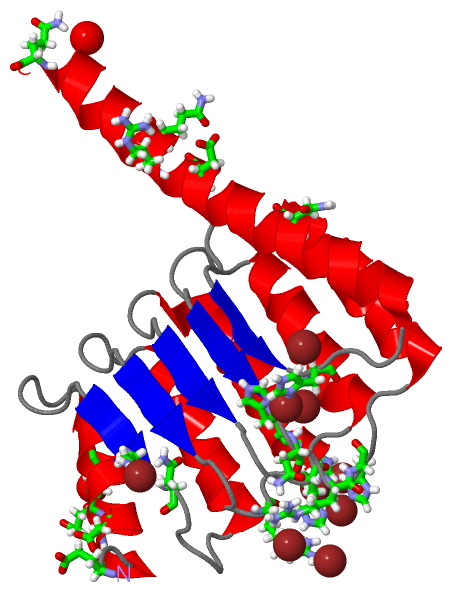
(-) Ligands, Modified Residues, Ions  (1, 9)

Asymmetric/Biological Unit (1, 9)
No.NameCountTypeFull Name
1NI9Ligand/IonNICKEL (II) ION

(-) Sites  (9, 9)

Asymmetric Unit (9, 9)
No.NameEvidenceResiduesDescription
1AC1SOFTWAREHIS A:423 , HIS A:453 , ASP A:472 , NI A:507binding site for residue NI A 501
2AC2SOFTWAREGLU A:185 , GLU A:188 , HIS A:398 , HIS A:427binding site for residue NI A 502
3AC3SOFTWAREASP A:182 , ASP A:372 , HIS A:374 , HOH A:697binding site for residue NI A 503
4AC4SOFTWAREGLU A:177 , ASP A:182 , HIS A:374 , GLN A:486 , HOH A:608binding site for residue NI A 504
5AC5SOFTWAREHIS A:398 , ARG A:426binding site for residue NI A 505
6AC6SOFTWAREGLN A:476 , HOH A:692binding site for residue NI A 506
7AC7SOFTWAREHIS A:423 , ASN A:424 , HIS A:453 , ASP A:472 , ARG A:475 , NI A:501binding site for residue NI A 507
8AC8SOFTWAREARG A:370 , HOH A:681binding site for residue NI A 508
9AC9SOFTWAREASN A:201 , ASN A:203 , ASN A:465binding site for residue NI A 509

(-) SS Bonds  (0, 0)

(no "SS Bond" information available for 4Z8G)

(-) Cis Peptide Bonds  (0, 0)

(no "Cis Peptide Bond" information available for 4Z8G)

 Sequence-Structure Mapping

(-) SAPs(SNPs)/Variants  (0, 0)

(no "SAP(SNP)/Variant" information available for 4Z8G)

(-) PROSITE Motifs  (0, 0)

(no "PROSITE Motif" information available for 4Z8G)

(-) Exons   (0, 0)

(no "Exon" information available for 4Z8G)

(-) Sequences/Alignments

Asymmetric/Biological Unit
   Reformat: Number of residues per line =  ('0' or empty: single-line sequence representation)
  Number of residues per labelling interval =   
  UniProt sequence: complete  aligned part    
   Show mapping: SCOP domains CATH domains Pfam domains Secondary structure (by author)
SAPs(SNPs) PROSITE motifs Exons
(details for a mapped element are shown in a popup box when the mouse pointer rests over it)
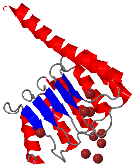
Chain A from PDB  Type:PROTEIN  Length:175
                                                                                                                                                                                                               
               SCOP domains ------------------------------------------------------------------------------------------------------------------------------------------------------------------------------- SCOP domains
               CATH domains ------------------------------------------------------------------------------------------------------------------------------------------------------------------------------- CATH domains
               Pfam domains ------------------------------------------------------------------------------------------------------------------------------------------------------------------------------- Pfam domains
         Sec.struct. author ..hhhhhhhhhhhhhh......eee.......hhhhhhhhhhhhh......eee......hhhhhhhhhhhhhhh....eee......hhhhhhhhhhhhh......eee........hhhhhhhhhhhhhh.....eee....hhhhhhhhhhhhhhhhhhhhhhhhhhhhhh. Sec.struct. author
                 SAPs(SNPs) ------------------------------------------------------------------------------------------------------------------------------------------------------------------------------- SAPs(SNPs)
                    PROSITE ------------------------------------------------------------------------------------------------------------------------------------------------------------------------------- PROSITE
                 Transcript ------------------------------------------------------------------------------------------------------------------------------------------------------------------------------- Transcript
                 4z8g A 177 EPNSTDVEETLERIKNNDPKLEEVNLNNIRNIPIPTLKAYAEALKENSYVKKFALANTRADDHVAFAIAIMLKANKTITSLNLDSNHITGKGILAIFRALLQNNTLTELRFHNQRHICGGKTEMEIAKLLKENTTLLKLGYHFELAGPRMTVTNLLSRNMDKQRQKRLQEQRQAQ 486
                                   186       196       206       216       226  ||   371       381       391       401       411       421       431       441       451       461       471       481     
                                                                              229|                                                                                                                         
                                                                               365                                                                                                                         

   Legend:   → Mismatch (orange background)
  - → Gap (green background, '-', border residues have a numbering label)
    → Modified Residue (blue background, lower-case, 'x' indicates undefined single-letter code, labelled with number + name)
  x → Chemical Group (purple background, 'x', labelled with number + name, e.g. ACE or NH2)
  extra numbering lines below/above indicate numbering irregularities and modified residue names etc., number ends below/above '|'

 Classification and Annotation

(-) SCOP Domains  (0, 0)

(no "SCOP Domain" information available for 4Z8G)

(-) CATH Domains  (0, 0)

(no "CATH Domain" information available for 4Z8G)

(-) Pfam Domains  (0, 0)

(no "Pfam Domain" information available for 4Z8G)

(-) Gene Ontology  (20, 31)

Asymmetric/Biological Unit(hide GO term definitions)

 Visualization

(-) Interactive Views

Asymmetric/Biological Unit
  Complete Structure
    Jena3D(integrated viewing of ligand, site, SAP, PROSITE, SCOP information)
    WebMol | AstexViewer[tm]@PDBe
(Java Applets, require no local installation except for Java; loading may be slow)
    STRAP
(Java WebStart application, automatic local installation, requires Java; full application with system access!)
    RasMol
(require local installation)
    Molscript (VRML)
(requires installation of a VRML viewer; select preferred view via VRML and generate a mono or stereo PDF format file)
 
  Ligands, Modified Residues, Ions
    NI  [ RasMol | Jena3D ]  +environment [ RasMol | Jena3D ]
 
  Sites
    AC1  [ RasMol ]  +environment [ RasMol ]
    AC2  [ RasMol ]  +environment [ RasMol ]
    AC3  [ RasMol ]  +environment [ RasMol ]
    AC4  [ RasMol ]  +environment [ RasMol ]
    AC5  [ RasMol ]  +environment [ RasMol ]
    AC6  [ RasMol ]  +environment [ RasMol ]
    AC7  [ RasMol ]  +environment [ RasMol ]
    AC8  [ RasMol ]  +environment [ RasMol ]
    AC9  [ RasMol ]  +environment [ RasMol ]
 
  Cis Peptide Bonds
(no "Cis Peptide Bonds" information available for 4z8g)
 

(-) Still Images

Jmol
  protein: cartoon or spacefill or dots and stick; nucleic acid: cartoon and stick; ligands: spacefill; active site: stick
Molscript
  protein, nucleic acid: cartoon; ligands: spacefill; active site: ball and stick

 Databases and Analysis Tools

(-) Databases

Access by PDB/NDB ID
  4z8g
    Family and Domain InformationProDom | SYSTERS
    General Structural InformationGlycoscienceDB | MMDB | NDB | OCA | PDB | PDBe | PDBj | PDBsum | PDBWiki | PQS | PROTEOPEDIA
    Orientation in MembranesOPM
    Protein SurfaceSURFACE
    Secondary StructureDSSP (structure derived) | HSSP (homology derived)
    Structural GenomicsGeneCensus
    Structural NeighboursCE | VAST
    Structure ClassificationCATH | Dali | SCOP
    Validation and Original DataBMRB Data View | BMRB Restraints Grid | EDS | PROCHECK | RECOORD | WHAT_CHECK
 
Access by UniProt ID/Accession number
  LMOD1_HUMAN | P29536
    Comparative Protein Structure ModelsModBase
    Genomic InformationEnsembl
    Protein-protein InteractionDIP
    Sequence, Family and Domain InformationInterPro | Pfam | SMART | UniProtKB/SwissProt
  TMOD1_HUMAN | P28289
    Comparative Protein Structure ModelsModBase
    Genomic InformationEnsembl
    Protein-protein InteractionDIP
    Sequence, Family and Domain InformationInterPro | Pfam | SMART | UniProtKB/SwissProt
 
Access by Enzyme Classificator   (EC Number)
  (no 'Enzyme Classificator' available)
    General Enzyme InformationBRENDA | EC-PDB | Enzyme | IntEnz
    PathwayKEGG | MetaCyc
 
Access by Disease Identifier   (MIM ID)
  (no 'MIM ID' available)
    Disease InformationOMIM
 
Access by GenAge ID
  (no 'GenAge ID' available)
    Age Related InformationGenAge

(-) Analysis Tools

Access by PDB/NDB ID
    Domain InformationXDom
    Interatomic Contacts of Structural UnitsCSU
    Ligand-protein ContactsLPC
    Protein CavitiescastP
    Sequence and Secondary StructurePDBCartoon
    Structure AlignmentSTRAP(Java WebStart application, automatic local installation, requires Java; full application with system access!)
    Structure and Sequence BrowserSTING
 
Access by UniProt ID/Accession number
  LMOD1_HUMAN | P29536
    Protein Disorder PredictionDisEMBL | FoldIndex | GLOBPLOT (for more information see DisProt)
  TMOD1_HUMAN | P28289
    Protein Disorder PredictionDisEMBL | FoldIndex | GLOBPLOT (for more information see DisProt)

 Related Entries

(-) Entries Sharing at Least One Protein Chain (UniProt ID)

UniProtKB/Swiss-Prot
        LMOD1_HUMAN | P295364z79 4z94
        TMOD1_HUMAN | P282894pkg 4pkh 4pki 4z94

(-) Related Entries Specified in the PDB File

4z94