Show PDB file:   
         Plain Text   HTML   (compressed file size)
QuickSearch:   
by PDB,NDB,UniProt,PROSITE Code or Search Term(s)  
(-)Asym./Biol. Unit
(-)Asym./Biol. Unit - sites
collapse expand < >
Image Asym./Biol. Unit
Asym./Biol. Unit  (Jmol Viewer)
Image Asym./Biol. Unit - sites
Asym./Biol. Unit - sites  (Jmol Viewer)

(-) Description

Title :  CRYSTAL STRUCTURE OF HUMAN ADAMTS-4 IN COMPLEX WITH INHIBITOR 5-CHLORO-N-{[(4R)-2,5-DIOXO-4-(1,3-THIAZOL-2-YL)IMIDAZOLIDIN-4-YL]METHYL}-1-BENZOFURAN-2-CARBOXAMIDE (COMPOUND 10)
 
Authors :  J. D. Durbin
Date :  02 Oct 14  (Deposition) - 10 Dec 14  (Release) - 14 Jan 15  (Revision)
Method :  X-RAY DIFFRACTION
Resolution :  1.62
Chains :  Asym./Biol. Unit :  A
Keywords :  Metalloprotease, Osteoarthritis, Inhibitor (Keyword Search: [Gene Ontology, PubMed, Web (Google))
 
Reference :  T. B. Durham, V. J. Klimkowski, C. J. Rito, J. Marimuthu, J. L. Toth, C. Liu, J. D. Durbin, S. L. Stout, L. Adams, C. Swearingen, C. Lin, M. G. Chambers, K. Thirunavukkarasu, M. R. Wiley
Identification Of Potent And Selective Hydantoin Inhibitors Of Aggrecanase-1 And Aggrecanase-2 That Are Efficacious In Both Chemical And Surgical Models Of Osteoarthritis.
J. Med. Chem. V. 57 10476 2014
PubMed-ID: 25415648  |  Reference-DOI: 10.1021/JM501522N

(-) Compounds

Molecule 1 - A DISINTEGRIN AND METALLOPROTEINASE WITH THROMBOSPONDIN MOTIFS 4
    ChainsA
    EC Number3.4.24.82
    EngineeredYES
    Expression SystemHOMO SAPIENS
    Expression System Cell LineHEK293
    Expression System Taxid9606
    FragmentUNP RESIDUES 213-439
    GeneADAMTS4, KIAA0688, UNQ769/PRO1563
    Organism CommonHUMAN
    Organism ScientificHOMO SAPIENS
    Organism Taxid9606
    SynonymADAMTS-4,ADMP-1,AGGRECANASE-1

 Structural Features

(-) Chains, Units

  1
Asymmetric/Biological Unit A

Summary Information (see also Sequences/Alignments below)
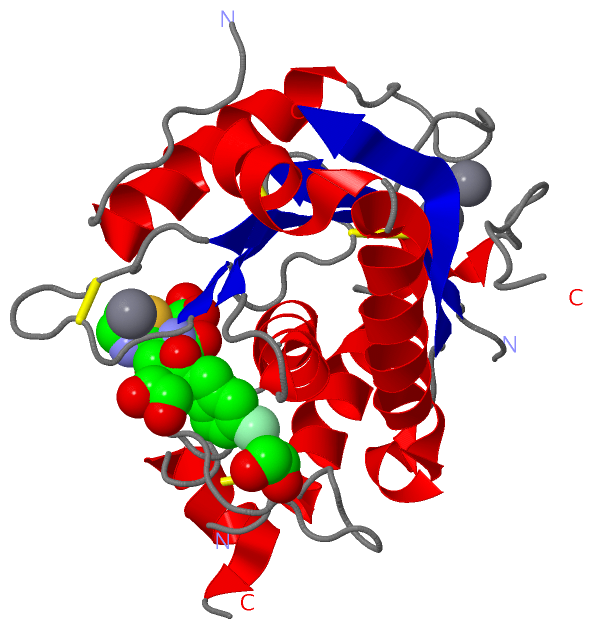
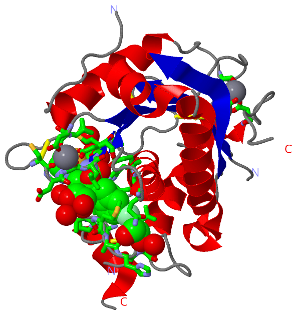
(-) Ligands, Modified Residues, Ions  (4, 8)

Asymmetric/Biological Unit (4, 8)
No.NameCountTypeFull Name
13PU1Ligand/Ion5-CHLORO-N-{[(4R)-2,5-DIOXO-4-(1,3-THIAZOL-2-YL)IMIDAZOLIDIN-4-YL]METHYL}-1-BENZOFURAN-2-CARBOXAMIDE
2CA3Ligand/IonCALCIUM ION
3EDO3Ligand/Ion1,2-ETHANEDIOL
4ZN1Ligand/IonZINC ION

(-) Sites  (8, 8)

Asymmetric Unit (8, 8)
No.NameEvidenceResiduesDescription
1AC1SOFTWAREHIS A:361 , HIS A:365 , HIS A:371 , 3PU A:508binding site for residue ZN A 501
2AC2SOFTWAREGLU A:221 , ASP A:304 , ASP A:311 , CYS A:423 , ASP A:426 , HOH A:672binding site for residue CA A 502
3AC3SOFTWAREGLU A:221 , ASP A:304 , ASP A:426 , HOH A:681 , HOH A:690binding site for residue CA A 503
4AC4SOFTWAREASP A:320 , LEU A:321 , CYS A:327 , THR A:329 , GLU A:349 , HOH A:674binding site for residue CA A 504
5AC5SOFTWAREGLN A:354 , ALA A:396 , HIS A:397 , VAL A:398 , HOH A:606binding site for residue EDO A 505
6AC6SOFTWAREMET A:332 , ALA A:333 , GLU A:362 , HIS A:365 , 3PU A:508 , HOH A:725binding site for residue EDO A 506
7AC7SOFTWAREASP A:328 , VAL A:394 , MET A:395 , 3PU A:508 , HOH A:732 , HOH A:758 , HOH A:763binding site for residue EDO A 507
8AC8SOFTWARETHR A:329 , LEU A:330 , GLY A:331 , MET A:332 , HIS A:361 , GLU A:362 , HIS A:365 , HIS A:371 , VAL A:390 , ALA A:392 , PRO A:393 , VAL A:394 , MET A:395 , ZN A:501 , EDO A:506 , EDO A:507binding site for residue 3PU A 508

(-) SS Bonds  (4, 4)

Asymmetric/Biological Unit
No.Residues
1A:293 -A:345
2A:322 -A:327
3A:339 -A:423
4A:377 -A:407

(-) Cis Peptide Bonds  (0, 0)

(no "Cis Peptide Bond" information available for 4WKE)

 Sequence-Structure Mapping

(-) SAPs(SNPs)/Variants  (0, 0)

(no "SAP(SNP)/Variant" information available for 4WKE)

(-) PROSITE Motifs  (0, 0)

(no "PROSITE Motif" information available for 4WKE)

(-) Exons   (0, 0)

(no "Exon" information available for 4WKE)

(-) Sequences/Alignments

Asymmetric/Biological Unit
   Reformat: Number of residues per line =  ('0' or empty: single-line sequence representation)
  Number of residues per labelling interval =   
  UniProt sequence: complete  aligned part    
   Show mapping: SCOP domains CATH domains Pfam domains Secondary structure (by author)
SAPs(SNPs) PROSITE motifs Exons
(details for a mapped element are shown in a popup box when the mouse pointer rests over it)
Chain A from PDB  Type:PROTEIN  Length:214
                                                                                                                                                                                                                                                      
               SCOP domains ---------------------------------------------------------------------------------------------------------------------------------------------------------------------------------------------------------------------- SCOP domains
               CATH domains ---------------------------------------------------------------------------------------------------------------------------------------------------------------------------------------------------------------------- CATH domains
               Pfam domains ---------------------------------------------------------------------------------------------------------------------------------------------------------------------------------------------------------------------- Pfam domains
         Sec.struct. author ...eeeeeeeehhhhhhhhh.hhhhhhhhhhhhhhhhhhhhhhh...eeeeeeeee........hhhhhhhhhhhhhhh.............eeeeee.............eee...........eeeee....hhhhhhhhhhhhhh......hhhhhhhhh....................hhhhhhhhhhhhhh..hhhhh.......... Sec.struct. author
                 SAPs(SNPs) ---------------------------------------------------------------------------------------------------------------------------------------------------------------------------------------------------------------------- SAPs(SNPs)
                    PROSITE ---------------------------------------------------------------------------------------------------------------------------------------------------------------------------------------------------------------------- PROSITE
                 Transcript ---------------------------------------------------------------------------------------------------------------------------------------------------------------------------------------------------------------------- Transcript
                 4wke A 216 LSRFVETLVVADDKMAAFHGAGLKRYLLTVMAAAAKAFKHPSIRNPVSLVVTRLVILEGPQVGPSAAQTLRSFCAWQRGLNTPEDSDPDHFDTAILFTRQDLCGVSTCDTLGMADVGTVCDPARSCAIVEDDGLQSAFTAAHELGHVFNMLHDNSKPCISLNGPLSRHVMAPVMAHVDPEEPWSPCSARFITDFLDNGYGHCLLDKPEAPLHLP 435
                                   225       235       245       255       265      |279       289       299       309       319       329       339       349       359       369       379    || 391       401       411       421       431    
                                                                                  272|                                                                                                        384|                                                
                                                                                   277                                                                                                         387                                                

   Legend:   → Mismatch (orange background)
  - → Gap (green background, '-', border residues have a numbering label)
    → Modified Residue (blue background, lower-case, 'x' indicates undefined single-letter code, labelled with number + name)
  x → Chemical Group (purple background, 'x', labelled with number + name, e.g. ACE or NH2)
  extra numbering lines below/above indicate numbering irregularities and modified residue names etc., number ends below/above '|'

 Classification and Annotation

(-) SCOP Domains  (0, 0)

(no "SCOP Domain" information available for 4WKE)

(-) CATH Domains  (0, 0)

(no "CATH Domain" information available for 4WKE)

(-) Pfam Domains  (0, 0)

(no "Pfam Domain" information available for 4WKE)

(-) Gene Ontology  (15, 15)

Asymmetric/Biological Unit(hide GO term definitions)

 Visualization

(-) Interactive Views

Asymmetric/Biological Unit
  Complete Structure
    Jena3D(integrated viewing of ligand, site, SAP, PROSITE, SCOP information)
    WebMol | AstexViewer[tm]@PDBe
(Java Applets, require no local installation except for Java; loading may be slow)
    STRAP
(Java WebStart application, automatic local installation, requires Java; full application with system access!)
    RasMol
(require local installation)
    Molscript (VRML)
(requires installation of a VRML viewer; select preferred view via VRML and generate a mono or stereo PDF format file)
 
  Ligands, Modified Residues, Ions
    3PU  [ RasMol | Jena3D ]  +environment [ RasMol | Jena3D ]
    CA  [ RasMol | Jena3D ]  +environment [ RasMol | Jena3D ]
    EDO  [ RasMol | Jena3D ]  +environment [ RasMol | Jena3D ]
    ZN  [ RasMol | Jena3D ]  +environment [ RasMol | Jena3D ]
 
  Sites
    AC1  [ RasMol ]  +environment [ RasMol ]
    AC2  [ RasMol ]  +environment [ RasMol ]
    AC3  [ RasMol ]  +environment [ RasMol ]
    AC4  [ RasMol ]  +environment [ RasMol ]
    AC5  [ RasMol ]  +environment [ RasMol ]
    AC6  [ RasMol ]  +environment [ RasMol ]
    AC7  [ RasMol ]  +environment [ RasMol ]
    AC8  [ RasMol ]  +environment [ RasMol ]
 
  Cis Peptide Bonds
(no "Cis Peptide Bonds" information available for 4wke)
 

(-) Still Images

Jmol
  protein: cartoon or spacefill or dots and stick; nucleic acid: cartoon and stick; ligands: spacefill; active site: stick
Molscript
  protein, nucleic acid: cartoon; ligands: spacefill; active site: ball and stick

 Databases and Analysis Tools

(-) Databases

Access by PDB/NDB ID
  4wke
    Family and Domain InformationProDom | SYSTERS
    General Structural InformationGlycoscienceDB | MMDB | NDB | OCA | PDB | PDBe | PDBj | PDBsum | PDBWiki | PQS | PROTEOPEDIA
    Orientation in MembranesOPM
    Protein SurfaceSURFACE
    Secondary StructureDSSP (structure derived) | HSSP (homology derived)
    Structural GenomicsGeneCensus
    Structural NeighboursCE | VAST
    Structure ClassificationCATH | Dali | SCOP
    Validation and Original DataBMRB Data View | BMRB Restraints Grid | EDS | PROCHECK | RECOORD | WHAT_CHECK
 
Access by UniProt ID/Accession number
  ATS4_HUMAN | O75173
    Comparative Protein Structure ModelsModBase
    Genomic InformationEnsembl
    Protein-protein InteractionDIP
    Sequence, Family and Domain InformationInterPro | Pfam | SMART | UniProtKB/SwissProt
 
Access by Enzyme Classificator   (EC Number)
  3.4.24.82
    General Enzyme InformationBRENDA | EC-PDB | Enzyme | IntEnz
    PathwayKEGG | MetaCyc
 
Access by Disease Identifier   (MIM ID)
  (no 'MIM ID' available)
    Disease InformationOMIM
 
Access by GenAge ID
  (no 'GenAge ID' available)
    Age Related InformationGenAge

(-) Analysis Tools

Access by PDB/NDB ID
    Domain InformationXDom
    Interatomic Contacts of Structural UnitsCSU
    Ligand-protein ContactsLPC
    Protein CavitiescastP
    Sequence and Secondary StructurePDBCartoon
    Structure AlignmentSTRAP(Java WebStart application, automatic local installation, requires Java; full application with system access!)
    Structure and Sequence BrowserSTING
 
Access by UniProt ID/Accession number
  ATS4_HUMAN | O75173
    Protein Disorder PredictionDisEMBL | FoldIndex | GLOBPLOT (for more information see DisProt)

 Related Entries

(-) Entries Sharing at Least One Protein Chain (UniProt ID)

UniProtKB/Swiss-Prot
        ATS4_HUMAN | O751732rjp 3b2z 4wk7 4wki

(-) Related Entries Specified in the PDB File

4wk7 4wki