Show PDB file:   
         Plain Text   HTML   (compressed file size)
QuickSearch:   
by PDB,NDB,UniProt,PROSITE Code or Search Term(s)  
(-)Asym./Biol. Unit
(-)Asym./Biol. Unit - sites
collapse expand < >
Image Asym./Biol. Unit
Asym./Biol. Unit  (Jmol Viewer)
Image Asym./Biol. Unit - sites
Asym./Biol. Unit - sites  (Jmol Viewer)

(-) Description

Title :  CRYSTAL STRUCTURE OF HCV NS3/4A PROTEASE VARIANT R155K IN COMPLEX WITH ASUNAPREVIR
 
Authors :  D. I. Soumana, A. Ali, C. A. Schiffer
Date :  20 Sep 14  (Deposition) - 08 Oct 14  (Release) - 25 Nov 15  (Revision)
Method :  X-RAY DIFFRACTION
Resolution :  1.99
Chains :  Asym./Biol. Unit :  A
Keywords :  Hcv Drug Resistant, Protease-Inhibitor Complex, Asunaprevir, Hydrolase-Hydrolase Inhibitor Complex (Keyword Search: [Gene Ontology, PubMed, Web (Google))
 
Reference :  D. I. Soumana, A. Ali, C. A. Schiffer
Structural Analysis Of Asunaprevir Resistance In Hcv Ns3/4A Protease.
Acs Chem. Biol. V. 9 2485 2014
PubMed-ID: 25243902  |  Reference-DOI: 10.1021/CB5006118

(-) Compounds

Molecule 1 - GENOME POLYPROTEIN
    ChainsA
    EngineeredYES
    Expression SystemESCHERICHIA COLI
    Expression System PlasmidPET28A
    Expression System Taxid562
    Organism ScientificHEPATITIS C VIRUS SUBTYPE 1A
    Organism Taxid31646

 Structural Features

(-) Chains, Units

  1
Asymmetric/Biological Unit A

Summary Information (see also Sequences/Alignments below)
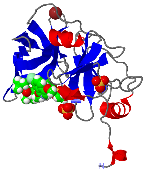
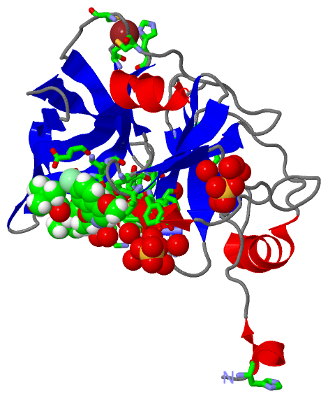
(-) Ligands, Modified Residues, Ions  (3, 4)

Asymmetric/Biological Unit (3, 4)
No.NameCountTypeFull Name
12R91Ligand/IonN-(TERT-BUTOXYCARBONYL)-3-METHYL-L-VALYL-(4R)-4-[(7-CHLORO-4-METHOXYISOQUINOLIN-1-YL)OXY]-N-{(1R,2S)-1-[(CYCLOPROPYLSULFONYL)CARBAMOYL]-2-ETHENYLCYCLOPROPYL}-L-PROLINAMIDE
2SO42Ligand/IonSULFATE ION
3ZN1Ligand/IonZINC ION

(-) Sites  (4, 4)

Asymmetric Unit (4, 4)
No.NameEvidenceResiduesDescription
1AC1SOFTWARETYR A:1006 , GLN A:1008 , TYR A:1056 , HOH A:1321 , HOH A:1354 , HOH A:1364 , HOH A:1378 , HOH A:1382 , HOH A:1430binding site for residue SO4 A 1201
2AC2SOFTWAREARG A:1109 , HIS A:1110 , HOH A:1316 , HOH A:1365 , HOH A:1374 , HOH A:1400 , HOH A:1406binding site for residue SO4 A 1202
3AC3SOFTWARECYS A:1097 , CYS A:1099 , CYS A:1145 , HIS A:1149binding site for residue ZN A 1203
4AC4SOFTWAREHIS A:982 , GLN A:1041 , PHE A:1043 , HIS A:1057 , GLY A:1058 , VAL A:1078 , ASP A:1081 , ILE A:1132 , LEU A:1135 , LYS A:1136 , GLY A:1137 , SER A:1138 , SER A:1139 , PHE A:1154 , LYS A:1155 , ALA A:1156 , ALA A:1157 , ASP A:1168 , HOH A:1388 , HOH A:1390 , HOH A:1397 , HOH A:1404 , HOH A:1411binding site for residue 2R9 A 1204

(-) SS Bonds  (0, 0)

(no "SS Bond" information available for 4WH6)

(-) Cis Peptide Bonds  (0, 0)

(no "Cis Peptide Bond" information available for 4WH6)

 Sequence-Structure Mapping

(-) SAPs(SNPs)/Variants  (0, 0)

(no "SAP(SNP)/Variant" information available for 4WH6)

(-) PROSITE Motifs  (0, 0)

(no "PROSITE Motif" information available for 4WH6)

(-) Exons   (0, 0)

(no "Exon" information available for 4WH6)

(-) Sequences/Alignments

Asymmetric/Biological Unit
   Reformat: Number of residues per line =  ('0' or empty: single-line sequence representation)
  Number of residues per labelling interval =   
  UniProt sequence: complete  aligned part    
   Show mapping: SCOP domains CATH domains Pfam domains Secondary structure (by author)
SAPs(SNPs) PROSITE motifs Exons
(details for a mapped element are shown in a popup box when the mouse pointer rests over it)
Chain A from PDB  Type:PROTEIN  Length:200
                                                                                                                                                                                                                                         
               SCOP domains -------------------------------------------------------------------------------------------------------------------------------------------------------------------------------------------------------- SCOP domains
               CATH domains -------------------------------------------------------------------------------------------------------------------------------------------------------------------------------------------------------- CATH domains
               Pfam domains -------------------------------------------------------------------------------------------------------------------------------------------------------------------------------------------------------- Pfam domains
         Sec.struct. author .hhhhh......eeeeeee......eeee..hhhhhhhhhhhh.........eeeee....eeeeeee..eeeeehhhhh..............eee....eeeee................eeeee.....eeeeee....eeeeeeeeehhhhh.....eee.....eeeeeeeeeee..eeeeeeeeehhhhhhhh. Sec.struct. author
                 SAPs(SNPs) -------------------------------------------------------------------------------------------------------------------------------------------------------------------------------------------------------- SAPs(SNPs)
                    PROSITE -------------------------------------------------------------------------------------------------------------------------------------------------------------------------------------------------------- PROSITE
                 Transcript -------------------------------------------------------------------------------------------------------------------------------------------------------------------------------------------------------- Transcript
                4wh6 A  981 SHMASMKKKGSVVIVGRINLSGDTAYAQQTRGEEGCQETSQTGRDKNQVEGEVQIVSTATQTFLATSINGVLWTVYHGAGTRTIASPKGPVTQMYTNVDKDLVGWQAPQGSRSLTPCTCGSSDLYLVTRHADVIPVRRRGDSRGSLLSPRPISYLKGSSGGPLLCPAGHAVGIFKAAVSTRGVAKAVDFIPVESLETTMR 1180
                                   990      1000      1010      1020      1030      1040      1050      1060      1070      1080      1090      1100      1110      1120      1130      1140      1150      1160      1170      1180

   Legend:   → Mismatch (orange background)
  - → Gap (green background, '-', border residues have a numbering label)
    → Modified Residue (blue background, lower-case, 'x' indicates undefined single-letter code, labelled with number + name)
  x → Chemical Group (purple background, 'x', labelled with number + name, e.g. ACE or NH2)
  extra numbering lines below/above indicate numbering irregularities and modified residue names etc., number ends below/above '|'

 Classification and Annotation

(-) SCOP Domains  (0, 0)

(no "SCOP Domain" information available for 4WH6)

(-) CATH Domains  (0, 0)

(no "CATH Domain" information available for 4WH6)

(-) Pfam Domains  (0, 0)

(no "Pfam Domain" information available for 4WH6)

(-) Gene Ontology  (31, 31)

Asymmetric/Biological Unit(hide GO term definitions)

 Visualization

(-) Interactive Views

Asymmetric/Biological Unit
  Complete Structure
    Jena3D(integrated viewing of ligand, site, SAP, PROSITE, SCOP information)
    WebMol | AstexViewer[tm]@PDBe
(Java Applets, require no local installation except for Java; loading may be slow)
    STRAP
(Java WebStart application, automatic local installation, requires Java; full application with system access!)
    RasMol
(require local installation)
    Molscript (VRML)
(requires installation of a VRML viewer; select preferred view via VRML and generate a mono or stereo PDF format file)
 
  Ligands, Modified Residues, Ions
    2R9  [ RasMol | Jena3D ]  +environment [ RasMol | Jena3D ]
    SO4  [ RasMol | Jena3D ]  +environment [ RasMol | Jena3D ]
    ZN  [ RasMol | Jena3D ]  +environment [ RasMol | Jena3D ]
 
  Sites
    AC1  [ RasMol ]  +environment [ RasMol ]
    AC2  [ RasMol ]  +environment [ RasMol ]
    AC3  [ RasMol ]  +environment [ RasMol ]
    AC4  [ RasMol ]  +environment [ RasMol ]
 
  Cis Peptide Bonds
(no "Cis Peptide Bonds" information available for 4wh6)
 

(-) Still Images

Jmol
  protein: cartoon or spacefill or dots and stick; nucleic acid: cartoon and stick; ligands: spacefill; active site: stick
Molscript
  protein, nucleic acid: cartoon; ligands: spacefill; active site: ball and stick

 Databases and Analysis Tools

(-) Databases

Access by PDB/NDB ID
  4wh6
    Family and Domain InformationProDom | SYSTERS
    General Structural InformationGlycoscienceDB | MMDB | NDB | OCA | PDB | PDBe | PDBj | PDBsum | PDBWiki | PQS | PROTEOPEDIA
    Orientation in MembranesOPM
    Protein SurfaceSURFACE
    Secondary StructureDSSP (structure derived) | HSSP (homology derived)
    Structural GenomicsGeneCensus
    Structural NeighboursCE | VAST
    Structure ClassificationCATH | Dali | SCOP
    Validation and Original DataBMRB Data View | BMRB Restraints Grid | EDS | PROCHECK | RECOORD | WHAT_CHECK
 
Access by UniProt ID/Accession number
  A8DG50_9HEPC | A8DG50
    Comparative Protein Structure ModelsModBase
    Genomic InformationEnsembl
    Protein-protein InteractionDIP
    Sequence, Family and Domain InformationInterPro | Pfam | SMART | UniProtKB/TrEMBL
 
Access by Enzyme Classificator   (EC Number)
  (no 'Enzyme Classificator' available)
    General Enzyme InformationBRENDA | EC-PDB | Enzyme | IntEnz
    PathwayKEGG | MetaCyc
 
Access by Disease Identifier   (MIM ID)
  (no 'MIM ID' available)
    Disease InformationOMIM
 
Access by GenAge ID
  (no 'GenAge ID' available)
    Age Related InformationGenAge

(-) Analysis Tools

Access by PDB/NDB ID
    Domain InformationXDom
    Interatomic Contacts of Structural UnitsCSU
    Ligand-protein ContactsLPC
    Protein CavitiescastP
    Sequence and Secondary StructurePDBCartoon
    Structure AlignmentSTRAP(Java WebStart application, automatic local installation, requires Java; full application with system access!)
    Structure and Sequence BrowserSTING
 
Access by UniProt ID/Accession number
  A8DG50_9HEPC | A8DG50
    Protein Disorder PredictionDisEMBL | FoldIndex | GLOBPLOT (for more information see DisProt)

 Related Entries

(-) Entries Sharing at Least One Protein Chain (UniProt ID)

UniProtKB/Swiss-Prot
        A8DG50_9HEPC | A8DG505voj 5vp9
UniProtKB/TrEMBL
        A8DG50_9HEPC | A8DG503m5l 3m5m 3m5n 3m5o 3rc6 3su0 3su1 3su2 3su3 3su4 3su5 3su6 3sud 3sue 3suf 3sug 3sv6 3sv7 3sv8 3sv9 4nwk 4nwl 4wh8

(-) Related Entries Specified in the PDB File

4wf8 4WF8 CONTAINS THE SAME NS3/4A PROTEASE-INHIBITOR (ASUNAPREVIR) COMPLEX. THE DIFFERENCE R155K MUTATION ID UNIQUE TO THIS STRUCTURE.