|
|
|
|
 Description
|
|
Compounds
|
||||||||||||||||||||||||||||||||||||||||||||||||||||||||||||||||||||||||||||||||||||||||||||||||||||||||||||||
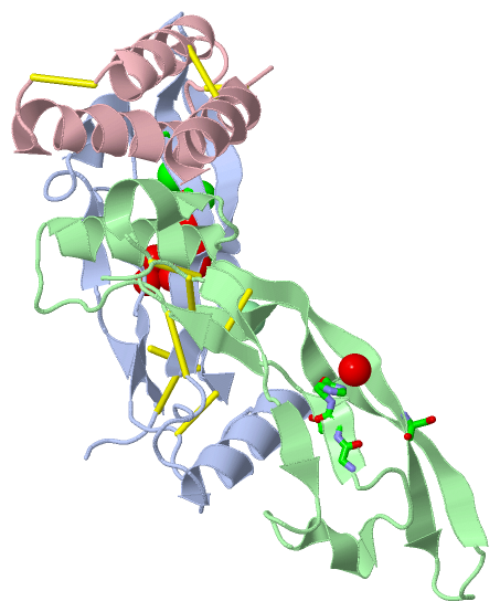
Chains, Units
Summary Information (see also Sequences/Alignments below) |
Ligands, Modified Residues, Ions (3, 3)| Asymmetric Unit (3, 3) Biological Unit 1 (2, 2) |
Sites (2, 2)
Asymmetric Unit (2, 2)
|
SS Bonds (10, 10)
Asymmetric Unit
|
||||||||||||||||||||||||||||||||||||||||||||
Cis Peptide Bonds (4, 4)
Asymmetric Unit
|
||||||||||||||||||||
SAPs(SNPs)/Variants (0, 0)| (no "SAP(SNP)/Variant" information available for 4UI0) |
PROSITE Motifs (0, 0)| (no "PROSITE Motif" information available for 4UI0) |
Exons (0, 0)| (no "Exon" information available for 4UI0) |
Sequences/Alignments
Asymmetric Unit
Chain A from PDB Type:PROTEIN Length:101
SCOP domains ----------------------------------------------------------------------------------------------------- SCOP domains
CATH domains ----------------------------------------------------------------------------------------------------- CATH domains
Pfam domains ----------------------------------------------------------------------------------------------------- Pfam domains
SAPs(SNPs) ----------------------------------------------------------------------------------------------------- SAPs(SNPs)
PROSITE ----------------------------------------------------------------------------------------------------- PROSITE
Transcript ----------------------------------------------------------------------------------------------------- Transcript
4ui0 A 293 KSSCKRHPLYVDFSDVGWNDWIVAPPGYHAFYCHGECPFPHLNSTNHAIVQTLVNSVNSKIPKACCVPTELSAISMLYLDENEKVVLKNYQDMVVEGCGCR 396
302 312 322 332| 345 355 365 375 385 395
332|
336
Chain B from PDB Type:PROTEIN Length:104
SCOP domains -------------------------------------------------------------------------------------------------------- SCOP domains
CATH domains -------------------------------------------------------------------------------------------------------- CATH domains
Pfam domains -------------------------------------------------------------------------------------------------------- Pfam domains
SAPs(SNPs) -------------------------------------------------------------------------------------------------------- SAPs(SNPs)
PROSITE -------------------------------------------------------------------------------------------------------- PROSITE
Transcript -------------------------------------------------------------------------------------------------------- Transcript
4ui0 B 293 KSSCKRHPLYVDFSDVGWNDWIVAPPGYHAFYCHGECPFPLADHLNSTNHAIVQTLVNSVNSKIPKACCVPTELSAISMLYLDENEKVVLKNYQDMVVEGCGCR 396
302 312 322 332 342 352 362 372 382 392
Chain C from PDB Type:PROTEIN Length:56
SCOP domains -------------------------------------------------------- SCOP domains
CATH domains -------------------------------------------------------- CATH domains
Pfam domains -------------------------------------------------------- Pfam domains
SAPs(SNPs) -------------------------------------------------------- SAPs(SNPs)
PROSITE -------------------------------------------------------- PROSITE
Transcript -------------------------------------------------------- Transcript
4ui0 C 53 QCRIQKCTTDFVSLTSCKALRAYAGCTQRTSKACRGNLVYHSAVLGISDLMSQRNC 121
62 || 85 95 105 115
68|
82
|
||||||||||||||||||||
SCOP Domains (0, 0)| (no "SCOP Domain" information available for 4UI0) |
CATH Domains (0, 0)| (no "CATH Domain" information available for 4UI0) |
Pfam Domains (0, 0)| (no "Pfam Domain" information available for 4UI0) |
Gene Ontology (117, 119)|
Asymmetric Unit(hide GO term definitions) |
Interactive Views
|
|||||||||||||||||||||||||||||||||||||||||||||||||||||||||||||||||||||||||||||||||||||||||||||||||||||||||||||||||||||||||||||||||||||||||||||||||||||||||||||||||||||||||||||||||||
Still Images
|
||||||||||||||||
Databases
|
||||||||||||||||||||||||||||||||||||||||||||||||||||||||||||||||||||||||||||||||||||||||||||||||||||||||||||||||||||||||||||||||||||||||||||||||||||||||||||||||||||||||||||||||||||||||||
Analysis Tools
|
||||||||||||||||||||||||||||||||||||||||||||||||||||||||||||||||||||||||
Entries Sharing at Least One Protein Chain (UniProt ID)
Related Entries Specified in the PDB File
|
|