Show PDB file:   
         Plain Text   HTML   (compressed file size)
QuickSearch:   
by PDB,NDB,UniProt,PROSITE Code or Search Term(s)  
(-)Asymmetric Unit
(-)Asym. Unit - sites
(-)Biological Unit 1
(-)Biol. Unit 1 - sites
(-)Biological Unit 2
collapse expand < >
Image Asymmetric Unit
Asymmetric Unit  (Jmol Viewer)
Image Asym. Unit - sites
Asym. Unit - sites  (Jmol Viewer)
Image Biological Unit 1
Biological Unit 1  (Jmol Viewer)
Image Biol. Unit 1 - sites
Biol. Unit 1 - sites  (Jmol Viewer)
Image Biological Unit 2
Biological Unit 2  (Jmol Viewer)

(-) Description

Title :  CFTR ASSOCIATED LIGAND (CAL) PDZ BOUND TO BIOTINYLATED PEPTIDE BT-L-ICAL36
 
Authors :  J. F. Amacher, D. R. Madden
Date :  23 Apr 14  (Deposition) - 29 Apr 15  (Release) - 29 Apr 15  (Revision)
Method :  X-RAY DIFFRACTION
Resolution :  1.45
Chains :  Asym. Unit :  A,B,C,D
Biol. Unit 1:  A,C  (1x)
Biol. Unit 2:  B,D  (1x)
Keywords :  Pdz-Peptide Complex, Transport Protein (Keyword Search: [Gene Ontology, PubMed, Web (Google))
 
Reference :  J. F. Amacher, P. R. Cushing, S. N. Cullati, S. A. Gerber, D. R. Madden
Modulator Preferences Are Critical Co-Determinants Of Pdz Network Wiring
To Be Published
PubMed: search

(-) Compounds

Molecule 1 - GOLGI-ASSOCIATED PDZ AND COILED-COIL MOTIF-CONTAINING PROTEIN
    ChainsA, B
    EngineeredYES
    Expression SystemESCHERICHIA COLI
    Expression System StrainBL21 (DE3) RIL
    Expression System Taxid562
    Expression System Vector TypePET16B
    FragmentUNP RESIDUES 284-370
    GeneGOPC, CAL, FIG
    Organism CommonHUMAN
    Organism ScientificHOMO SAPIENS
    Organism Taxid9606
    SynonymCFTR-ASSOCIATED LIGAND, FUSED IN GLIOBLASTOMA, PDZ PROTEIN INTERACTING SPECIFICALLY WITH TC10, PIST
 
Molecule 2 - BT-L-ICAL36 PEPTIDE
    ChainsC, D
    EngineeredYES
    Other DetailsPEPTIDE SYNTHESIS
    SyntheticYES

 Structural Features

(-) Chains, Units

  1234
Asymmetric Unit ABCD
Biological Unit 1 (1x)A C 
Biological Unit 2 (1x) B D

Summary Information (see also Sequences/Alignments below)

(-) Ligands, Modified Residues, Ions  (2, 4)

Asymmetric Unit (2, 4)
No.NameCountTypeFull Name
1BTN2Mod. Amino AcidBIOTIN
2DAR2Mod. Amino AcidD-ARGININE
Biological Unit 1 (2, 2)
No.NameCountTypeFull Name
1BTN1Mod. Amino AcidBIOTIN
2DAR1Mod. Amino AcidD-ARGININE
Biological Unit 2 (2, 2)
No.NameCountTypeFull Name
1BTN1Mod. Amino AcidBIOTIN
2DAR1Mod. Amino AcidD-ARGININE

(-) Sites  (2, 2)

Asymmetric Unit (2, 2)
No.NameEvidenceResiduesDescription
1AC1SOFTWAREARG A:326 , ASN B:343 , ARG B:345 , ASP B:346 , TRP C:1 , PHE C:3 , HOH C:110 , HOH C:113BINDING SITE FOR RESIDUE DAR C 2
2AC2SOFTWAREGLY A:298 , VAL B:370 , BTN C:0 , HOH C:101 , BTN D:0 , TRP D:1 , PHE D:3 , HOH D:104 , HOH D:107BINDING SITE FOR RESIDUE DAR D 2

(-) SS Bonds  (0, 0)

(no "SS Bond" information available for 4Q6S)

(-) Cis Peptide Bonds  (1, 1)

Asymmetric Unit
No.Residues
1Ala D:6 -Asn D:7

 Sequence-Structure Mapping

(-) SAPs(SNPs)/Variants  (0, 0)

(no "SAP(SNP)/Variant" information available for 4Q6S)

(-) PROSITE Motifs  (0, 0)

(no "PROSITE Motif" information available for 4Q6S)

(-) Exons   (0, 0)

(no "Exon" information available for 4Q6S)

(-) Sequences/Alignments

Asymmetric Unit
   Reformat: Number of residues per line =  ('0' or empty: single-line sequence representation)
  Number of residues per labelling interval =   
  UniProt sequence: complete  aligned part    
   Show mapping: SCOP domains CATH domains Pfam domains Secondary structure (by author)
SAPs(SNPs) PROSITE motifs Exons
(details for a mapped element are shown in a popup box when the mouse pointer rests over it)
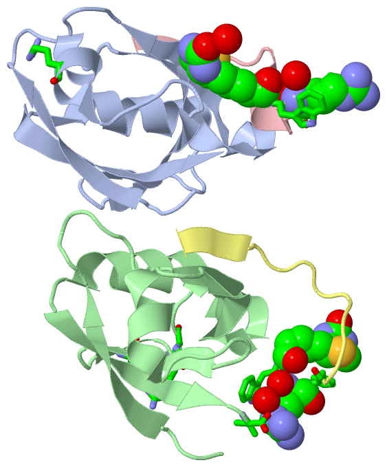
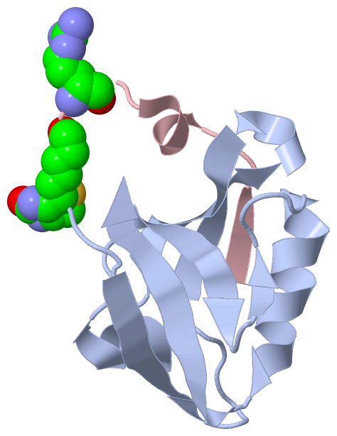
Chain A from PDB  Type:PROTEIN  Length:87
                                                                                                                       
               SCOP domains --------------------------------------------------------------------------------------- SCOP domains
               CATH domains --------------------------------------------------------------------------------------- CATH domains
               Pfam domains --------------------------------------------------------------------------------------- Pfam domains
         Sec.struct. author ...eeeeeee.......eeeeeehhhheeeeeeee...hhhhhhh.....eeeeee..ee....hhhhhhhhhhhh.eeeeeeeee. Sec.struct. author
                 SAPs(SNPs) --------------------------------------------------------------------------------------- SAPs(SNPs)
                    PROSITE --------------------------------------------------------------------------------------- PROSITE
                 Transcript --------------------------------------------------------------------------------------- Transcript
                 4q6s A 284 GPIRKVLLLKEDHEGLGISITGGKEHGVPILISEIHPGQPADRCGGLHVGDAILAVNGVNLRDTKHKEAVTILSQQRGEIEFEVVYV 370
                                   293       303       313       323       333       343       353       363       

Chain B from PDB  Type:PROTEIN  Length:87
                                                                                                                       
               SCOP domains --------------------------------------------------------------------------------------- SCOP domains
               CATH domains --------------------------------------------------------------------------------------- CATH domains
               Pfam domains --------------------------------------------------------------------------------------- Pfam domains
         Sec.struct. author ...eeeeee........eeeeeehhhheeeeeeee...hhhhhhh......eeeee..ee....hhhhhhhhhhhh..eeeeeee.. Sec.struct. author
                 SAPs(SNPs) --------------------------------------------------------------------------------------- SAPs(SNPs)
                    PROSITE --------------------------------------------------------------------------------------- PROSITE
                 Transcript --------------------------------------------------------------------------------------- Transcript
                 4q6s B 284 GPIRKVLLLKEDHEGLGISITGGKEHGVPILISEIHPGQPADRCGGLHVGDAILAVNGVNLRDTKHKEAVTILSQQRGEIEFEVVYV 370
                                   293       303       313       323       333       343       353       363       

Chain C from PDB  Type:PROTEIN  Length:16
                                                
               SCOP domains ---------------- SCOP domains
               CATH domains ---------------- CATH domains
               Pfam domains ---------------- Pfam domains
         Sec.struct. author ....hhhhh...eeee Sec.struct. author
                 SAPs(SNPs) ---------------- SAPs(SNPs)
                    PROSITE ---------------- PROSITE
                 Transcript ---------------- Transcript
                 4q6s C   0 xWxFKKANSRLPTSII  15
                            | |      9      
                            0-BTN           
                              2-DAR         

Chain D from PDB  Type:PROTEIN  Length:16
                                                
               SCOP domains ---------------- SCOP domains
               CATH domains ---------------- CATH domains
               Pfam domains ---------------- Pfam domains
         Sec.struct. author ............eeee Sec.struct. author
                 SAPs(SNPs) ---------------- SAPs(SNPs)
                    PROSITE ---------------- PROSITE
                 Transcript ---------------- Transcript
                 4q6s D   0 xWxFKKANSRLPTSII  15
                            | |      9      
                            0-BTN           
                              2-DAR         

   Legend:   → Mismatch (orange background)
  - → Gap (green background, '-', border residues have a numbering label)
    → Modified Residue (blue background, lower-case, 'x' indicates undefined single-letter code, labelled with number + name)
  x → Chemical Group (purple background, 'x', labelled with number + name, e.g. ACE or NH2)
  extra numbering lines below/above indicate numbering irregularities and modified residue names etc., number ends below/above '|'

 Classification and Annotation

(-) SCOP Domains  (0, 0)

(no "SCOP Domain" information available for 4Q6S)

(-) CATH Domains  (0, 0)

(no "CATH Domain" information available for 4Q6S)

(-) Pfam Domains  (0, 0)

(no "Pfam Domain" information available for 4Q6S)

(-) Gene Ontology  (24, 24)

Asymmetric Unit(hide GO term definitions)

 Visualization

(-) Interactive Views

Asymmetric Unit
  Complete Structure
    Jena3D(integrated viewing of ligand, site, SAP, PROSITE, SCOP information)
    WebMol | AstexViewer[tm]@PDBe
(Java Applets, require no local installation except for Java; loading may be slow)
    STRAP
(Java WebStart application, automatic local installation, requires Java; full application with system access!)
    RasMol
(require local installation)
    Molscript (VRML)
(requires installation of a VRML viewer; select preferred view via VRML and generate a mono or stereo PDF format file)
 
  Ligands, Modified Residues, Ions
    BTN  [ RasMol | Jena3D ]  +environment [ RasMol | Jena3D ]
    DAR  [ RasMol | Jena3D ]  +environment [ RasMol | Jena3D ]
 
  Sites
    AC1  [ RasMol ]  +environment [ RasMol ]
    AC2  [ RasMol ]  +environment [ RasMol ]
 
  Cis Peptide Bonds
    Ala D:6 - Asn D:7   [ RasMol ]  
 
Biological Units
  Complete Structure
    Biological Unit 1  [ Jena3D ]
    Biological Unit 2  [ Jena3D ]

(-) Still Images

Jmol
  protein: cartoon or spacefill or dots and stick; nucleic acid: cartoon and stick; ligands: spacefill; active site: stick
Molscript
  protein, nucleic acid: cartoon; ligands: spacefill; active site: ball and stick

 Databases and Analysis Tools

(-) Databases

Access by PDB/NDB ID
  4q6s
    Family and Domain InformationProDom | SYSTERS
    General Structural InformationGlycoscienceDB | MMDB | NDB | OCA | PDB | PDBe | PDBj | PDBsum | PDBWiki | PQS | PROTEOPEDIA
    Orientation in MembranesOPM
    Protein SurfaceSURFACE
    Secondary StructureDSSP (structure derived) | HSSP (homology derived)
    Structural GenomicsGeneCensus
    Structural NeighboursCE | VAST
    Structure ClassificationCATH | Dali | SCOP
    Validation and Original DataBMRB Data View | BMRB Restraints Grid | EDS | PROCHECK | RECOORD | WHAT_CHECK
 
Access by UniProt ID/Accession number
  GOPC_HUMAN | Q9HD26
    Comparative Protein Structure ModelsModBase
    Genomic InformationEnsembl
    Protein-protein InteractionDIP
    Sequence, Family and Domain InformationInterPro | Pfam | SMART | UniProtKB/SwissProt
 
Access by Enzyme Classificator   (EC Number)
  (no 'Enzyme Classificator' available)
    General Enzyme InformationBRENDA | EC-PDB | Enzyme | IntEnz
    PathwayKEGG | MetaCyc
 
Access by Disease Identifier   (MIM ID)
  (no 'MIM ID' available)
    Disease InformationOMIM
 
Access by GenAge ID
  (no 'GenAge ID' available)
    Age Related InformationGenAge

(-) Analysis Tools

Access by PDB/NDB ID
    Domain InformationXDom
    Interatomic Contacts of Structural UnitsCSU
    Ligand-protein ContactsLPC
    Protein CavitiescastP
    Sequence and Secondary StructurePDBCartoon
    Structure AlignmentSTRAP(Java WebStart application, automatic local installation, requires Java; full application with system access!)
    Structure and Sequence BrowserSTING
 
Access by UniProt ID/Accession number
  GOPC_HUMAN | Q9HD26
    Protein Disorder PredictionDisEMBL | FoldIndex | GLOBPLOT (for more information see DisProt)

 Related Entries

(-) Entries Sharing at Least One Protein Chain (UniProt ID)

UniProtKB/Swiss-Prot
        GOPC_HUMAN | Q9HD262dc2 2lob 4e34 4e35 4joe 4jof 4jog 4joh 4joj 4jok 4jop 4jor 4k6y 4k72 4k75 4k76 4k78 4nmo 4nmp 4nmq 4nmr 4nms 4nmt 4nmv 4q6h

(-) Related Entries Specified in the PDB File

4jof CAL PDZ BOUND TO L-ICAL36