Show PDB file:   
         Plain Text   HTML   (compressed file size)
QuickSearch:   
by PDB,NDB,UniProt,PROSITE Code or Search Term(s)  
(-)Asymmetric Unit
(-)Asym. Unit - sites
(-)Biological Unit 1
(-)Biol. Unit 1 - sites
(-)Biological Unit 2
(-)Biological Unit 3
collapse expand < >
Image Asymmetric Unit
Asymmetric Unit  (Jmol Viewer)
Image Asym. Unit - sites
Asym. Unit - sites  (Jmol Viewer)
Image Biological Unit 1
Biological Unit 1  (Jmol Viewer)
Image Biol. Unit 1 - sites
Biol. Unit 1 - sites  (Jmol Viewer)
Image Biological Unit 2
Biological Unit 2  (Jmol Viewer)
Image Biological Unit 3
Biological Unit 3  (Jmol Viewer)

(-) Description

Title :  CRYSTAL STRUCTURE OF THE C1QTNF5 GLOBULAR DOMAIN IN SPACE GROUP P63
 
Authors :  X. Tu, K. Palczewski
Date :  15 Nov 13  (Deposition) - 26 Mar 14  (Release) - 23 Apr 14  (Revision)
Method :  X-RAY DIFFRACTION
Resolution :  1.42
Chains :  Asym. Unit :  A,B,C
Biol. Unit 1:  A  (3x)
Biol. Unit 2:  B  (3x)
Biol. Unit 3:  C  (3x)
Keywords :  L-Ormd, Late Onset Retinal Macular Degeneration, S163R, 10-Strand Jelly-Roll Fold, Cellular Adhesion, Cell Adhesion (Keyword Search: [Gene Ontology, PubMed, Web (Google))
 
Reference :  X. Tu, K. Palczewski
The Macular Degeneration-Linked C1Qtnf5 (S163) Mutation Causes Higher-Order Structural Rearrangements.
J. Struct. Biol. V. 186 86 2014
PubMed-ID: 24531000  |  Reference-DOI: 10.1016/J.JSB.2014.02.001

(-) Compounds

Molecule 1 - COMPLEMENT C1Q TUMOR NECROSIS FACTOR-RELATED PROTEIN 5
    ChainsA, B, C
    EngineeredYES
    Expression SystemESCHERICHIA COLI
    Expression System Taxid562
    FragmentGLOBULAR DOMAIN (UNP RESIDUES 103-243)
    GeneC1QTNF5, CTRP5, UNQ303/PRO344
    Organism CommonHUMAN
    Organism ScientificHOMO SAPIENS
    Organism Taxid9606

 Structural Features

(-) Chains, Units

  123
Asymmetric Unit ABC
Biological Unit 1 (3x)A  
Biological Unit 2 (3x) B 
Biological Unit 3 (3x)  C

Summary Information (see also Sequences/Alignments below)

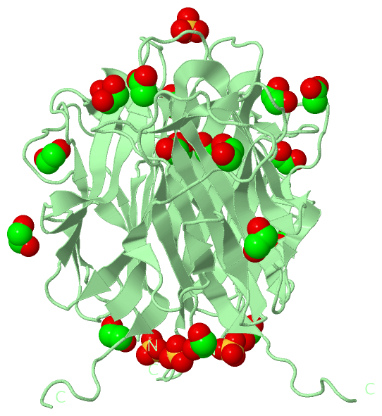
(-) Ligands, Modified Residues, Ions  (3, 23)

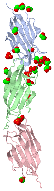
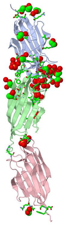
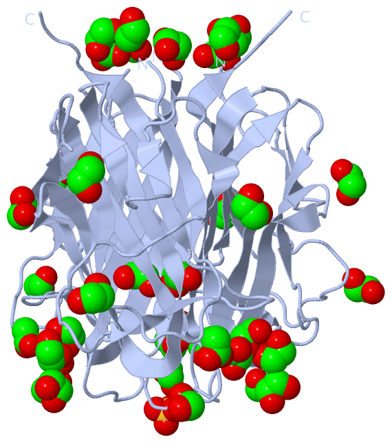
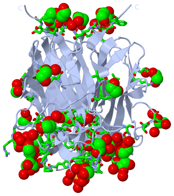
Asymmetric Unit (3, 23)
No.NameCountTypeFull Name
1ACT2Ligand/IonACETATE ION
2EDO18Ligand/Ion1,2-ETHANEDIOL
3SO43Ligand/IonSULFATE ION
Biological Unit 1 (3, 42)
No.NameCountTypeFull Name
1ACT3Ligand/IonACETATE ION
2EDO36Ligand/Ion1,2-ETHANEDIOL
3SO43Ligand/IonSULFATE ION
Biological Unit 2 (3, 24)
No.NameCountTypeFull Name
1ACT3Ligand/IonACETATE ION
2EDO15Ligand/Ion1,2-ETHANEDIOL
3SO46Ligand/IonSULFATE ION
Biological Unit 3 (1, 3)
No.NameCountTypeFull Name
1ACT-1Ligand/IonACETATE ION
2EDO3Ligand/Ion1,2-ETHANEDIOL
3SO4-1Ligand/IonSULFATE ION

(-) Sites  (23, 23)

Asymmetric Unit (23, 23)
No.NameEvidenceResiduesDescription
01AC1SOFTWAREALA A:157 , SER A:190 , LEU A:191 , SER A:192 , HOH A:514BINDING SITE FOR RESIDUE EDO A 301
02AC2SOFTWAREPHE A:179 , PRO A:188 , EDO A:305 , EDO A:309 , HOH A:416BINDING SITE FOR RESIDUE EDO A 302
03AC3SOFTWARELYS A:187 , EDO A:305 , HOH A:459 , HOH A:520 , TYR B:214 , EDO B:301BINDING SITE FOR RESIDUE EDO A 303
04AC4SOFTWAREPRO A:118 , SER A:119 , ASP A:120 , HOH A:460 , HOH A:470BINDING SITE FOR RESIDUE EDO A 304
05AC5SOFTWARETYR A:214 , EDO A:302 , EDO A:303 , HOH A:517 , LYS B:187BINDING SITE FOR RESIDUE EDO A 305
06AC6SOFTWARELYS A:142 , GLN A:204 , HOH A:458BINDING SITE FOR RESIDUE EDO A 306
07AC7SOFTWAREALA A:102 , TRP A:236 , SER C:234 , ASP C:235 , TRP C:236BINDING SITE FOR RESIDUE EDO A 307
08AC8SOFTWAREALA A:102 , ARG A:103 , HOH A:489 , EDO C:301BINDING SITE FOR RESIDUE EDO A 308
09AC9SOFTWARETYR A:160 , PRO A:186 , PRO A:188 , TYR A:218 , EDO A:302BINDING SITE FOR RESIDUE EDO A 309
10BC1SOFTWARELYS A:109 , VAL A:128BINDING SITE FOR RESIDUE EDO A 310
11BC2SOFTWARESER A:234 , ASP A:235 , TRP A:236 , ALA C:102 , TRP C:236BINDING SITE FOR RESIDUE EDO A 311
12BC3SOFTWAREARG A:161 , HOH A:421 , ARG B:161 , TYR B:218 , HOH B:512BINDING SITE FOR RESIDUE EDO A 312
13BC4SOFTWARETRP A:185 , HOH A:427 , HOH A:429 , HOH A:432BINDING SITE FOR RESIDUE SO4 A 313
14BC5SOFTWARESER A:113 , ARG A:161 , TYR A:218 , HOH A:526 , HOH A:533 , ARG B:114BINDING SITE FOR RESIDUE ACT A 314
15BC6SOFTWAREEDO A:303 , HOH B:420 , HOH B:516BINDING SITE FOR RESIDUE EDO B 301
16BC7SOFTWAREALA B:102 , ARG B:103BINDING SITE FOR RESIDUE EDO B 302
17BC8SOFTWAREALA B:157 , SER B:190 , LEU B:191 , SER B:192 , HOH B:503BINDING SITE FOR RESIDUE EDO B 303
18BC9SOFTWAREPRO B:118 , ALA B:121 , PRO B:122 , LEU B:123 , PRO B:124 , HOH B:414 , HOH B:470 , HOH B:493BINDING SITE FOR RESIDUE EDO B 304
19CC1SOFTWARELYS B:142 , GLN B:204 , HOH B:457BINDING SITE FOR RESIDUE EDO B 305
20CC2SOFTWARETRP B:185 , HOH B:427 , HOH B:429 , HOH B:435BINDING SITE FOR RESIDUE SO4 B 306
21CC3SOFTWAREALA B:102 , ARG B:103 , ARG C:114BINDING SITE FOR RESIDUE SO4 B 307
22CC4SOFTWAREARG A:114 , SER B:113 , ARG B:161 , TYR B:218 , HOH B:525 , HOH B:526BINDING SITE FOR RESIDUE ACT B 308
23CC5SOFTWAREEDO A:308 , ALA C:102 , ARG C:103 , HOH C:410BINDING SITE FOR RESIDUE EDO C 301

(-) SS Bonds  (0, 0)

(no "SS Bond" information available for 4NN0)

(-) Cis Peptide Bonds  (0, 0)

(no "Cis Peptide Bond" information available for 4NN0)

 Sequence-Structure Mapping

(-) SAPs(SNPs)/Variants  (0, 0)

(no "SAP(SNP)/Variant" information available for 4NN0)

(-) PROSITE Motifs  (0, 0)

(no "PROSITE Motif" information available for 4NN0)

(-) Exons   (0, 0)

(no "Exon" information available for 4NN0)

(-) Sequences/Alignments

Asymmetric Unit
   Reformat: Number of residues per line =  ('0' or empty: single-line sequence representation)
  Number of residues per labelling interval =   
  UniProt sequence: complete  aligned part    
   Show mapping: SCOP domains CATH domains Pfam domains Secondary structure (by author)
SAPs(SNPs) PROSITE motifs Exons
(details for a mapped element are shown in a popup box when the mouse pointer rests over it)
Chain A from PDB  Type:PROTEIN  Length:137
                                                                                                                                                                         
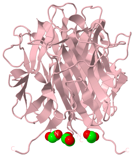
               SCOP domains d4nn0a_ A: automated matches                                                                                                              SCOP domains
               CATH domains ----------------------------------------------------------------------------------------------------------------------------------------- CATH domains
               Pfam domains ----------------------------------------------------------------------------------------------------------------------------------------- Pfam domains
         Sec.struct. author ...eeeee.................eeee.....ee....eee....eeeeeeeeeeeeeeeeeeeeee..eeeeeeee........eeeeeeeeeee....eeeee......eeee.......eeeeeeeee.... Sec.struct. author
                 SAPs(SNPs) ----------------------------------------------------------------------------------------------------------------------------------------- SAPs(SNPs)
                    PROSITE ----------------------------------------------------------------------------------------------------------------------------------------- PROSITE
                 Transcript ----------------------------------------------------------------------------------------------------------------------------------------- Transcript
                 4nn0 A 102 ARSAFSAKRSESRVPPPSDAPLPFDRVLVNEQGHYDAVTGKFTCQVPGVYYFAVHATVYRASLQFDLVKNGESIASFFQFFGGWPKPASLSGGAMVRLEPEDQVWVQVGVGDYIGIYASIKTDSTFSGFLVYSDWHS 238
                                   111       121       131       141       151       161       171       181       191       201       211       221       231       

Chain B from PDB  Type:PROTEIN  Length:141
                                                                                                                                                                             
               SCOP domains d4nn0b_ B: automated matches                                                                                                                  SCOP domains
               CATH domains --------------------------------------------------------------------------------------------------------------------------------------------- CATH domains
               Pfam domains --------------------------------------------------------------------------------------------------------------------------------------------- Pfam domains
         Sec.struct. author ...eeeee.................eeee.....ee....eee....eeeeeeeeeeeeeeeeeeeeee..eeeeeeee........eeeeeeeeeee....eeeee......eeee.......eeeeeeeee........ Sec.struct. author
                 SAPs(SNPs) --------------------------------------------------------------------------------------------------------------------------------------------- SAPs(SNPs)
                    PROSITE --------------------------------------------------------------------------------------------------------------------------------------------- PROSITE
                 Transcript --------------------------------------------------------------------------------------------------------------------------------------------- Transcript
                 4nn0 B 102 ARSAFSAKRSESRVPPPSDAPLPFDRVLVNEQGHYDAVTGKFTCQVPGVYYFAVHATVYRASLQFDLVKNGESIASFFQFFGGWPKPASLSGGAMVRLEPEDQVWVQVGVGDYIGIYASIKTDSTFSGFLVYSDWHSSPVF 242
                                   111       121       131       141       151       161       171       181       191       201       211       221       231       241 

Chain C from PDB  Type:PROTEIN  Length:138
                                                                                                                                                                          
               SCOP domains d4nn0c_ C: automated matches                                                                                                               SCOP domains
               CATH domains ------------------------------------------------------------------------------------------------------------------------------------------ CATH domains
               Pfam domains ------------------------------------------------------------------------------------------------------------------------------------------ Pfam domains
         Sec.struct. author ...eeeee.................eeee............ee....eeeeeeeeeeeeeeeeeeeeee..eeeeeeee........eeeeeeeeeee....eeeee......eeee.......eeeeeeeee..... Sec.struct. author
                 SAPs(SNPs) ------------------------------------------------------------------------------------------------------------------------------------------ SAPs(SNPs)
                    PROSITE ------------------------------------------------------------------------------------------------------------------------------------------ PROSITE
                 Transcript ------------------------------------------------------------------------------------------------------------------------------------------ Transcript
                 4nn0 C 102 ARSAFSAKRSESRVPPPSDAPLPFDRVLVNEQGHYDAVTGKFTCQVPGVYYFAVHATVYRASLQFDLVKNGESIASFFQFFGGWPKPASLSGGAMVRLEPEDQVWVQVGVGDYIGIYASIKTDSTFSGFLVYSDWHSS 239
                                   111       121       131       141       151       161       171       181       191       201       211       221       231        

   Legend:   → Mismatch (orange background)
  - → Gap (green background, '-', border residues have a numbering label)
    → Modified Residue (blue background, lower-case, 'x' indicates undefined single-letter code, labelled with number + name)
  x → Chemical Group (purple background, 'x', labelled with number + name, e.g. ACE or NH2)
  extra numbering lines below/above indicate numbering irregularities and modified residue names etc., number ends below/above '|'

 Classification and Annotation

(-) SCOP Domains  (1, 3)

Asymmetric Unit

(-) CATH Domains  (0, 0)

(no "CATH Domain" information available for 4NN0)

(-) Pfam Domains  (0, 0)

(no "Pfam Domain" information available for 4NN0)

(-) Gene Ontology  (3, 3)

Asymmetric Unit(hide GO term definitions)

 Visualization

(-) Interactive Views

Asymmetric Unit
  Complete Structure
    Jena3D(integrated viewing of ligand, site, SAP, PROSITE, SCOP information)
    WebMol | AstexViewer[tm]@PDBe
(Java Applets, require no local installation except for Java; loading may be slow)
    STRAP
(Java WebStart application, automatic local installation, requires Java; full application with system access!)
    RasMol
(require local installation)
    Molscript (VRML)
(requires installation of a VRML viewer; select preferred view via VRML and generate a mono or stereo PDF format file)
 
  Ligands, Modified Residues, Ions
    ACT  [ RasMol | Jena3D ]  +environment [ RasMol | Jena3D ]
    EDO  [ RasMol | Jena3D ]  +environment [ RasMol | Jena3D ]
    SO4  [ RasMol | Jena3D ]  +environment [ RasMol | Jena3D ]
 
  Sites
    AC1  [ RasMol ]  +environment [ RasMol ]
    AC2  [ RasMol ]  +environment [ RasMol ]
    AC3  [ RasMol ]  +environment [ RasMol ]
    AC4  [ RasMol ]  +environment [ RasMol ]
    AC5  [ RasMol ]  +environment [ RasMol ]
    AC6  [ RasMol ]  +environment [ RasMol ]
    AC7  [ RasMol ]  +environment [ RasMol ]
    AC8  [ RasMol ]  +environment [ RasMol ]
    AC9  [ RasMol ]  +environment [ RasMol ]
    BC1  [ RasMol ]  +environment [ RasMol ]
    BC2  [ RasMol ]  +environment [ RasMol ]
    BC3  [ RasMol ]  +environment [ RasMol ]
    BC4  [ RasMol ]  +environment [ RasMol ]
    BC5  [ RasMol ]  +environment [ RasMol ]
    BC6  [ RasMol ]  +environment [ RasMol ]
    BC7  [ RasMol ]  +environment [ RasMol ]
    BC8  [ RasMol ]  +environment [ RasMol ]
    BC9  [ RasMol ]  +environment [ RasMol ]
    CC1  [ RasMol ]  +environment [ RasMol ]
    CC2  [ RasMol ]  +environment [ RasMol ]
    CC3  [ RasMol ]  +environment [ RasMol ]
    CC4  [ RasMol ]  +environment [ RasMol ]
    CC5  [ RasMol ]  +environment [ RasMol ]
 
  Cis Peptide Bonds
(no "Cis Peptide Bonds" information available for 4nn0)
 
Biological Units
  Complete Structure
    Biological Unit 1  [ Jena3D ]
    Biological Unit 2  [ Jena3D ]
    Biological Unit 3  [ Jena3D ]

(-) Still Images

Jmol
  protein: cartoon or spacefill or dots and stick; nucleic acid: cartoon and stick; ligands: spacefill; active site: stick
Molscript
  protein, nucleic acid: cartoon; ligands: spacefill; active site: ball and stick

 Databases and Analysis Tools

(-) Databases

Access by PDB/NDB ID
  4nn0
    Family and Domain InformationProDom | SYSTERS
    General Structural InformationGlycoscienceDB | MMDB | NDB | OCA | PDB | PDBe | PDBj | PDBsum | PDBWiki | PQS | PROTEOPEDIA
    Orientation in MembranesOPM
    Protein SurfaceSURFACE
    Secondary StructureDSSP (structure derived) | HSSP (homology derived)
    Structural GenomicsGeneCensus
    Structural NeighboursCE | VAST
    Structure ClassificationCATH | Dali | SCOP
    Validation and Original DataBMRB Data View | BMRB Restraints Grid | EDS | PROCHECK | RECOORD | WHAT_CHECK
 
Access by UniProt ID/Accession number
  C1QT5_HUMAN | Q9BXJ0
    Comparative Protein Structure ModelsModBase
    Genomic InformationEnsembl
    Protein-protein InteractionDIP
    Sequence, Family and Domain InformationInterPro | Pfam | SMART | UniProtKB/SwissProt
 
Access by Enzyme Classificator   (EC Number)
  (no 'Enzyme Classificator' available)
    General Enzyme InformationBRENDA | EC-PDB | Enzyme | IntEnz
    PathwayKEGG | MetaCyc
 
Access by Disease Identifier   (MIM ID)
  (no 'MIM ID' available)
    Disease InformationOMIM
 
Access by GenAge ID
  (no 'GenAge ID' available)
    Age Related InformationGenAge

(-) Analysis Tools

Access by PDB/NDB ID
    Domain InformationXDom
    Interatomic Contacts of Structural UnitsCSU
    Ligand-protein ContactsLPC
    Protein CavitiescastP
    Sequence and Secondary StructurePDBCartoon
    Structure AlignmentSTRAP(Java WebStart application, automatic local installation, requires Java; full application with system access!)
    Structure and Sequence BrowserSTING
 
Access by UniProt ID/Accession number
  C1QT5_HUMAN | Q9BXJ0
    Protein Disorder PredictionDisEMBL | FoldIndex | GLOBPLOT (for more information see DisProt)

 Related Entries

(-) Entries Sharing at Least One Protein Chain (UniProt ID)

UniProtKB/Swiss-Prot
        C1QT5_HUMAN | Q9BXJ04f3j

(-) Related Entries Specified in the PDB File

4f3j CRYSTAL STRUCTURE OF C1QTNF5 GLOBULAR DOMAIN IN SPACE GROUP OF H3