|
|
|
|
 Description
|
|
Compounds
|
||||||||||||||||||||||||||||||||||||||||
Chains, Units
Summary Information (see also Sequences/Alignments below) |
Ligands, Modified Residues, Ions (0, 0)| (no "Ligand,Modified Residues,Ions" information available for 4F3J) |
Sites (0, 0)| (no "Site" information available for 4F3J) |
SS Bonds (0, 0)| (no "SS Bond" information available for 4F3J) |
Cis Peptide Bonds (0, 0)| (no "Cis Peptide Bond" information available for 4F3J) |
SAPs(SNPs)/Variants (0, 0)| (no "SAP(SNP)/Variant" information available for 4F3J) |
PROSITE Motifs (0, 0)| (no "PROSITE Motif" information available for 4F3J) |
Exons (0, 0)| (no "Exon" information available for 4F3J) |
Sequences/Alignments
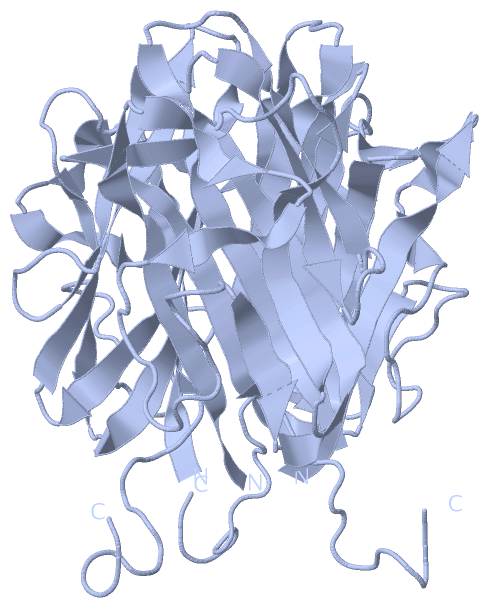
Asymmetric Unit
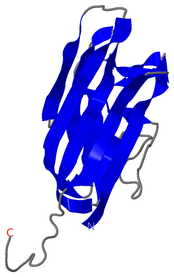
Chain A from PDB Type:PROTEIN Length:143
SCOP domains d4f3ja_ A: automated matches SCOP domains
CATH domains ----------------------------------------------------------------------------------------------------------------------------------------------- CATH domains
Pfam domains ----------------------------------------------------------------------------------------------------------------------------------------------- Pfam domains
SAPs(SNPs) ----------------------------------------------------------------------------------------------------------------------------------------------- SAPs(SNPs)
PROSITE ----------------------------------------------------------------------------------------------------------------------------------------------- PROSITE
Transcript ----------------------------------------------------------------------------------------------------------------------------------------------- Transcript
4f3j A 102 ARSAFSAKRSESRVPPPSDAPLPFDRVLVNEQGHYDAVTGKFTCQVPGVYYFAVHATVYRASLQFDLVKNGESIASFFQFFGGWPKPASLSGGAMVRLEPEDQVWVQVGVGDYIGIYASIKTDSTFSGFLVYSDWHSSPVFAH 244
111 121 131 141 151 161 171 181 191 201 211 221 231 241
|
||||||||||||||||||||
SCOP Domains (1, 1)
Asymmetric Unit
 |
CATH Domains (0, 0)| (no "CATH Domain" information available for 4F3J) |
Pfam Domains (0, 0)| (no "Pfam Domain" information available for 4F3J) |
Gene Ontology (3, 3)|
Asymmetric Unit(hide GO term definitions) |
Interactive Views
|
||||||||||||||||||||||||||||||||||||||||||||||||||||||||||||||||||||||||||||||||||||||||||||||||||||||||||||||||||||||||||||||||||||||
Still Images
|
||||||||||||||||
Databases
|
||||||||||||||||||||||||||||||||||||||||||||||||||||||||||||||||||||||||||||||||||||||||||||||||||||||||||||||||||||||||||||||||||||||||||||||||||||||||||||||||
Analysis Tools
|
|||||||||||||||||||||||||||||||||||||||||||||||||||||||||||||
Entries Sharing at Least One Protein Chain (UniProt ID)
Related Entries Specified in the PDB File
|
|