Show PDB file:   
         Plain Text   HTML   (compressed file size)
QuickSearch:   
by PDB,NDB,UniProt,PROSITE Code or Search Term(s)  
(-)Asym./Biol. Unit
(-)Asym./Biol. Unit - sites
collapse expand < >
Image Asym./Biol. Unit
Asym./Biol. Unit  (Jmol Viewer)
Image Asym./Biol. Unit - sites
Asym./Biol. Unit - sites  (Jmol Viewer)

(-) Description

Title :  CRYSTAL STRUCTURE OF THE JCV LARGE T-ANTIGEN ORIGIN BINDING DOMAIN
 
Authors :  G. Meinke, A. Bohm, P. Bullock
Date :  23 Oct 13  (Deposition) - 05 Mar 14  (Release) - 02 Apr 14  (Revision)
Method :  X-RAY DIFFRACTION
Resolution :  1.31
Chains :  Asym./Biol. Unit :  A
Keywords :  Origin Binding Domain, Dna Replication, Binds Dna, Hydrolase (Keyword Search: [Gene Ontology, PubMed, Web (Google))
 
Reference :  G. Meinke, P. J. Phelan, R. Kalekar, J. Shin, J. Archambault, A. Bohm, P. A. Bullock
Insights Into The Initiation Of Jc Virus Dna Replication Derived From The Crystal Structure Of The T-Antigen Origin Binding Domain.
Plos Pathog. V. 10 03966 2014
PubMed-ID: 24586168  |  Reference-DOI: 10.1371/JOURNAL.PPAT.1003966

(-) Compounds

Molecule 1 - LARGE T ANTIGEN
    ChainsA
    EC Number3.6.4.-
    EngineeredYES
    Expression SystemESCHERICHIA COLI
    Expression System PlasmidPGEX
    Expression System StrainBL21
    Expression System Taxid511693
    Expression System Vector TypePLASMID
    FragmentORIGIN BINDING DOMAIN
    GeneLARGE T ANTIGEN
    Organism CommonJCPYV
    Organism ScientificJC POLYOMAVIRUS
    Organism Taxid10632
    SynonymLT, LT-AG

 Structural Features

(-) Chains, Units

  1
Asymmetric/Biological Unit A

Summary Information (see also Sequences/Alignments below)

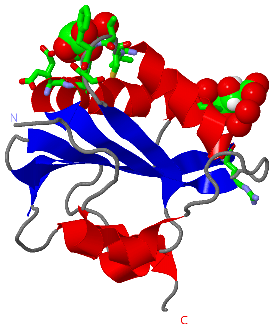
(-) Ligands, Modified Residues, Ions  (1, 2)

Asymmetric/Biological Unit (1, 2)
No.NameCountTypeFull Name
1TLA2Ligand/IonL(+)-TARTARIC ACID

(-) Sites  (2, 2)

Asymmetric Unit (2, 2)
No.NameEvidenceResiduesDescription
1AC1SOFTWAREGLU A:167 , LYS A:168 , LEU A:216 , CYS A:217 , THR A:218 , PHE A:219 , SER A:220 , HOH A:591BINDING SITE FOR RESIDUE TLA A 301
2AC2SOFTWAREHIS A:202 , ARG A:203 , HIS A:204 , ALA A:208 , HOH A:409 , HOH A:418 , HOH A:437 , HOH A:487BINDING SITE FOR RESIDUE TLA A 302

(-) SS Bonds  (0, 0)

(no "SS Bond" information available for 4NBP)

(-) Cis Peptide Bonds  (1, 1)

Asymmetric/Biological Unit
No.Residues
1Gln A:240 -Pro A:241

 Sequence-Structure Mapping

(-) SAPs(SNPs)/Variants  (0, 0)

(no "SAP(SNP)/Variant" information available for 4NBP)

(-) PROSITE Motifs  (0, 0)

(no "PROSITE Motif" information available for 4NBP)

(-) Exons   (0, 0)

(no "Exon" information available for 4NBP)

(-) Sequences/Alignments

Asymmetric/Biological Unit
   Reformat: Number of residues per line =  ('0' or empty: single-line sequence representation)
  Number of residues per labelling interval =   
  UniProt sequence: complete  aligned part    
   Show mapping: SCOP domains CATH domains Pfam domains Secondary structure (by author)
SAPs(SNPs) PROSITE motifs Exons
(details for a mapped element are shown in a popup box when the mouse pointer rests over it)
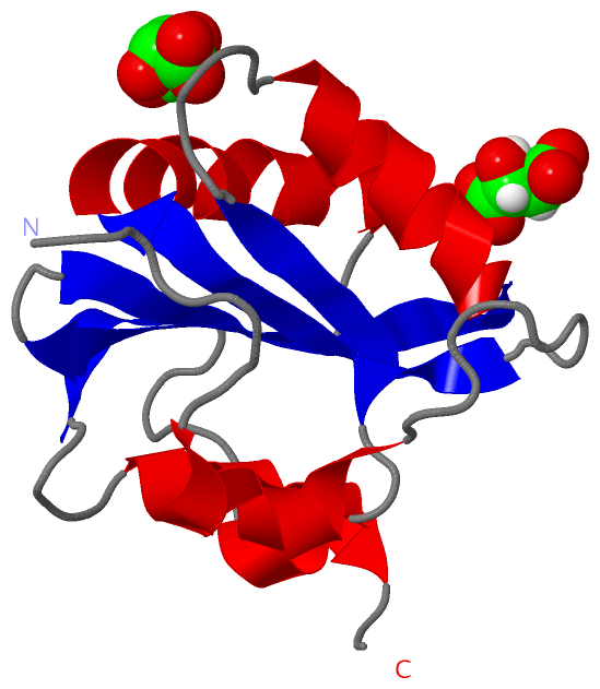
Chain A from PDB  Type:PROTEIN  Length:129
                                                                                                                                                                 
               SCOP domains d4nbpa_ A: The origin DNA-binding domain of SV40 T-antigen                                                                        SCOP domains
               CATH domains --------------------------------------------------------------------------------------------------------------------------------- CATH domains
               Pfam domains --------------------------------------------------------------------------------------------------------------------------------- Pfam domains
         Sec.struct. author .......hhhhhhhh.........eeeeeeeeehhhhhhhhhhhhhhh...eeeeeee..eeeeeeeeeeeehhhhhhhhhhh......eeeee..hhhhhhhhh.....eeeee......hhhhh... Sec.struct. author
                 SAPs(SNPs) --------------------------------------------------------------------------------------------------------------------------------- SAPs(SNPs)
                    PROSITE --------------------------------------------------------------------------------------------------------------------------------- PROSITE
                 Transcript --------------------------------------------------------------------------------------------------------------------------------- Transcript
                 4nbp A 133 VEDPKDFPVDLHAFLSQAVFSNRTVASFAVYTTKEKAQILYKKLMEKYSVTFISRHGFGGHNILFFLTPHRHRVSAINNYCQKLCTFSFLICKGVNKEYLFYSALCRQPYAVVEESIQGGLKEHDFNPE 261
                                   142       152       162       172       182       192       202       212       222       232       242       252         

   Legend:   → Mismatch (orange background)
  - → Gap (green background, '-', border residues have a numbering label)
    → Modified Residue (blue background, lower-case, 'x' indicates undefined single-letter code, labelled with number + name)
  x → Chemical Group (purple background, 'x', labelled with number + name, e.g. ACE or NH2)
  extra numbering lines below/above indicate numbering irregularities and modified residue names etc., number ends below/above '|'

 Classification and Annotation

(-) SCOP Domains  (1, 1)

Asymmetric/Biological Unit

(-) CATH Domains  (0, 0)

(no "CATH Domain" information available for 4NBP)

(-) Pfam Domains  (0, 0)

(no "Pfam Domain" information available for 4NBP)

(-) Gene Ontology  (16, 16)

Asymmetric/Biological Unit(hide GO term definitions)

 Visualization

(-) Interactive Views

Asymmetric/Biological Unit
  Complete Structure
    Jena3D(integrated viewing of ligand, site, SAP, PROSITE, SCOP information)
    WebMol | AstexViewer[tm]@PDBe
(Java Applets, require no local installation except for Java; loading may be slow)
    STRAP
(Java WebStart application, automatic local installation, requires Java; full application with system access!)
    RasMol
(require local installation)
    Molscript (VRML)
(requires installation of a VRML viewer; select preferred view via VRML and generate a mono or stereo PDF format file)
 
  Ligands, Modified Residues, Ions
    TLA  [ RasMol | Jena3D ]  +environment [ RasMol | Jena3D ]
 
  Sites
    AC1  [ RasMol ]  +environment [ RasMol ]
    AC2  [ RasMol ]  +environment [ RasMol ]
 
  Cis Peptide Bonds
    Gln A:240 - Pro A:241   [ RasMol ]  
 

(-) Still Images

Jmol
  protein: cartoon or spacefill or dots and stick; nucleic acid: cartoon and stick; ligands: spacefill; active site: stick
Molscript
  protein, nucleic acid: cartoon; ligands: spacefill; active site: ball and stick

 Databases and Analysis Tools

(-) Databases

Access by PDB/NDB ID
  4nbp
    Family and Domain InformationProDom | SYSTERS
    General Structural InformationGlycoscienceDB | MMDB | NDB | OCA | PDB | PDBe | PDBj | PDBsum | PDBWiki | PQS | PROTEOPEDIA
    Orientation in MembranesOPM
    Protein SurfaceSURFACE
    Secondary StructureDSSP (structure derived) | HSSP (homology derived)
    Structural GenomicsGeneCensus
    Structural NeighboursCE | VAST
    Structure ClassificationCATH | Dali | SCOP
    Validation and Original DataBMRB Data View | BMRB Restraints Grid | EDS | PROCHECK | RECOORD | WHAT_CHECK
 
Access by UniProt ID/Accession number
  LT_POVJC | P03072
    Comparative Protein Structure ModelsModBase
    Genomic InformationEnsembl
    Protein-protein InteractionDIP
    Sequence, Family and Domain InformationInterPro | Pfam | SMART | UniProtKB/SwissProt
 
Access by Enzyme Classificator   (EC Number)
  3.6.4.-
    General Enzyme InformationBRENDA | EC-PDB | Enzyme | IntEnz
    PathwayKEGG | MetaCyc
 
Access by Disease Identifier   (MIM ID)
  (no 'MIM ID' available)
    Disease InformationOMIM
 
Access by GenAge ID
  (no 'GenAge ID' available)
    Age Related InformationGenAge

(-) Analysis Tools

Access by PDB/NDB ID
    Domain InformationXDom
    Interatomic Contacts of Structural UnitsCSU
    Ligand-protein ContactsLPC
    Protein CavitiescastP
    Sequence and Secondary StructurePDBCartoon
    Structure AlignmentSTRAP(Java WebStart application, automatic local installation, requires Java; full application with system access!)
    Structure and Sequence BrowserSTING
 
Access by UniProt ID/Accession number
  LT_POVJC | P03072
    Protein Disorder PredictionDisEMBL | FoldIndex | GLOBPLOT (for more information see DisProt)

 Related Entries

(-) Entries Sharing at Least One Protein Chain (UniProt ID)

UniProtKB/Swiss-Prot
        LT_POVJC | P030724lif 4lmd 5cyn 5j40 5j47 5j4v 5j4y

(-) Related Entries Specified in the PDB File

4lif JCV OBD
4lmd JCV OBD