|
|
|
|
 Description
|
|
Compounds
|
||||||||||||||||||||||||||||||||||||||||||||||||||||||||||||
Chains, Units
Summary Information (see also Sequences/Alignments below) |
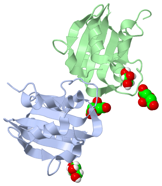
Ligands, Modified Residues, Ions (2, 4)| Asymmetric Unit (2, 4) Biological Unit 1 (1, 1) Biological Unit 2 (2, 3) |
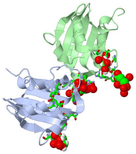
Sites (4, 4)
Asymmetric Unit (4, 4)
|
SS Bonds (0, 0)| (no "SS Bond" information available for 4LMD) |
Cis Peptide Bonds (3, 3)
Asymmetric Unit
|
||||||||||||||||
SAPs(SNPs)/Variants (0, 0)| (no "SAP(SNP)/Variant" information available for 4LMD) |
PROSITE Motifs (0, 0)| (no "PROSITE Motif" information available for 4LMD) |
Exons (0, 0)| (no "Exon" information available for 4LMD) |
Sequences/Alignments
Asymmetric Unit
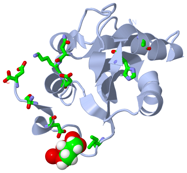
Chain A from PDB Type:PROTEIN Length:128
SCOP domains d4lmda_ A: The origin DNA-binding domain of SV40 T-antigen SCOP domains
CATH domains -------------------------------------------------------------------------------------------------------------------------------- CATH domains
Pfam domains -------------------------------------------------------------------------------------------------------------------------------- Pfam domains
SAPs(SNPs) -------------------------------------------------------------------------------------------------------------------------------- SAPs(SNPs)
PROSITE -------------------------------------------------------------------------------------------------------------------------------- PROSITE
Transcript -------------------------------------------------------------------------------------------------------------------------------- Transcript
4lmd A 134 EDPKDFPVDLHAFLSQAVFSNRTVASFAVYTTKEKAQILYKKLMEKYSVTFISRHGFGGHNILFFLTPHRHRVSAINNYCQKLCTFSFLICKGVNKEYLFYSALCRQPYAVVEESIQGGLKEHDFNPE 261
143 153 163 173 183 193 203 213 223 233 243 253
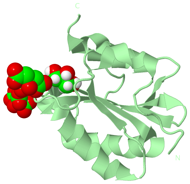
Chain B from PDB Type:PROTEIN Length:130
SCOP domains d4lmdb_ B: The origin DNA-binding domain of SV40 T-antigen SCOP domains
CATH domains ---------------------------------------------------------------------------------------------------------------------------------- CATH domains
Pfam domains ---------------------------------------------------------------------------------------------------------------------------------- Pfam domains
SAPs(SNPs) ---------------------------------------------------------------------------------------------------------------------------------- SAPs(SNPs)
PROSITE ---------------------------------------------------------------------------------------------------------------------------------- PROSITE
Transcript ---------------------------------------------------------------------------------------------------------------------------------- Transcript
4lmd B 132 KVEDPKDFPVDLHAFLSQAVFSNRTVASFAVYTTKEKAQILYKKLMEKYSVTFISRHGFGGHNILFFLTPHRHRVSAINNYCQKLCTFSFLICKGVNKEYLFYSALCRQPYAVVEESIQGGLKEHDFNPE 261
141 151 161 171 181 191 201 211 221 231 241 251 261
|
||||||||||||||||||||
SCOP Domains (1, 2)
Asymmetric Unit
 |
CATH Domains (0, 0)| (no "CATH Domain" information available for 4LMD) |
Pfam Domains (0, 0)| (no "Pfam Domain" information available for 4LMD) |
Gene Ontology (16, 16)|
Asymmetric Unit(hide GO term definitions) |
Interactive Views
|
||||||||||||||||||||||||||||||||||||||||||||||||||||||||||||||||||||||||||||||||||||||||||||||||||||||||||||||||||||||||||||||||||||||||||||||||||||||||||||||||||||||||||||||||||||||||
Still Images
|
||||||||||||||||
Databases
|
||||||||||||||||||||||||||||||||||||||||||||||||||||||||||||||||||||||||||||||||||||||||||||||||||||||||||||||||||||||||||||||||||||||||||||||||||||||||||||||||
Analysis Tools
|
|||||||||||||||||||||||||||||||||||||||||||||||||||||||||||||
Entries Sharing at Least One Protein Chain (UniProt ID)
Related Entries Specified in the PDB File
|
|