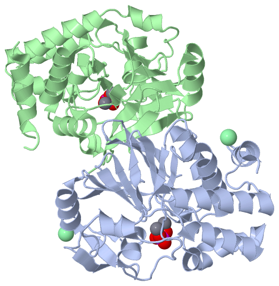
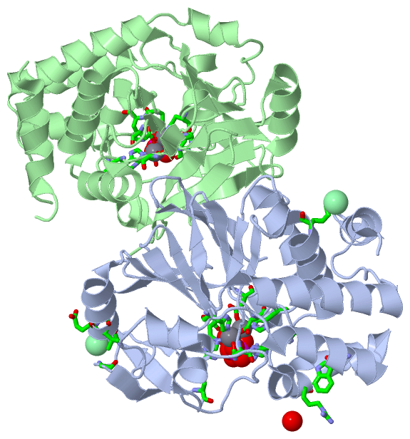
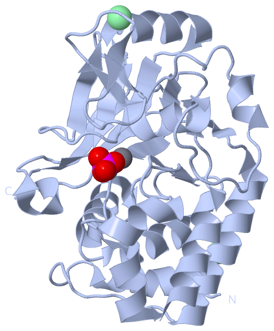
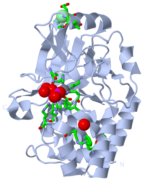
Show PDB file:   
         Plain Text   HTML   (compressed file size)
QuickSearch:   
by PDB,NDB,UniProt,PROSITE Code or Search Term(s)  
(-)Asymmetric Unit
(-)Asym. Unit - sites
(-)Biological Unit 1
(-)Biol. Unit 1 - sites
(-)Biological Unit 2
collapse expand < >
Image Asymmetric Unit
Asymmetric Unit  (Jmol Viewer)
Image Asym. Unit - sites
Asym. Unit - sites  (Jmol Viewer)
Image Biological Unit 1
Biological Unit 1  (Jmol Viewer)
Image Biol. Unit 1 - sites
Biol. Unit 1 - sites  (Jmol Viewer)
Image Biological Unit 2
Biological Unit 2  (Jmol Viewer)

(-) Description

Title :  1.45 A RESOLUTION CRYSTAL STRUCTURE OF PROTEIN PHOSPHATASE 1
 
Authors :  M. S. Choy, W. Peti, R. Page
Date :  12 Sep 13  (Deposition) - 26 Mar 14  (Release) - 23 Apr 14  (Revision)
Method :  X-RAY DIFFRACTION
Resolution :  1.45
Chains :  Asym. Unit :  A,B
Biol. Unit 1:  A  (1x)
Biol. Unit 2:  B  (1x)
Keywords :  Catalytic Subunit, Serine/Threonine Phosphatase, Nucleus, Hydrolase (Keyword Search: [Gene Ontology, PubMed, Web (Google))
 
Reference :  M. S. Choy, M. Hieke, G. S. Kumar, G. R. Lewis, K. R. Gonzalez-Dewhitt, R. P. Kessler, B. J. Stein, M. Hessenberger, A. C. Nairn, W. Peti, R. Pag
Understanding The Antagonism Of Retinoblastoma Protein Dephosphorylation By Pnuts Provides Insights Into The Pp1 Regulatory Code.
Proc. Natl. Acad. Sci. Usa V. 111 4097 2014
PubMed-ID: 24591642  |  Reference-DOI: 10.1073/PNAS.1317395111

(-) Compounds

Molecule 1 - SERINE/THREONINE-PROTEIN PHOSPHATASE PP1-ALPHA CATALYTIC SUBUNIT
    ChainsA, B
    EC Number3.1.3.16
    EngineeredYES
    Expression SystemESCHERICHIA COLI
    Expression System Taxid562
    GenePPP1CA, PPP1A
    Organism CommonHUMAN
    Organism ScientificHOMO SAPIENS
    Organism Taxid9606
    SynonymPP-1A

 Structural Features

(-) Chains, Units

  12
Asymmetric Unit AB
Biological Unit 1 (1x)A 
Biological Unit 2 (1x) B

Summary Information (see also Sequences/Alignments below)

(-) Ligands, Modified Residues, Ions  (3, 8)

Asymmetric Unit (3, 8)
No.NameCountTypeFull Name
1CL2Ligand/IonCHLORIDE ION
2MN4Ligand/IonMANGANESE (II) ION
3PO42Ligand/IonPHOSPHATE ION
Biological Unit 1 (1, 1)
No.NameCountTypeFull Name
1CL-1Ligand/IonCHLORIDE ION
2MN-1Ligand/IonMANGANESE (II) ION
3PO41Ligand/IonPHOSPHATE ION
Biological Unit 2 (1, 1)
No.NameCountTypeFull Name
1CL-1Ligand/IonCHLORIDE ION
2MN-1Ligand/IonMANGANESE (II) ION
3PO41Ligand/IonPHOSPHATE ION

(-) Sites  (8, 8)

Asymmetric Unit (8, 8)
No.NameEvidenceResiduesDescription
1AC1SOFTWAREASP A:92 , ASN A:124 , HIS A:173 , HIS A:248 , MN A:402 , PO4 A:403 , HOH A:737BINDING SITE FOR RESIDUE MN A 401
2AC2SOFTWAREASP A:64 , HIS A:66 , ASP A:92 , MN A:401 , PO4 A:403 , HOH A:737 , HOH A:746BINDING SITE FOR RESIDUE MN A 402
3AC3SOFTWAREHIS A:66 , ASP A:92 , ARG A:96 , ASN A:124 , HIS A:125 , ARG A:221 , HIS A:248 , MN A:401 , MN A:402 , HOH A:737 , HOH A:746 , HOH A:754 , HOH A:791BINDING SITE FOR RESIDUE PO4 A 403
4AC4SOFTWARELYS A:113 , ARG A:132 , TRP A:149 , HOH A:806BINDING SITE FOR RESIDUE CL A 404
5AC5SOFTWAREGLY A:215 , GLY A:228 , ALA A:229 , GLU A:230 , VAL A:231BINDING SITE FOR RESIDUE CL A 405
6AC6SOFTWAREASP B:92 , ASN B:124 , HIS B:173 , HIS B:248 , MN B:402 , PO4 B:403 , HOH B:508BINDING SITE FOR RESIDUE MN B 401
7AC7SOFTWAREASP B:64 , HIS B:66 , ASP B:92 , MN B:401 , PO4 B:403 , HOH B:508 , HOH B:685BINDING SITE FOR RESIDUE MN B 402
8AC8SOFTWAREHIS B:66 , ASP B:92 , ARG B:96 , ASN B:124 , HIS B:125 , ARG B:221 , HIS B:248 , MN B:401 , MN B:402 , HOH B:508 , HOH B:685 , HOH B:688BINDING SITE FOR RESIDUE PO4 B 403

(-) SS Bonds  (0, 0)

(no "SS Bond" information available for 4MOV)

(-) Cis Peptide Bonds  (6, 6)

Asymmetric Unit
No.Residues
1Ala A:57 -Pro A:58
2Pro A:82 -Pro A:83
3Arg A:191 -Pro A:192
4Ala B:57 -Pro B:58
5Pro B:82 -Pro B:83
6Arg B:191 -Pro B:192

 Sequence-Structure Mapping

(-) SAPs(SNPs)/Variants  (0, 0)

(no "SAP(SNP)/Variant" information available for 4MOV)

(-) PROSITE Motifs  (0, 0)

(no "PROSITE Motif" information available for 4MOV)

(-) Exons   (0, 0)

(no "Exon" information available for 4MOV)

(-) Sequences/Alignments

Asymmetric Unit
   Reformat: Number of residues per line =  ('0' or empty: single-line sequence representation)
  Number of residues per labelling interval =   
  UniProt sequence: complete  aligned part    
   Show mapping: SCOP domains CATH domains Pfam domains Secondary structure (by author)
SAPs(SNPs) PROSITE motifs Exons
(details for a mapped element are shown in a popup box when the mouse pointer rests over it)
Chain A from PDB  Type:PROTEIN  Length:293
                                                                                                                                                                                                                                                                                                                                     
               SCOP domains d4mova_ A: Protein phosphatase-1 (PP-1)                                                                                                                                                                                                                                                               SCOP domains
               CATH domains ----------------------------------------------------------------------------------------------------------------------------------------------------------------------------------------------------------------------------------------------------------------------------------------------------- CATH domains
               Pfam domains ----------------------------------------------------------------------------------------------------------------------------------------------------------------------------------------------------------------------------------------------------------------------------------------------------- Pfam domains
         Sec.struct. author .hhhhhhhhhhhh...........hhhhhhhhhhhhhhhhhhh..eeee...eeee.....hhhhhhhhhhhhh......eee.........hhhhhhhhhhhhhhhh...eee......hhhhhhhhhhhhhhhhhhhhhhhhhhhhhhh....eeee...eee...........hhhhhh..........hhhhhhhhhee......eee......eeehhhhhhhhhhhhh..eeee........eeee....eeee....hhhhh....eeeeee.....eeeeee... Sec.struct. author
                 SAPs(SNPs) ----------------------------------------------------------------------------------------------------------------------------------------------------------------------------------------------------------------------------------------------------------------------------------------------------- SAPs(SNPs)
                    PROSITE ----------------------------------------------------------------------------------------------------------------------------------------------------------------------------------------------------------------------------------------------------------------------------------------------------- PROSITE
                 Transcript ----------------------------------------------------------------------------------------------------------------------------------------------------------------------------------------------------------------------------------------------------------------------------------------------------- Transcript
                 4mov A   7 LNLDSIIGRLLEVQGSRPGKNVQLTENEIRGLCLKSREIFLSQPILLELEAPLKICGDIHGQYYDLLRLFEYGGFPPESNYLFLGDYVDRGKQSLETICLLLAYKIKYPENFFLLRGNHECASINRIYGFYDECKRRYNIKLWKTFTDCFNCLPIAAIVDEKIFCCHGGLSPDLQSMEQIRRIMRPTDVPDQGLLCDLLWSDPDKDVQGWGENDRGVSFTFGAEVVAKFLHKHDLDLICRAHQVVEDGYEFFAKRQLVTLFSAPNYCGEFDNAGAMMSVDETLMCSFQILKPA 299
                                    16        26        36        46        56        66        76        86        96       106       116       126       136       146       156       166       176       186       196       206       216       226       236       246       256       266       276       286       296   

Chain B from PDB  Type:PROTEIN  Length:291
                                                                                                                                                                                                                                                                                                                                   
               SCOP domains d4movb_ B: Protein phosphatase-1 (PP-1)                                                                                                                                                                                                                                                             SCOP domains
               CATH domains --------------------------------------------------------------------------------------------------------------------------------------------------------------------------------------------------------------------------------------------------------------------------------------------------- CATH domains
               Pfam domains --------------------------------------------------------------------------------------------------------------------------------------------------------------------------------------------------------------------------------------------------------------------------------------------------- Pfam domains
         Sec.struct. author .hhhhhhhhhhhh.........hhhhhhhhhhhhhhhhhhh..eeee...eeee.....hhhhhhhhhhhhh......eee.........hhhhhhhhhhhhhhhh...eee......hhhhhhhhhhhhhhhhhhhhhhhhhhhhhhh....eeee...eee...........hhhhhh..........hhhhhhhhhee......eee......eeehhhhhhhhhhhhh..eeee........eeee....eeee....hhhhh....eeeeee.....eeeeee... Sec.struct. author
                 SAPs(SNPs) --------------------------------------------------------------------------------------------------------------------------------------------------------------------------------------------------------------------------------------------------------------------------------------------------- SAPs(SNPs)
                    PROSITE --------------------------------------------------------------------------------------------------------------------------------------------------------------------------------------------------------------------------------------------------------------------------------------------------- PROSITE
                 Transcript --------------------------------------------------------------------------------------------------------------------------------------------------------------------------------------------------------------------------------------------------------------------------------------------------- Transcript
                 4mov B   7 LNLDSIIGRLLEVQGSGKNVQLTENEIRGLCLKSREIFLSQPILLELEAPLKICGDIHGQYYDLLRLFEYGGFPPESNYLFLGDYVDRGKQSLETICLLLAYKIKYPENFFLLRGNHECASINRIYGFYDECKRRYNIKLWKTFTDCFNCLPIAAIVDEKIFCCHGGLSPDLQSMEQIRRIMRPTDVPDQGLLCDLLWSDPDKDVQGWGENDRGVSFTFGAEVVAKFLHKHDLDLICRAHQVVEDGYEFFAKRQLVTLFSAPNYCGEFDNAGAMMSVDETLMCSFQILKPA 299
                                    16     || 28        38        48        58        68        78        88        98       108       118       128       138       148       158       168       178       188       198       208       218       228       238       248       258       268       278       288       298 
                                          22|                                                                                                                                                                                                                                                                                  
                                           25                                                                                                                                                                                                                                                                                  

   Legend:   → Mismatch (orange background)
  - → Gap (green background, '-', border residues have a numbering label)
    → Modified Residue (blue background, lower-case, 'x' indicates undefined single-letter code, labelled with number + name)
  x → Chemical Group (purple background, 'x', labelled with number + name, e.g. ACE or NH2)
  extra numbering lines below/above indicate numbering irregularities and modified residue names etc., number ends below/above '|'

 Classification and Annotation

(-) SCOP Domains  (1, 2)

Asymmetric Unit

(-) CATH Domains  (0, 0)

(no "CATH Domain" information available for 4MOV)

(-) Pfam Domains  (0, 0)

(no "Pfam Domain" information available for 4MOV)

(-) Gene Ontology  (43, 43)

Asymmetric Unit(hide GO term definitions)

 Visualization

(-) Interactive Views

Asymmetric Unit
  Complete Structure
    Jena3D(integrated viewing of ligand, site, SAP, PROSITE, SCOP information)
    WebMol | AstexViewer[tm]@PDBe
(Java Applets, require no local installation except for Java; loading may be slow)
    STRAP
(Java WebStart application, automatic local installation, requires Java; full application with system access!)
    RasMol
(require local installation)
    Molscript (VRML)
(requires installation of a VRML viewer; select preferred view via VRML and generate a mono or stereo PDF format file)
 
  Ligands, Modified Residues, Ions
    CL  [ RasMol | Jena3D ]  +environment [ RasMol | Jena3D ]
    MN  [ RasMol | Jena3D ]  +environment [ RasMol | Jena3D ]
    PO4  [ RasMol | Jena3D ]  +environment [ RasMol | Jena3D ]
 
  Sites
    AC1  [ RasMol ]  +environment [ RasMol ]
    AC2  [ RasMol ]  +environment [ RasMol ]
    AC3  [ RasMol ]  +environment [ RasMol ]
    AC4  [ RasMol ]  +environment [ RasMol ]
    AC5  [ RasMol ]  +environment [ RasMol ]
    AC6  [ RasMol ]  +environment [ RasMol ]
    AC7  [ RasMol ]  +environment [ RasMol ]
    AC8  [ RasMol ]  +environment [ RasMol ]
 
  Cis Peptide Bonds
    Ala A:57 - Pro A:58   [ RasMol ]  
    Ala B:57 - Pro B:58   [ RasMol ]  
    Arg A:191 - Pro A:192   [ RasMol ]  
    Arg B:191 - Pro B:192   [ RasMol ]  
    Pro A:82 - Pro A:83   [ RasMol ]  
    Pro B:82 - Pro B:83   [ RasMol ]  
 
Biological Units
  Complete Structure
    Biological Unit 1  [ Jena3D ]
    Biological Unit 2  [ Jena3D ]

(-) Still Images

Jmol
  protein: cartoon or spacefill or dots and stick; nucleic acid: cartoon and stick; ligands: spacefill; active site: stick
Molscript
  protein, nucleic acid: cartoon; ligands: spacefill; active site: ball and stick

 Databases and Analysis Tools

(-) Databases

Access by PDB/NDB ID
  4mov
    Family and Domain InformationProDom | SYSTERS
    General Structural InformationGlycoscienceDB | MMDB | NDB | OCA | PDB | PDBe | PDBj | PDBsum | PDBWiki | PQS | PROTEOPEDIA
    Orientation in MembranesOPM
    Protein SurfaceSURFACE
    Secondary StructureDSSP (structure derived) | HSSP (homology derived)
    Structural GenomicsGeneCensus
    Structural NeighboursCE | VAST
    Structure ClassificationCATH | Dali | SCOP
    Validation and Original DataBMRB Data View | BMRB Restraints Grid | EDS | PROCHECK | RECOORD | WHAT_CHECK
 
Access by UniProt ID/Accession number
  PP1A_HUMAN | P62136
    Comparative Protein Structure ModelsModBase
    Genomic InformationEnsembl
    Protein-protein InteractionDIP
    Sequence, Family and Domain InformationInterPro | Pfam | SMART | UniProtKB/SwissProt
 
Access by Enzyme Classificator   (EC Number)
  3.1.3.16
    General Enzyme InformationBRENDA | EC-PDB | Enzyme | IntEnz
    PathwayKEGG | MetaCyc
 
Access by Disease Identifier   (MIM ID)
  (no 'MIM ID' available)
    Disease InformationOMIM
 
Access by GenAge ID
  (no 'GenAge ID' available)
    Age Related InformationGenAge

(-) Analysis Tools

Access by PDB/NDB ID
    Domain InformationXDom
    Interatomic Contacts of Structural UnitsCSU
    Ligand-protein ContactsLPC
    Protein CavitiescastP
    Sequence and Secondary StructurePDBCartoon
    Structure AlignmentSTRAP(Java WebStart application, automatic local installation, requires Java; full application with system access!)
    Structure and Sequence BrowserSTING
 
Access by UniProt ID/Accession number
  PP1A_HUMAN | P62136
    Protein Disorder PredictionDisEMBL | FoldIndex | GLOBPLOT (for more information see DisProt)

 Related Entries

(-) Entries Sharing at Least One Protein Chain (UniProt ID)

UniProtKB/Swiss-Prot
        PP1A_HUMAN | P621361fjm 3e7a 3e7b 3egg 3egh 3hvq 3n5u 3v4y 4g9j 4moy 4mp0 4xpn 5ioh

(-) Related Entries Specified in the PDB File

3e7a PP1 COMPLEXED WITH NODULARIN R
3egg PP1:SPINOPHILIN CRYSTAL STRUCTURE
3v4y PP1:NIPP1 CRYSTAL STRUCTURE