Show PDB file:   
         Plain Text   HTML   (compressed file size)
QuickSearch:   
by PDB,NDB,UniProt,PROSITE Code or Search Term(s)  
(-)Asymmetric Unit
(-)Biological Unit 1
(-)Biological Unit 2
collapse expand < >
Image Asymmetric Unit
Asymmetric Unit  (Jmol Viewer)
Image Biological Unit 1
Biological Unit 1  (Jmol Viewer)
Image Biological Unit 2
Biological Unit 2  (Jmol Viewer)

(-) Description

Title :  CRYSTAL STRUCTURE OF THE SHROOM-BINDING DOMAIN OF HUMAN ROCK1
 
Authors :  S. Mohan, A. P. Vandemark
Date :  04 Jun 13  (Deposition) - 16 Apr 14  (Release) - 16 Apr 14  (Revision)
Method :  X-RAY DIFFRACTION
Resolution :  2.49
Chains :  Asym. Unit :  A,B,C,D
Biol. Unit 1:  C,D  (1x)
Biol. Unit 2:  A,B  (1x)
Keywords :  Coiled-Coil, Shroom Sd2, Kinase, Myosin, Protein Binding (Keyword Search: [Gene Ontology, PubMed, Web (Google))
 
Reference :  S. Mohan, D. Das, R. J. Bauer, A. Heroux, J. K. Zalewski, S. Heber, A. M. Dosunmu-Ogunbi, M. A. Trakselis, J. D. Hildebrand, A. P. Vandemark
Structure Of A Highly Conserved Domain Of Rock1 Required Fo Shroom-Mediated Regulation Of Cell Morphology.
Plos One V. 8 81075 2013
PubMed-ID: 24349032  |  Reference-DOI: 10.1371/JOURNAL.PONE.0081075

(-) Compounds

Molecule 1 - RHO-ASSOCIATED PROTEIN KINASE 1
    ChainsC, D, A, B
    EngineeredYES
    Expression SystemESCHERICHIA COLI
    Expression System PlasmidPET151 D-TOPO
    Expression System StrainBL21(DE3)
    Expression System Taxid469008
    Expression System Vector TypePLASMID
    FragmentSHROOM BINDING DOMAIN (UNP RESIDUES 834-914)
    GeneROCK1
    MutationYES
    Organism CommonHUMAN
    Organism ScientificHOMO SAPIENS
    Organism Taxid9606

 Structural Features

(-) Chains, Units

  1234
Asymmetric Unit ABCD
Biological Unit 1 (1x)  CD
Biological Unit 2 (1x)AB  

Summary Information (see also Sequences/Alignments below)

(-) Ligands, Modified Residues, Ions  (0, 0)

(no "Ligand,Modified Residues,Ions" information available for 4L2W)

(-) Sites  (0, 0)

(no "Site" information available for 4L2W)

(-) SS Bonds  (0, 0)

(no "SS Bond" information available for 4L2W)

(-) Cis Peptide Bonds  (0, 0)

(no "Cis Peptide Bond" information available for 4L2W)

 Sequence-Structure Mapping

(-) SAPs(SNPs)/Variants  (0, 0)

(no "SAP(SNP)/Variant" information available for 4L2W)

(-) PROSITE Motifs  (0, 0)

(no "PROSITE Motif" information available for 4L2W)

(-) Exons   (0, 0)

(no "Exon" information available for 4L2W)

(-) Sequences/Alignments

Asymmetric Unit
   Reformat: Number of residues per line =  ('0' or empty: single-line sequence representation)
  Number of residues per labelling interval =   
  UniProt sequence: complete  aligned part    
   Show mapping: SCOP domains CATH domains Pfam domains Secondary structure (by author)
SAPs(SNPs) PROSITE motifs Exons
(details for a mapped element are shown in a popup box when the mouse pointer rests over it)
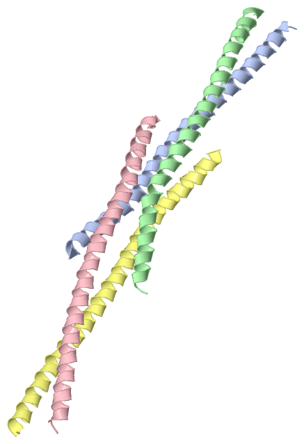
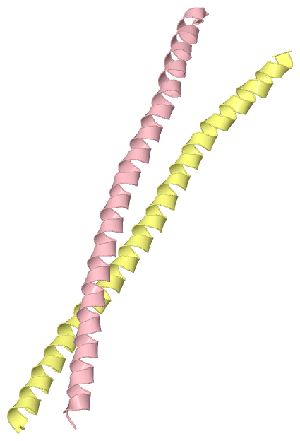
Chain A from PDB  Type:PROTEIN  Length:67
                                                                                                   
               SCOP domains ------------------------------------------------------------------- SCOP domains
               CATH domains ------------------------------------------------------------------- CATH domains
               Pfam domains ------------------------------------------------------------------- Pfam domains
         Sec.struct. author .hhhhhhhhhhhhhhhhhhhhhhhhhhhhhhhhhhhhhhhhhhhhhhhhhhhhhhhhhhhhhhhhhh Sec.struct. author
                 SAPs(SNPs) ------------------------------------------------------------------- SAPs(SNPs)
                    PROSITE ------------------------------------------------------------------- PROSITE
                 Transcript ------------------------------------------------------------------- Transcript
                 4l2w A 837 GQMRELQDQLEAEQYFSTLYKTQVKELKEEIEEKNRENLKKIQELQNAAATLATQLDLAETKAESEQ 903
                                   846       856       866       876       886       896       

Chain B from PDB  Type:PROTEIN  Length:67
                                                                                                   
               SCOP domains ------------------------------------------------------------------- SCOP domains
               CATH domains ------------------------------------------------------------------- CATH domains
               Pfam domains ------------------------------------------------------------------- Pfam domains
         Sec.struct. author .hhhhhhhhhhhhhhhhhhhhhhhhhhhhhhhhhhhhhhhhhhhhhhhhhhhhhhhhhhhhhhhh.. Sec.struct. author
                 SAPs(SNPs) ------------------------------------------------------------------- SAPs(SNPs)
                    PROSITE ------------------------------------------------------------------- PROSITE
                 Transcript ------------------------------------------------------------------- Transcript
                 4l2w B 838 QMRELQDQLEAEQYFSTLYKTQVKELKEEIEEKNRENLKKIQELQNAAATLATQLDLAETKAESEQL 904
                                   847       857       867       877       887       897       

Chain C from PDB  Type:PROTEIN  Length:67
                                                                                                   
               SCOP domains ------------------------------------------------------------------- SCOP domains
               CATH domains ------------------------------------------------------------------- CATH domains
               Pfam domains ------------------------------------------------------------------- Pfam domains
         Sec.struct. author .hhhhhhhhhhhhhhhhhhhhhhhhhhhhhhhhhhhhhhhhhhhhhhhhhhhhhhhhhhhhhhhhh. Sec.struct. author
                 SAPs(SNPs) ------------------------------------------------------------------- SAPs(SNPs)
                    PROSITE ------------------------------------------------------------------- PROSITE
                 Transcript ------------------------------------------------------------------- Transcript
                 4l2w C 838 QMRELQDQLEAEQYFSTLYKTQVKELKEEIEEKNRENLKKIQELQNAAATLATQLDLAETKAESEQL 904
                                   847       857       867       877       887       897       

Chain D from PDB  Type:PROTEIN  Length:68
                                                                                                    
               SCOP domains -------------------------------------------------------------------- SCOP domains
               CATH domains -------------------------------------------------------------------- CATH domains
               Pfam domains -------------------------------------------------------------------- Pfam domains
         Sec.struct. author .hhhhhhhhhhhhhhhhhhhhhhhhhhhhhhhhhhhhhhhhhhhhhhhhhhhhhhhhhhhhhhhhhhh Sec.struct. author
                 SAPs(SNPs) -------------------------------------------------------------------- SAPs(SNPs)
                    PROSITE -------------------------------------------------------------------- PROSITE
                 Transcript -------------------------------------------------------------------- Transcript
                 4l2w D 835 NEGQMRELQDQLEAEQYFSTLYKTQVKELKEEIEEKNRENLKKIQELQNAAATLATQLDLAETKAESE 902
                                   844       854       864       874       884       894        

   Legend:   → Mismatch (orange background)
  - → Gap (green background, '-', border residues have a numbering label)
    → Modified Residue (blue background, lower-case, 'x' indicates undefined single-letter code, labelled with number + name)
  x → Chemical Group (purple background, 'x', labelled with number + name, e.g. ACE or NH2)
  extra numbering lines below/above indicate numbering irregularities and modified residue names etc., number ends below/above '|'

 Classification and Annotation

(-) SCOP Domains  (0, 0)

(no "SCOP Domain" information available for 4L2W)

(-) CATH Domains  (0, 0)

(no "CATH Domain" information available for 4L2W)

(-) Pfam Domains  (0, 0)

(no "Pfam Domain" information available for 4L2W)

(-) Gene Ontology  (59, 59)

Asymmetric Unit(hide GO term definitions)

 Visualization

(-) Interactive Views

Asymmetric Unit
  Complete Structure
    Jena3D(integrated viewing of ligand, site, SAP, PROSITE, SCOP information)
    WebMol | AstexViewer[tm]@PDBe
(Java Applets, require no local installation except for Java; loading may be slow)
    STRAP
(Java WebStart application, automatic local installation, requires Java; full application with system access!)
    RasMol
(require local installation)
    Molscript (VRML)
(requires installation of a VRML viewer; select preferred view via VRML and generate a mono or stereo PDF format file)
 
  Ligands, Modified Residues, Ions
(no "Ligands, Modified Residues, Ions" information available for 4l2w)
 
  Sites
(no "Sites" information available for 4l2w)
 
  Cis Peptide Bonds
(no "Cis Peptide Bonds" information available for 4l2w)
 
Biological Units
  Complete Structure
    Biological Unit 1  [ Jena3D ]
    Biological Unit 2  [ Jena3D ]

(-) Still Images

Jmol
  protein: cartoon or spacefill or dots and stick; nucleic acid: cartoon and stick; ligands: spacefill; active site: stick
Molscript
  protein, nucleic acid: cartoon; ligands: spacefill; active site: ball and stick

 Databases and Analysis Tools

(-) Databases

Access by PDB/NDB ID
  4l2w
    Family and Domain InformationProDom | SYSTERS
    General Structural InformationGlycoscienceDB | MMDB | NDB | OCA | PDB | PDBe | PDBj | PDBsum | PDBWiki | PQS | PROTEOPEDIA
    Orientation in MembranesOPM
    Protein SurfaceSURFACE
    Secondary StructureDSSP (structure derived) | HSSP (homology derived)
    Structural GenomicsGeneCensus
    Structural NeighboursCE | VAST
    Structure ClassificationCATH | Dali | SCOP
    Validation and Original DataBMRB Data View | BMRB Restraints Grid | EDS | PROCHECK | RECOORD | WHAT_CHECK
 
Access by UniProt ID/Accession number
  ROCK1_HUMAN | Q13464
    Comparative Protein Structure ModelsModBase
    Genomic InformationEnsembl
    Protein-protein InteractionDIP
    Sequence, Family and Domain InformationInterPro | Pfam | SMART | UniProtKB/SwissProt
 
Access by Enzyme Classificator   (EC Number)
  (no 'Enzyme Classificator' available)
    General Enzyme InformationBRENDA | EC-PDB | Enzyme | IntEnz
    PathwayKEGG | MetaCyc
 
Access by Disease Identifier   (MIM ID)
  (no 'MIM ID' available)
    Disease InformationOMIM
 
Access by GenAge ID
  (no 'GenAge ID' available)
    Age Related InformationGenAge

(-) Analysis Tools

Access by PDB/NDB ID
    Domain InformationXDom
    Interatomic Contacts of Structural UnitsCSU
    Ligand-protein ContactsLPC
    Protein CavitiescastP
    Sequence and Secondary StructurePDBCartoon
    Structure AlignmentSTRAP(Java WebStart application, automatic local installation, requires Java; full application with system access!)
    Structure and Sequence BrowserSTING
 
Access by UniProt ID/Accession number
  ROCK1_HUMAN | Q13464
    Protein Disorder PredictionDisEMBL | FoldIndex | GLOBPLOT (for more information see DisProt)

 Related Entries

(-) Entries Sharing at Least One Protein Chain (UniProt ID)

UniProtKB/Swiss-Prot
        ROCK1_HUMAN | Q134641s1c 2esm 2etk 2etr 2v55 3d9v 3ncz 3ndm 3o0z 3tv7 3twj 3v8s 4w7p 4yvc 4yve 5bml 5f5p 5hvu 5kks 5kkt

(-) Related Entries Specified in the PDB File

(no "Related Entries Specified in the PDB File" available for 4L2W)