Show PDB file:   
         Plain Text   HTML   (compressed file size)
QuickSearch:   
by PDB,NDB,UniProt,PROSITE Code or Search Term(s)  
(-)Asymmetric Unit
(-)Asym. Unit - sites
(-)Biological Unit 1
(-)Biol. Unit 1 - sites
(-)Biological Unit 2
(-)Biological Unit 3
collapse expand < >
Image Asymmetric Unit
Asymmetric Unit  (Jmol Viewer)
Image Asym. Unit - sites
Asym. Unit - sites  (Jmol Viewer)
Image Biological Unit 1
Biological Unit 1  (Jmol Viewer)
Image Biol. Unit 1 - sites
Biol. Unit 1 - sites  (Jmol Viewer)
Image Biological Unit 2
Biological Unit 2  (Jmol Viewer)
Image Biological Unit 3
Biological Unit 3  (Jmol Viewer)

(-) Description

Title :  CRYSTAL STRUCTURE OF THE K463A MUTANT OF THE RRM DOMAIN OF RNA HELICASE HERA FROM T. THERMOPHILUS
 
Authors :  M. G. Rudolph, D. Klostermeier
Date :  29 Nov 12  (Deposition) - 24 Apr 13  (Release) - 26 Nov 14  (Revision)
Method :  X-RAY DIFFRACTION
Resolution :  1.79
Chains :  Asym. Unit :  A,B,C
Biol. Unit 1:  A  (1x)
Biol. Unit 2:  B  (1x)
Biol. Unit 3:  C  (1x)
Keywords :  Rna Recognition Motif, Atp Binding, Nucleotide Binding, Rna Binding Protein, Dead Box Protein, Hydrolase (Keyword Search: [Gene Ontology, PubMed, Web (Google))
 
Reference :  L. Steimer, J. P. Wurm, M. H. Linden, M. G. Rudolph, J. Wohnert, D. Klostermeier
Recognition Of Two Distinct Elements In The Rna Substrate B The Rna-Binding Domain Of The T. Thermophilus Dead Box Helicase Hera.
Nucleic Acids Res. V. 41 6259 2013
PubMed-ID: 23625962  |  Reference-DOI: 10.1093/NAR/GKT323

(-) Compounds

Molecule 1 - HEAT RESISTANT RNA DEPENDENT ATPASE
    ChainsA, B, C
    EC Number3.6.4.13
    EngineeredYES
    Expression SystemESCHERICHIA COLI
    Expression System Taxid562
    FragmentRRM DOMAIN (UNP RESIDUES 431-517)
    GeneTT_C1895
    MutationYES
    Organism ScientificTHERMUS THERMOPHILUS
    Organism Taxid274
    SynonymHERA RNA HELICASE

 Structural Features

(-) Chains, Units

  123
Asymmetric Unit ABC
Biological Unit 1 (1x)A  
Biological Unit 2 (1x) B 
Biological Unit 3 (1x)  C

Summary Information (see also Sequences/Alignments below)

(-) Ligands, Modified Residues, Ions  (1, 4)

Asymmetric Unit (1, 4)
No.NameCountTypeFull Name
1CL4Ligand/IonCHLORIDE ION
Biological Unit 1 (0, 0)
No.NameCountTypeFull Name
1CL-1Ligand/IonCHLORIDE ION
Biological Unit 2 (0, 0)
No.NameCountTypeFull Name
1CL-1Ligand/IonCHLORIDE ION
Biological Unit 3 (0, 0)
No.NameCountTypeFull Name
1CL-1Ligand/IonCHLORIDE ION

(-) Sites  (4, 4)

Asymmetric Unit (4, 4)
No.NameEvidenceResiduesDescription
1AC1SOFTWAREARG A:480 , GLU B:460BINDING SITE FOR RESIDUE CL A 601
2AC2SOFTWAREARG B:426 , ARG B:449 , HOH B:723BINDING SITE FOR RESIDUE CL B 601
3AC3SOFTWAREARG B:493BINDING SITE FOR RESIDUE CL B 602
4AC4SOFTWAREARG A:487 , ARG C:444 , ARG C:503BINDING SITE FOR RESIDUE CL C 601

(-) SS Bonds  (0, 0)

(no "SS Bond" information available for 4I69)

(-) Cis Peptide Bonds  (0, 0)

(no "Cis Peptide Bond" information available for 4I69)

 Sequence-Structure Mapping

(-) SAPs(SNPs)/Variants  (0, 0)

(no "SAP(SNP)/Variant" information available for 4I69)

(-) PROSITE Motifs  (0, 0)

(no "PROSITE Motif" information available for 4I69)

(-) Exons   (0, 0)

(no "Exon" information available for 4I69)

(-) Sequences/Alignments

Asymmetric Unit
   Reformat: Number of residues per line =  ('0' or empty: single-line sequence representation)
  Number of residues per labelling interval =   
  UniProt sequence: complete  aligned part    
   Show mapping: SCOP domains CATH domains Pfam domains Secondary structure (by author)
SAPs(SNPs) PROSITE motifs Exons
(details for a mapped element are shown in a popup box when the mouse pointer rests over it)
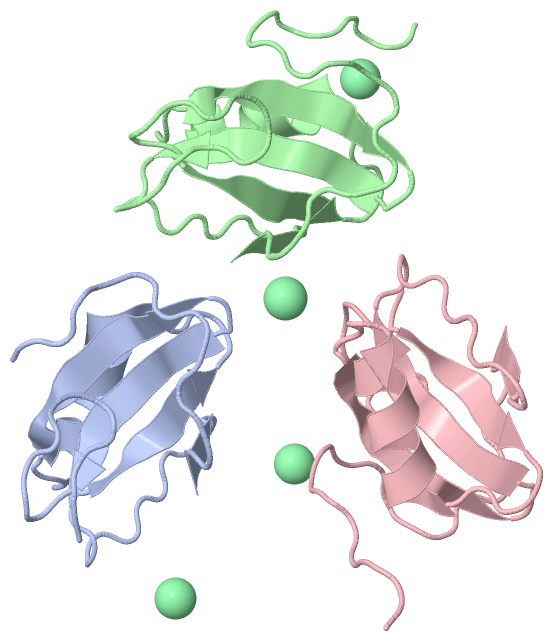
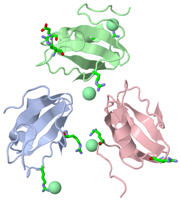
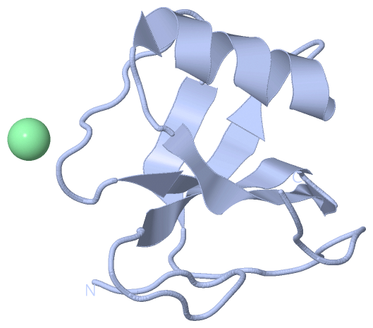
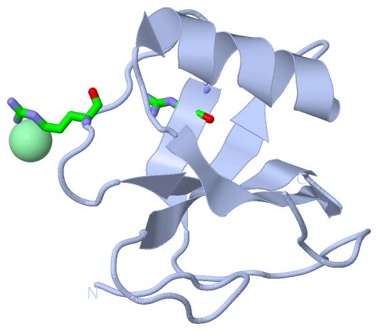
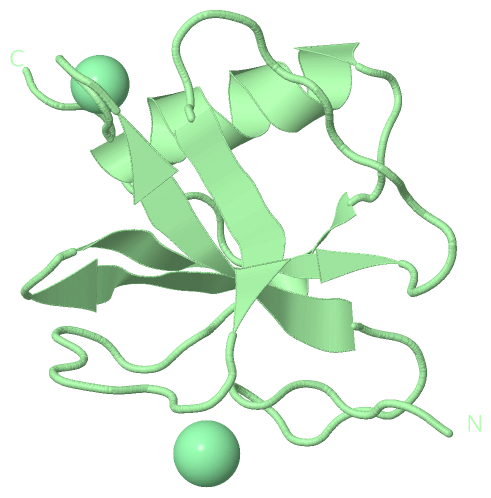
Chain A from PDB  Type:PROTEIN  Length:79
                                                                                                               
               SCOP domains ------------------------------------------------------------------------------- SCOP domains
               CATH domains ------------------------------------------------------------------------------- CATH domains
               Pfam domains ------------------------------------------------------------------------------- Pfam domains
         Sec.struct. author ...........eeeeee....hhhhhhhhhhhh...eeeeeee..eeeeee...........eeee............. Sec.struct. author
                 SAPs(SNPs) ------------------------------------------------------------------------------- SAPs(SNPs)
                    PROSITE ------------------------------------------------------------------------------- PROSITE
                 Transcript ------------------------------------------------------------------------------- Transcript
                 4i69 A 425 ERSLLTGEEGWRTYKATGPRLSLPRLVALLKGQGLEVGAVAEAEGGFYVDLRPEARPEVAGLRLEPARRVEGLLEIPSR 503
                                   434       444       454       464       474       484       494         

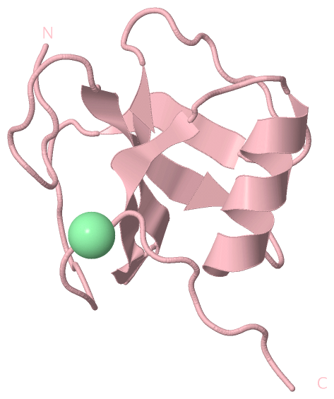
Chain B from PDB  Type:PROTEIN  Length:87
                                                                                                                       
               SCOP domains --------------------------------------------------------------------------------------- SCOP domains
               CATH domains --------------------------------------------------------------------------------------- CATH domains
               Pfam domains --------------------------------------------------------------------------------------- Pfam domains
         Sec.struct. author ............eeeeee....hhhhhhhhhhh....eeeeeee..eeeeee...........eeee.................... Sec.struct. author
                 SAPs(SNPs) --------------------------------------------------------------------------------------- SAPs(SNPs)
                    PROSITE --------------------------------------------------------------------------------------- PROSITE
                 Transcript --------------------------------------------------------------------------------------- Transcript
                 4i69 B 424 AERSLLTGEEGWRTYKATGPRLSLPRLVALLKGQGLEVGAVAEAEGGFYVDLRPEARPEVAGLRLEPARRVEGLLEIPSRTRRPARA 510
                                   433       443       453       463       473       483       493       503       

Chain C from PDB  Type:PROTEIN  Length:86
                                                                                                                      
               SCOP domains -------------------------------------------------------------------------------------- SCOP domains
               CATH domains -------------------------------------------------------------------------------------- CATH domains
               Pfam domains -------------------------------------------------------------------------------------- Pfam domains
         Sec.struct. author ...........eeeeee....hhhhhhhhhhh....eeeeeee..eeeeee...........eeee.................... Sec.struct. author
                 SAPs(SNPs) -------------------------------------------------------------------------------------- SAPs(SNPs)
                    PROSITE -------------------------------------------------------------------------------------- PROSITE
                 Transcript -------------------------------------------------------------------------------------- Transcript
                 4i69 C 425 ERSLLTGEEGWRTYKATGPRLSLPRLVALLKGQGLEVGAVAEAEGGFYVDLRPEARPEVAGLRLEPARRVEGLLEIPSRTRRPARA 510
                                   434       444       454       464       474       484       494       504      

   Legend:   → Mismatch (orange background)
  - → Gap (green background, '-', border residues have a numbering label)
    → Modified Residue (blue background, lower-case, 'x' indicates undefined single-letter code, labelled with number + name)
  x → Chemical Group (purple background, 'x', labelled with number + name, e.g. ACE or NH2)
  extra numbering lines below/above indicate numbering irregularities and modified residue names etc., number ends below/above '|'

 Classification and Annotation

(-) SCOP Domains  (0, 0)

(no "SCOP Domain" information available for 4I69)

(-) CATH Domains  (0, 0)

(no "CATH Domain" information available for 4I69)

(-) Pfam Domains  (0, 0)

(no "Pfam Domain" information available for 4I69)

(-) Gene Ontology  (5, 5)

Asymmetric Unit(hide GO term definitions)

 Visualization

(-) Interactive Views

Asymmetric Unit
  Complete Structure
    Jena3D(integrated viewing of ligand, site, SAP, PROSITE, SCOP information)
    WebMol | AstexViewer[tm]@PDBe
(Java Applets, require no local installation except for Java; loading may be slow)
    STRAP
(Java WebStart application, automatic local installation, requires Java; full application with system access!)
    RasMol
(require local installation)
    Molscript (VRML)
(requires installation of a VRML viewer; select preferred view via VRML and generate a mono or stereo PDF format file)
 
  Ligands, Modified Residues, Ions
    CL  [ RasMol | Jena3D ]  +environment [ RasMol | Jena3D ]
 
  Sites
    AC1  [ RasMol ]  +environment [ RasMol ]
    AC2  [ RasMol ]  +environment [ RasMol ]
    AC3  [ RasMol ]  +environment [ RasMol ]
    AC4  [ RasMol ]  +environment [ RasMol ]
 
  Cis Peptide Bonds
(no "Cis Peptide Bonds" information available for 4i69)
 
Biological Units
  Complete Structure
    Biological Unit 1  [ Jena3D ]
    Biological Unit 2  [ Jena3D ]
    Biological Unit 3  [ Jena3D ]

(-) Still Images

Jmol
  protein: cartoon or spacefill or dots and stick; nucleic acid: cartoon and stick; ligands: spacefill; active site: stick
Molscript
  protein, nucleic acid: cartoon; ligands: spacefill; active site: ball and stick

 Databases and Analysis Tools

(-) Databases

Access by PDB/NDB ID
  4i69
    Family and Domain InformationProDom | SYSTERS
    General Structural InformationGlycoscienceDB | MMDB | NDB | OCA | PDB | PDBe | PDBj | PDBsum | PDBWiki | PQS | PROTEOPEDIA
    Orientation in MembranesOPM
    Protein SurfaceSURFACE
    Secondary StructureDSSP (structure derived) | HSSP (homology derived)
    Structural GenomicsGeneCensus
    Structural NeighboursCE | VAST
    Structure ClassificationCATH | Dali | SCOP
    Validation and Original DataBMRB Data View | BMRB Restraints Grid | EDS | PROCHECK | RECOORD | WHAT_CHECK
 
Access by UniProt ID/Accession number
  Q72GF3_THET2 | Q72GF3
    Comparative Protein Structure ModelsModBase
    Genomic InformationEnsembl
    Protein-protein InteractionDIP
    Sequence, Family and Domain InformationInterPro | Pfam | SMART | UniProtKB/TrEMBL
 
Access by Enzyme Classificator   (EC Number)
  3.6.4.13
    General Enzyme InformationBRENDA | EC-PDB | Enzyme | IntEnz
    PathwayKEGG | MetaCyc
 
Access by Disease Identifier   (MIM ID)
  (no 'MIM ID' available)
    Disease InformationOMIM
 
Access by GenAge ID
  (no 'GenAge ID' available)
    Age Related InformationGenAge

(-) Analysis Tools

Access by PDB/NDB ID
    Domain InformationXDom
    Interatomic Contacts of Structural UnitsCSU
    Ligand-protein ContactsLPC
    Protein CavitiescastP
    Sequence and Secondary StructurePDBCartoon
    Structure AlignmentSTRAP(Java WebStart application, automatic local installation, requires Java; full application with system access!)
    Structure and Sequence BrowserSTING
 
Access by UniProt ID/Accession number
  Q72GF3_THET2 | Q72GF3
    Protein Disorder PredictionDisEMBL | FoldIndex | GLOBPLOT (for more information see DisProt)

 Related Entries

(-) Entries Sharing at Least One Protein Chain (UniProt ID)

UniProtKB/TrEMBL
        Q72GF3_THET2 | Q72GF33eaq 3ear 3eas 3i31 3i32 3mwj 3mwk 3mwl 3nbf 3nej 4i67 4i68 4kbf 4kbg 5mao

(-) Related Entries Specified in the PDB File

3i31 4i67 4i68