|
|
|
|
 Description
|
|
Compounds
|
||||||||||||||||||||||||||||||||||||||||
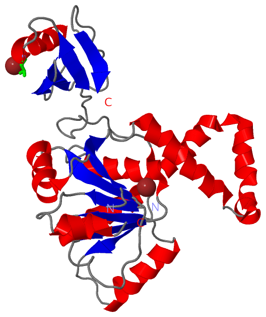
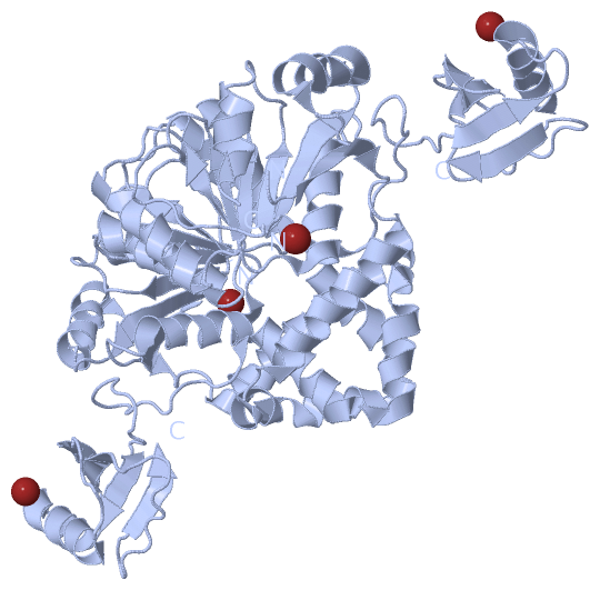
Chains, Units
Summary Information (see also Sequences/Alignments below) |
Ligands, Modified Residues, Ions (1, 2)
Asymmetric Unit (1, 2)
|
Sites (1, 1)
Asymmetric Unit (1, 1)
|
SS Bonds (0, 0)| (no "SS Bond" information available for 3I32) |
Cis Peptide Bonds (0, 0)| (no "Cis Peptide Bond" information available for 3I32) |
SAPs(SNPs)/Variants (0, 0)| (no "SAP(SNP)/Variant" information available for 3I32) |
PROSITE Motifs (0, 0)| (no "PROSITE Motif" information available for 3I32) |
Exons (0, 0)| (no "Exon" information available for 3I32) |
Sequences/Alignments
Asymmetric UnitChain A from PDB Type:PROTEIN Length:276 aligned with Q72GF3_THET2 | Q72GF3 from UniProtKB/TrEMBL Length:517 Alignment length:281 227 237 247 257 267 277 287 297 307 317 327 337 347 357 367 377 387 397 407 417 427 437 447 457 467 477 487 497 Q72GF3_THET2 218 VTYEEEAVPAPVRGRLEVLSDLLYVASPDRAMVFTRTKAETEEIAQGLLRLGHPAQALHGDLSQGERERVLGAFRQGEVRVLVATDVAARGLDIPQVDLVVHYRLPDRAEAYQHRSGRTGRAGRGGRVVLLYGPRERRDVEALERAVGRRFKRVNPPTPEEVLEAKWRHLLARLARVPEKDYRLYQDFAGRLFAEGRVEVVAALLALLLGGAPAERSLLTGEEGWRTYKATGPRLSLPRLVALLKGQGLEVGKVAEAEGGFYVDLRPEARPEVAGLRLEPA 498 SCOP domains ----------------------------------------------------------------------------------------------------------------------------------------------------------------------------------------------------------------------------------------------------------------------------------------- SCOP domains CATH domains ----------------------------------------------------------------------------------------------------------------------------------------------------------------------------------------------------------------------------------------------------------------------------------------- CATH domains Pfam domains ----------------------------------------------------------------------------------------------------------------------------------------------------------------------------------------------------------------------------------------------------------------------------------------- Pfam domains SAPs(SNPs) ----------------------------------------------------------------------------------------------------------------------------------------------------------------------------------------------------------------------------------------------------------------------------------------- SAPs(SNPs) PROSITE ----------------------------------------------------------------------------------------------------------------------------------------------------------------------------------------------------------------------------------------------------------------------------------------- PROSITE Transcript ----------------------------------------------------------------------------------------------------------------------------------------------------------------------------------------------------------------------------------------------------------------------------------------- Transcript 3i32 A 211 VTYEEEAVPAPVRGRLEVLSDLLYVASPDRAMVFTRTKAETEEIAQGLLRLGHPAQALHGDMSQGERERVMGAFRQGEVRVLVATDVAARGLDIPQVDLVVHYRMPDRAEAYQHRSGRT-----GGRVVLLYGPRERRDVEALERAVGRRFKRVNPPTPEEVLEAKWRHLLARLARVPEKDYRLYQDFAGRLFAEGRVEVVAALLALLLGGAPAERSLLTGEEGWRTYKATGPRLSLPRLVALLKGQGLEVGKVAEAEGGFYVDLRPEARPEVAGLRLEPA 491 220 230 240 250 260 270 280 290 300 310 320 |- | 340 350 360 370 380 390 400 410 420 430 440 450 460 470 480 490 329 335
|
||||||||||||||||||||
SCOP Domains (0, 0)| (no "SCOP Domain" information available for 3I32) |
CATH Domains (0, 0)| (no "CATH Domain" information available for 3I32) |
Pfam Domains (0, 0)| (no "Pfam Domain" information available for 3I32) |
Gene Ontology (5, 5)|
Asymmetric Unit(hide GO term definitions) Chain A (Q72GF3_THET2 | Q72GF3)
|
||||||||||||||||||||||||||||||||||||
Interactive Views
|
||||||||||||||||||||||||||||||||||||||||||||||||||||||||||||||||||||||||||||||||||||||||||||||||||||||||||||||||||||||||||||||||||||||||
Still Images
|
||||||||||||||||
Databases
|
||||||||||||||||||||||||||||||||||||||||||||||||||||||||||||||||||||||||||||||||||||||||||||||||||||||||||||||||||||||||||||||||||||||||||||||||||||||||||||||||
Analysis Tools
|
|||||||||||||||||||||||||||||||||||||||||||||||||||||||||||||
Entries Sharing at Least One Protein Chain (UniProt ID)
Related Entries Specified in the PDB File
|
|