|
|
|
|
 Description
|
|
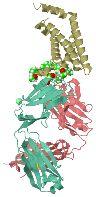
Compounds
|
||||||||||||||||||||||||||||||||||||||||||||||||||||||||||||||||||||||||||||||||||||||||||||||||||||||||||||||||||||||||||||||||||||||||
Chains, Units
Summary Information (see also Sequences/Alignments below) |
Ligands, Modified Residues, Ions (3, 5)| Asymmetric/Biological Unit (3, 5) |
Sites (5, 5)
Asymmetric Unit (5, 5)
|
SS Bonds (4, 4)
Asymmetric/Biological Unit
|
||||||||||||||||||||
Cis Peptide Bonds (5, 5)
Asymmetric/Biological Unit
|
||||||||||||||||||||||||
SAPs(SNPs)/Variants (0, 0)| (no "SAP(SNP)/Variant" information available for 4G7V) |
PROSITE Motifs (0, 0)| (no "PROSITE Motif" information available for 4G7V) |
Exons (0, 0)| (no "Exon" information available for 4G7V) |
Sequences/Alignments
Asymmetric/Biological Unit
Chain H from PDB Type:PROTEIN Length:215
SCOP domains ----------------------------------------------------------------------------------------------------------------------------------------------------------------------------------------------------------------------- SCOP domains
CATH domains ----------------------------------------------------------------------------------------------------------------------------------------------------------------------------------------------------------------------- CATH domains
Pfam domains ----------------------------------------------------------------------------------------------------------------------------------------------------------------------------------------------------------------------- Pfam domains
SAPs(SNPs) ----------------------------------------------------------------------------------------------------------------------------------------------------------------------------------------------------------------------- SAPs(SNPs)
PROSITE ----------------------------------------------------------------------------------------------------------------------------------------------------------------------------------------------------------------------- PROSITE
Transcript ----------------------------------------------------------------------------------------------------------------------------------------------------------------------------------------------------------------------- Transcript
4g7v H 1 EVQLVESGGGLVQPGGSLRLSCAASGFNVSSSSIHWVRQAPGKGLEWVASISSYYGYTSYADSVKGRFTISADTSKNTAYLQMNSLRAEDTAVYYCARSYSWSYAIDYWGQGTLVTVSSASTKGPSVFPLAPSSGGTAALGCLVKDYFPEPVTVSWNSGALTSGVHTFPAVLQSSGLYSLSSVVTVPSSSLGTQTYICNVNHKPSNTKVDKKVEP 219
10 20 30 40 50 60 70 80 90 100 110 120 130 || 144 154 164 174 184 194 204 214
134|
139
Chain L from PDB Type:PROTEIN Length:211
SCOP domains d4g7vl1 L:1-107 automated matches d4g7vl2 L:108-211 automated matches SCOP domains
CATH domains ------------------------------------------------------------------------------------------------------------------------------------------------------------------------------------------------------------------- CATH domains
Pfam domains ------------------------------------------------------------------------------------------------------------------------------------------------------------------------------------------------------------------- Pfam domains
SAPs(SNPs) ------------------------------------------------------------------------------------------------------------------------------------------------------------------------------------------------------------------- SAPs(SNPs)
PROSITE ------------------------------------------------------------------------------------------------------------------------------------------------------------------------------------------------------------------- PROSITE
Transcript ------------------------------------------------------------------------------------------------------------------------------------------------------------------------------------------------------------------- Transcript
4g7v L 1 DIQMTQSPSSLSASVGDRVTITCRASQSVSSAVAWYQQKPGKAPKLLIYSASSLYSGVPSRFSGSGSGTDFTLTISSLQPEDFATYYCQQYFYWPITFGQGTKVEIKRTVAAPSVFIFPPSDEQLKSGTASVVCLLNNFYPREAKVQWKVDNALQAGNSQESVTEQDSKDSTYSLSSTLTLSKADYEKHKVYACEVTHQGLSSPVTKSFNR 211
10 20 30 40 50 60 70 80 90 100 110 120 130 140 150 160 170 180 190 200 210
Chain S from PDB Type:PROTEIN Length:136
SCOP domains ---------------------------------------------------------------------------------------------------------------------------------------- SCOP domains
CATH domains ---------------------------------------------------------------------------------------------------------------------------------------- CATH domains
Pfam domains ---------------------------------------------------------------------------------------------------------------------------------------- Pfam domains
SAPs(SNPs) ---------------------------------------------------------------------------------------------------------------------------------------- SAPs(SNPs)
PROSITE ---------------------------------------------------------------------------------------------------------------------------------------- PROSITE
Transcript ---------------------------------------------------------------------------------------------------------------------------------------- Transcript
4g7v S 101 GVGRVQFRVRAVIDHLGMRVFGVFLIFLDIILMIIDLSLPGKSESSQSFYDGMALALSCYFMLDLGLRIFAYGPKNFFTNPWEVADGLIIVVTFVVTIFYTVLDEYVQETGADGLGELVVLARLLRVVRLARIFYS 236
110 120 130 140 150 160 170 180 190 200 210 220 230
|
||||||||||||||||||||
SCOP Domains (2, 2)
Asymmetric/Biological Unit

|
CATH Domains (0, 0)| (no "CATH Domain" information available for 4G7V) |
Pfam Domains (0, 0)| (no "Pfam Domain" information available for 4G7V) |
Gene Ontology (15, 30)|
Asymmetric/Biological Unit(hide GO term definitions) |
Interactive Views
|
|||||||||||||||||||||||||||||||||||||||||||||||||||||||||||||||||||||||||||||||||||||||||||||||||||||||||||||||||||||||||||||||||||||||||||||||||||||||||||||||||||||||||||||||||||||||||||||
Still Images
|
||||||||||||||||
Databases
|
||||||||||||||||||||||||||||||||||||||||||||||||||||||||||||||||||||||||||||||||||||||||||||||||||||||||||||||||||||||||||||||||||||||||||||||||||||||||||||||||||||||||||||||||||||||||||
Analysis Tools
|
||||||||||||||||||||||||||||||||||||||||||||||||||||||||||||||||||||||||
Entries Sharing at Least One Protein Chain (UniProt ID)
Related Entries Specified in the PDB File
|
|