|
|
|
|
 Description
|
|
Compounds
|
||||||||||||||||||||||||||||||||||||||||||||||||||||||||
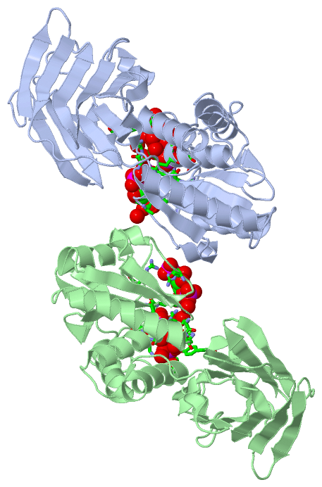
Chains, Units
Summary Information (see also Sequences/Alignments below) |
Ligands, Modified Residues, Ions (1, 4)
Asymmetric Unit (1, 4)
|
Sites (4, 4)
Asymmetric Unit (4, 4)
|
SS Bonds (0, 0)| (no "SS Bond" information available for 3V0H) |
Cis Peptide Bonds (2, 2)
Asymmetric Unit
|
||||||||||||
SAPs(SNPs)/Variants (0, 0)| (no "SAP(SNP)/Variant" information available for 3V0H) |
PROSITE Motifs (0, 0)| (no "PROSITE Motif" information available for 3V0H) |
Exons (0, 0)| (no "Exon" information available for 3V0H) |
Sequences/Alignments
Asymmetric UnitChain A from PDB Type:PROTEIN Length:326 aligned with Q4W8A1_CIOIN | Q4W8A1 from UniProtKB/TrEMBL Length:576 Alignment length:326 259 269 279 289 299 309 319 329 339 349 359 369 379 389 399 409 419 429 439 449 459 469 479 489 499 509 519 529 539 549 559 569 Q4W8A1_CIOIN 250 QNKRRYRKDGFDLDLTYVTDHVIAMSFPSSGRQSLFRNPIGEVSRFFKTKHPDKFRIYNLCSERGYDETKFDNHVYRVMIDDHNVPTLVDLLKFIDDAKVWMTSDPDHVIAIHCKGGKGRTGTLVSSWLLEDGKFDTAKEALEYFGSRRTDFEVGDVFQGVETASQIRYVGYFEKIKKNYGGQLPPMKKLKVTGVTITAIQGVGRGNGSDLSMQIVSERQEVLLCKFAEGYNCALQYDATDDCVTCEVKNCPVLAGDIKVRFMSTSKSLPRGYDNCPFYFWFNTSLVEGDHVTLKREEIDNPHKKKTWKIYRDNFTVKLTFSDAED 575 SCOP domains -------------------------------------------------------------------------------------------------------------------------------------------------------------------------------------------------------------------------------------------------------------------------------------------------------------------------------------- SCOP domains CATH domains -------------------------------------------------------------------------------------------------------------------------------------------------------------------------------------------------------------------------------------------------------------------------------------------------------------------------------------- CATH domains Pfam domains -------------------------------------------------------------------------------------------------------------------------------------------------------------------------------------------------------------------------------------------------------------------------------------------------------------------------------------- Pfam domains SAPs(SNPs) -------------------------------------------------------------------------------------------------------------------------------------------------------------------------------------------------------------------------------------------------------------------------------------------------------------------------------------- SAPs(SNPs) PROSITE -------------------------------------------------------------------------------------------------------------------------------------------------------------------------------------------------------------------------------------------------------------------------------------------------------------------------------------- PROSITE Transcript -------------------------------------------------------------------------------------------------------------------------------------------------------------------------------------------------------------------------------------------------------------------------------------------------------------------------------------- Transcript 3v0h A 250 QNKRRYRKDGFDLDLTYVTDHVIAMSFPSSGRQSLFRNPIGEVSRFFKTKHPDKFRIYNLCSERGYDETKFDNHVYRVMIDDHNVPTLVDLLKFIDDAKVWMTSDPDHVIAIHSKGGKGRTGTLVSSWLLEDGKFDTAKEALEYFGSRRTDFEVGDVFQGVETASQIRYVGYFEKIKKNYGGQLPPMKKLKVTGVTITAIQGVGRGNGSDLSMQIVSERQEVLLCKFAEGYNCALQYDATDDCVTCEVKNCPVLAGDIKVRFMSTSKSLPRGYDNCPFYFWFNTSLVEGDHVTLKREEIDNPHKKKTWKIYRDNFTVKLTFSDAED 575 259 269 279 289 299 309 319 329 339 349 359 369 379 389 399 409 419 429 439 449 459 469 479 489 499 509 519 529 539 549 559 569 Chain B from PDB Type:PROTEIN Length:327 aligned with Q4W8A1_CIOIN | Q4W8A1 from UniProtKB/TrEMBL Length:576 Alignment length:327 258 268 278 288 298 308 318 328 338 348 358 368 378 388 398 408 418 428 438 448 458 468 478 488 498 508 518 528 538 548 558 568 Q4W8A1_CIOIN 249 SQNKRRYRKDGFDLDLTYVTDHVIAMSFPSSGRQSLFRNPIGEVSRFFKTKHPDKFRIYNLCSERGYDETKFDNHVYRVMIDDHNVPTLVDLLKFIDDAKVWMTSDPDHVIAIHCKGGKGRTGTLVSSWLLEDGKFDTAKEALEYFGSRRTDFEVGDVFQGVETASQIRYVGYFEKIKKNYGGQLPPMKKLKVTGVTITAIQGVGRGNGSDLSMQIVSERQEVLLCKFAEGYNCALQYDATDDCVTCEVKNCPVLAGDIKVRFMSTSKSLPRGYDNCPFYFWFNTSLVEGDHVTLKREEIDNPHKKKTWKIYRDNFTVKLTFSDAED 575 SCOP domains --------------------------------------------------------------------------------------------------------------------------------------------------------------------------------------------------------------------------------------------------------------------------------------------------------------------------------------- SCOP domains CATH domains --------------------------------------------------------------------------------------------------------------------------------------------------------------------------------------------------------------------------------------------------------------------------------------------------------------------------------------- CATH domains Pfam domains --------------------------------------------------------------------------------------------------------------------------------------------------------------------------------------------------------------------------------------------------------------------------------------------------------------------------------------- Pfam domains SAPs(SNPs) --------------------------------------------------------------------------------------------------------------------------------------------------------------------------------------------------------------------------------------------------------------------------------------------------------------------------------------- SAPs(SNPs) PROSITE --------------------------------------------------------------------------------------------------------------------------------------------------------------------------------------------------------------------------------------------------------------------------------------------------------------------------------------- PROSITE Transcript --------------------------------------------------------------------------------------------------------------------------------------------------------------------------------------------------------------------------------------------------------------------------------------------------------------------------------------- Transcript 3v0h B 249 SQNKRRYRKDGFDLDLTYVTDHVIAMSFPSSGRQSLFRNPIGEVSRFFKTKHPDKFRIYNLCSERGYDETKFDNHVYRVMIDDHNVPTLVDLLKFIDDAKVWMTSDPDHVIAIHSKGGKGRTGTLVSSWLLEDGKFDTAKEALEYFGSRRTDFEVGDVFQGVETASQIRYVGYFEKIKKNYGGQLPPMKKLKVTGVTITAIQGVGRGNGSDLSMQIVSERQEVLLCKFAEGYNCALQYDATDDCVTCEVKNCPVLAGDIKVRFMSTSKSLPRGYDNCPFYFWFNTSLVEGDHVTLKREEIDNPHKKKTWKIYRDNFTVKLTFSDAED 575 258 268 278 288 298 308 318 328 338 348 358 368 378 388 398 408 418 428 438 448 458 468 478 488 498 508 518 528 538 548 558 568
|
||||||||||||||||||||
SCOP Domains (0, 0)| (no "SCOP Domain" information available for 3V0H) |
CATH Domains (0, 0)| (no "CATH Domain" information available for 3V0H) |
Pfam Domains (0, 0)| (no "Pfam Domain" information available for 3V0H) |
Gene Ontology (15, 15)|
Asymmetric Unit(hide GO term definitions) Chain A,B (Q4W8A1_CIOIN | Q4W8A1)
|
||||||||||||||||||||||||||||||||||||||||||||||||||||||||||||||||||||||||||||||||||||||||||||||||||||||||||||
Interactive Views
|
||||||||||||||||||||||||||||||||||||||||||||||||||||||||||||||||||||||||||||||||||||||||||||||||||||||||||||||||||||||||||||||||||||||||||||||||||||||||||||||||||||||||||
Still Images
|
||||||||||||||||
Databases
|
||||||||||||||||||||||||||||||||||||||||||||||||||||||||||||||||||||||||||||||||||||||||||||||||||||||||||||||||||||||||||||||||||||||||||||||||||||||||||||||||
Analysis Tools
|
|||||||||||||||||||||||||||||||||||||||||||||||||||||||||||||
Entries Sharing at Least One Protein Chain (UniProt ID)
Related Entries Specified in the PDB File
|
|