Show PDB file:   
         Plain Text   HTML   (compressed file size)
QuickSearch:   
by PDB,NDB,UniProt,PROSITE Code or Search Term(s)  
(-)Asymmetric Unit
(-)Asym. Unit - sites
(-)Biological Unit 1
(-)Biol. Unit 1 - sites
(-)Biological Unit 2
collapse expand < >
Image Asymmetric Unit
Asymmetric Unit  (Jmol Viewer)
Image Asym. Unit - sites
Asym. Unit - sites  (Jmol Viewer)
Image Biological Unit 1
Biological Unit 1  (Jmol Viewer)
Image Biol. Unit 1 - sites
Biol. Unit 1 - sites  (Jmol Viewer)
Image Biological Unit 2
Biological Unit 2  (Jmol Viewer)

(-) Description

Title :  CRYSTAL STRUCTURE OF HUMAN HISTIDINE TRIAD NUCLEOTIDE-BINDING PROTEIN 1 (HHINT1) REFINED TO 1.92A AT P21 SPACE GROUP
 
Authors :  R. M. Dolot, A. Seda, B. Nawrot
Date :  30 Apr 15  (Deposition) - 02 Mar 16  (Release) - 09 Mar 16  (Revision)
Method :  X-RAY DIFFRACTION
Resolution :  1.92
Chains :  Asym. Unit :  A,B,C,D
Biol. Unit 1:  A,B  (1x)
Biol. Unit 2:  C,D  (1x)
Keywords :  Hint, Hit, Histidine Triad, Hydrolase, Phosphoramidase (Keyword Search: [Gene Ontology, PubMed, Web (Google))
 
Reference :  R. Dolot, R. Kaczmarek, A. Seda, A. Krakowiak, J. Baraniak, B. Nawrot
Crystallographic Studies Of The Complex Of Human Hint1 Protein With A Non-Hydrolyzable Analog Of Ap4A.
Int. J. Biol. Macromol. V. 87 62 2016
PubMed-ID: 26905466  |  Reference-DOI: 10.1016/J.IJBIOMAC.2016.02.047

(-) Compounds

Molecule 1 - HISTIDINE TRIAD NUCLEOTIDE-BINDING PROTEIN 1
    ChainsA, B, C, D
    EC Number3.-.-.-
    EngineeredYES
    Expression SystemESCHERICHIA COLI BL21(DE3)
    Expression System PlasmidPSGA02
    Expression System Taxid469008
    Expression System Vector TypePLASMID
    GeneHINT1, HINT, PKCI1, PRKCNH1
    Organism CommonHUMAN
    Organism ScientificHOMO SAPIENS
    Organism Taxid9606
    SynonymADENOSINE 5'-MONOPHOSPHORAMIDASE,PROTEIN KINASE C INHIBITOR 1,PROTEIN KINASE C-INTERACTING PROTEIN 1,PKCI-1

 Structural Features

(-) Chains, Units

  1234
Asymmetric Unit ABCD
Biological Unit 1 (1x)AB  
Biological Unit 2 (1x)  CD

Summary Information (see also Sequences/Alignments below)

(-) Ligands, Modified Residues, Ions  (1, 2)

Asymmetric Unit (1, 2)
No.NameCountTypeFull Name
1SO42Ligand/IonSULFATE ION
Biological Unit 1 (1, 1)
No.NameCountTypeFull Name
1SO41Ligand/IonSULFATE ION
Biological Unit 2 (1, 1)
No.NameCountTypeFull Name
1SO41Ligand/IonSULFATE ION

(-) Sites  (2, 2)

Asymmetric Unit (2, 2)
No.NameEvidenceResiduesDescription
1AC1SOFTWAREASP B:43 , SER B:107 , HIS B:114 , HOH B:305 , HOH B:310 , HOH B:330binding site for residue SO4 B 201
2AC2SOFTWAREASP C:43 , SER C:107 , HIS C:114 , HOH C:343 , HOH C:367binding site for residue SO4 C 201

(-) SS Bonds  (0, 0)

(no "SS Bond" information available for 4ZKV)

(-) Cis Peptide Bonds  (4, 4)

Asymmetric Unit
No.Residues
1Trp A:123 -Pro A:124
2Trp B:123 -Pro B:124
3Trp C:123 -Pro C:124
4Trp D:123 -Pro D:124

 Sequence-Structure Mapping

(-) SAPs(SNPs)/Variants  (0, 0)

(no "SAP(SNP)/Variant" information available for 4ZKV)

(-) PROSITE Motifs  (0, 0)

(no "PROSITE Motif" information available for 4ZKV)

(-) Exons   (0, 0)

(no "Exon" information available for 4ZKV)

(-) Sequences/Alignments

Asymmetric Unit
   Reformat: Number of residues per line =  ('0' or empty: single-line sequence representation)
  Number of residues per labelling interval =   
  UniProt sequence: complete  aligned part    
   Show mapping: SCOP domains CATH domains Pfam domains Secondary structure (by author)
SAPs(SNPs) PROSITE motifs Exons
(details for a mapped element are shown in a popup box when the mouse pointer rests over it)
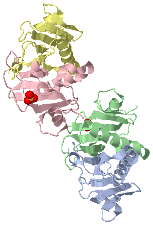
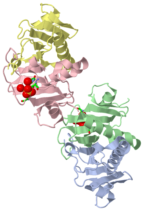
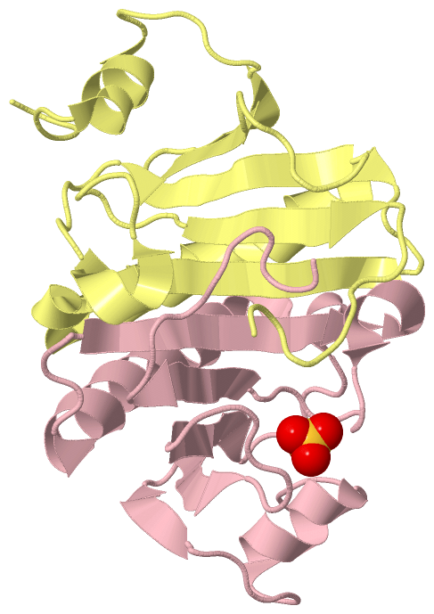
Chain A from PDB  Type:PROTEIN  Length:115
                                                                                                                                                   
               SCOP domains ------------------------------------------------------------------------------------------------------------------- SCOP domains
               CATH domains ------------------------------------------------------------------------------------------------------------------- CATH domains
               Pfam domains ------------------------------------------------------------------------------------------------------------------- Pfam domains
         Sec.struct. author .....hhhhhhhh......eeee...eeeee.......eeeeeee.....hhhhhhhhhhhhhhhhhhhhhhhhhhh....eeeeeeeehhhhh.......eeeee......... Sec.struct. author
                 SAPs(SNPs) ------------------------------------------------------------------------------------------------------------------- SAPs(SNPs)
                    PROSITE ------------------------------------------------------------------------------------------------------------------- PROSITE
                 Transcript ------------------------------------------------------------------------------------------------------------------- Transcript
                 4zkv A  12 RPGGDTIFGKIIRKEIPAKIIFEDDRCLAFHDISPQAPTHFLVIPKKHISQISVAEDDDESLLGHLMIVGKKCAADLGLNKGYRMVVNEGSDGGQSVYHVHLHVLGGRQMHWPPG 126
                                    21        31        41        51        61        71        81        91       101       111       121     

Chain B from PDB  Type:PROTEIN  Length:115
                                                                                                                                                   
               SCOP domains ------------------------------------------------------------------------------------------------------------------- SCOP domains
               CATH domains ------------------------------------------------------------------------------------------------------------------- CATH domains
               Pfam domains ------------------------------------------------------------------------------------------------------------------- Pfam domains
         Sec.struct. author .....hhhhhhhh......eeee...eeeee.......eeeeeee.....hhhhhhhhhhhhhhhhhhhhhhhhhhh.....eeeeeeehhhhh.......eeeee......... Sec.struct. author
                 SAPs(SNPs) ------------------------------------------------------------------------------------------------------------------- SAPs(SNPs)
                    PROSITE ------------------------------------------------------------------------------------------------------------------- PROSITE
                 Transcript ------------------------------------------------------------------------------------------------------------------- Transcript
                 4zkv B  12 RPGGDTIFGKIIRKEIPAKIIFEDDRCLAFHDISPQAPTHFLVIPKKHISQISVAEDDDESLLGHLMIVGKKCAADLGLNKGYRMVVNEGSDGGQSVYHVHLHVLGGRQMHWPPG 126
                                    21        31        41        51        61        71        81        91       101       111       121     

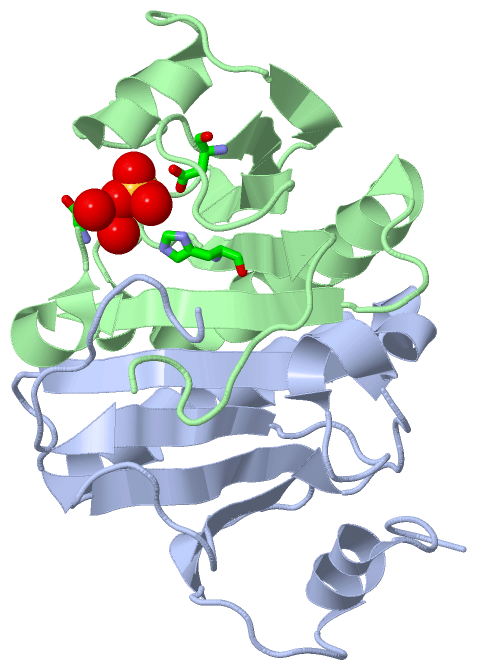
Chain C from PDB  Type:PROTEIN  Length:115
                                                                                                                                                   
               SCOP domains ------------------------------------------------------------------------------------------------------------------- SCOP domains
               CATH domains ------------------------------------------------------------------------------------------------------------------- CATH domains
               Pfam domains ------------------------------------------------------------------------------------------------------------------- Pfam domains
         Sec.struct. author .....hhhhhhhh......eeee...eeeee.......eeeeeee.....hhhhhhhhhhhhhhhhhhhhhhhhhhh....eeeeeeeehhhhh.......eeeee......... Sec.struct. author
                 SAPs(SNPs) ------------------------------------------------------------------------------------------------------------------- SAPs(SNPs)
                    PROSITE ------------------------------------------------------------------------------------------------------------------- PROSITE
                 Transcript ------------------------------------------------------------------------------------------------------------------- Transcript
                 4zkv C  12 RPGGDTIFGKIIRKEIPAKIIFEDDRCLAFHDISPQAPTHFLVIPKKHISQISVAEDDDESLLGHLMIVGKKCAADLGLNKGYRMVVNEGSDGGQSVYHVHLHVLGGRQMHWPPG 126
                                    21        31        41        51        61        71        81        91       101       111       121     

Chain D from PDB  Type:PROTEIN  Length:115
                                                                                                                                                   
               SCOP domains ------------------------------------------------------------------------------------------------------------------- SCOP domains
               CATH domains ------------------------------------------------------------------------------------------------------------------- CATH domains
               Pfam domains ------------------------------------------------------------------------------------------------------------------- Pfam domains
         Sec.struct. author .....hhhhhhhh......eeee...eeeee.......eeeeeee.....hhhhhhhhhhhhhhhhhhhhhhhhhhh....eeeeeeeehhhhh.......eeeee......... Sec.struct. author
                 SAPs(SNPs) ------------------------------------------------------------------------------------------------------------------- SAPs(SNPs)
                    PROSITE ------------------------------------------------------------------------------------------------------------------- PROSITE
                 Transcript ------------------------------------------------------------------------------------------------------------------- Transcript
                 4zkv D  12 RPGGDTIFGKIIRKEIPAKIIFEDDRCLAFHDISPQAPTHFLVIPKKHISQISVAEDDDESLLGHLMIVGKKCAADLGLNKGYRMVVNEGSDGGQSVYHVHLHVLGGRQMHWPPG 126
                                    21        31        41        51        61        71        81        91       101       111       121     

   Legend:   → Mismatch (orange background)
  - → Gap (green background, '-', border residues have a numbering label)
    → Modified Residue (blue background, lower-case, 'x' indicates undefined single-letter code, labelled with number + name)
  x → Chemical Group (purple background, 'x', labelled with number + name, e.g. ACE or NH2)
  extra numbering lines below/above indicate numbering irregularities and modified residue names etc., number ends below/above '|'

 Classification and Annotation

(-) SCOP Domains  (0, 0)

(no "SCOP Domain" information available for 4ZKV)

(-) CATH Domains  (0, 0)

(no "CATH Domain" information available for 4ZKV)

(-) Pfam Domains  (0, 0)

(no "Pfam Domain" information available for 4ZKV)

(-) Gene Ontology  (17, 17)

Asymmetric Unit(hide GO term definitions)

 Visualization

(-) Interactive Views

Asymmetric Unit
  Complete Structure
    Jena3D(integrated viewing of ligand, site, SAP, PROSITE, SCOP information)
    WebMol | AstexViewer[tm]@PDBe
(Java Applets, require no local installation except for Java; loading may be slow)
    STRAP
(Java WebStart application, automatic local installation, requires Java; full application with system access!)
    RasMol
(require local installation)
    Molscript (VRML)
(requires installation of a VRML viewer; select preferred view via VRML and generate a mono or stereo PDF format file)
 
  Ligands, Modified Residues, Ions
    SO4  [ RasMol | Jena3D ]  +environment [ RasMol | Jena3D ]
 
  Sites
    AC1  [ RasMol ]  +environment [ RasMol ]
    AC2  [ RasMol ]  +environment [ RasMol ]
 
  Cis Peptide Bonds
    Trp A:123 - Pro A:124   [ RasMol ]  
    Trp B:123 - Pro B:124   [ RasMol ]  
    Trp C:123 - Pro C:124   [ RasMol ]  
    Trp D:123 - Pro D:124   [ RasMol ]  
 
Biological Units
  Complete Structure
    Biological Unit 1  [ Jena3D ]
    Biological Unit 2  [ Jena3D ]

(-) Still Images

Jmol
  protein: cartoon or spacefill or dots and stick; nucleic acid: cartoon and stick; ligands: spacefill; active site: stick
Molscript
  protein, nucleic acid: cartoon; ligands: spacefill; active site: ball and stick

 Databases and Analysis Tools

(-) Databases

Access by PDB/NDB ID
  4zkv
    Family and Domain InformationProDom | SYSTERS
    General Structural InformationGlycoscienceDB | MMDB | NDB | OCA | PDB | PDBe | PDBj | PDBsum | PDBWiki | PQS | PROTEOPEDIA
    Orientation in MembranesOPM
    Protein SurfaceSURFACE
    Secondary StructureDSSP (structure derived) | HSSP (homology derived)
    Structural GenomicsGeneCensus
    Structural NeighboursCE | VAST
    Structure ClassificationCATH | Dali | SCOP
    Validation and Original DataBMRB Data View | BMRB Restraints Grid | EDS | PROCHECK | RECOORD | WHAT_CHECK
 
Access by UniProt ID/Accession number
  HINT1_HUMAN | P49773
    Comparative Protein Structure ModelsModBase
    Genomic InformationEnsembl
    Protein-protein InteractionDIP
    Sequence, Family and Domain InformationInterPro | Pfam | SMART | UniProtKB/SwissProt
 
Access by Enzyme Classificator   (EC Number)
  3.-.-.-
    General Enzyme InformationBRENDA | EC-PDB | Enzyme | IntEnz
    PathwayKEGG | MetaCyc
 
Access by Disease Identifier   (MIM ID)
  (no 'MIM ID' available)
    Disease InformationOMIM
 
Access by GenAge ID
  (no 'GenAge ID' available)
    Age Related InformationGenAge

(-) Analysis Tools

Access by PDB/NDB ID
    Domain InformationXDom
    Interatomic Contacts of Structural UnitsCSU
    Ligand-protein ContactsLPC
    Protein CavitiescastP
    Sequence and Secondary StructurePDBCartoon
    Structure AlignmentSTRAP(Java WebStart application, automatic local installation, requires Java; full application with system access!)
    Structure and Sequence BrowserSTING
 
Access by UniProt ID/Accession number
  HINT1_HUMAN | P49773
    Protein Disorder PredictionDisEMBL | FoldIndex | GLOBPLOT (for more information see DisProt)

 Related Entries

(-) Entries Sharing at Least One Protein Chain (UniProt ID)

UniProtKB/Swiss-Prot
        HINT1_HUMAN | P497731av5 1kpa 1kpb 1kpc 1kpe 1kpf 3tw2 4eqe 4eqg 4eqh 4zkl 5ed3 5ed6 5emt 5i2e 5i2f 5ipb 5ipc 5ipd 5ipe 5kly 5klz 5km0 5km1 5km2 5km3 5km4 5km6 5km7 5kma 5kmb 5kmc 5o8g 5o8i

(-) Related Entries Specified in the PDB File

1kpc 1KPC CONTAINS MODEL REFINED TO LOWER RESOLUTION AT THE SAME SPACE GROUP