Show PDB file:   
         Plain Text   HTML   (compressed file size)
QuickSearch:   
by PDB,NDB,UniProt,PROSITE Code or Search Term(s)  
(-)Asymmetric Unit
(-)Asym. Unit - sites
(-)Biological Unit 1
(-)Biol. Unit 1 - sites
(-)Biological Unit 2
collapse expand < >
Image Asymmetric Unit
Asymmetric Unit  (Jmol Viewer)
Image Asym. Unit - sites
Asym. Unit - sites  (Jmol Viewer)
Image Biological Unit 1
Biological Unit 1  (Jmol Viewer)
Image Biol. Unit 1 - sites
Biol. Unit 1 - sites  (Jmol Viewer)
Image Biological Unit 2
Biological Unit 2  (Jmol Viewer)

(-) Description

Title :  CRYSTAL STRUCTURE OF LARGE STOKES SHIFT FLUORESCENT PROTEIN LSSMORANGE
 
Authors :  S. Pletnev, Z. Dauter
Date :  25 Apr 14  (Deposition) - 09 Jul 14  (Release) - 09 Jul 14  (Revision)
Method :  X-RAY DIFFRACTION
Resolution :  1.40
Chains :  Asym. Unit :  A,B
Biol. Unit 1:  A  (1x)
Biol. Unit 2:  B  (1x)
Keywords :  Beta-Barrel, Large Stokes Shift Fluorescent Protein, Chromophore, Fluorescent Protein (Keyword Search: [Gene Ontology, PubMed, Web (Google))
 
Reference :  S. Pletnev, D. M. Shcherbakova, O. M. Subach, N. V. Pletneva, V. N. Malashkevich, S. C. Almo, Z. Dauter, V. V. Verkhusha
Orange Fluorescent Proteins: Structural Studies Of Lssmorange, Psmorange And Psmorange2.
Plos One V. 9 99136 2014
PubMed-ID: 24960050  |  Reference-DOI: 10.1371/JOURNAL.PONE.0099136

(-) Compounds

Molecule 1 - LSSMORANGE
    ChainsA, B
    EngineeredYES
    Expression SystemESCHERICHIA COLI
    Expression System StrainLMG194
    Expression System Taxid562
    MutationYES
    Organism CommonSEA ANEMONE
    Organism ScientificDISCOSOMA SP.
    Organism Taxid86600

 Structural Features

(-) Chains, Units

  12
Asymmetric Unit AB
Biological Unit 1 (1x)A 
Biological Unit 2 (1x) B

Summary Information (see also Sequences/Alignments below)

(-) Ligands, Modified Residues, Ions  (3, 28)

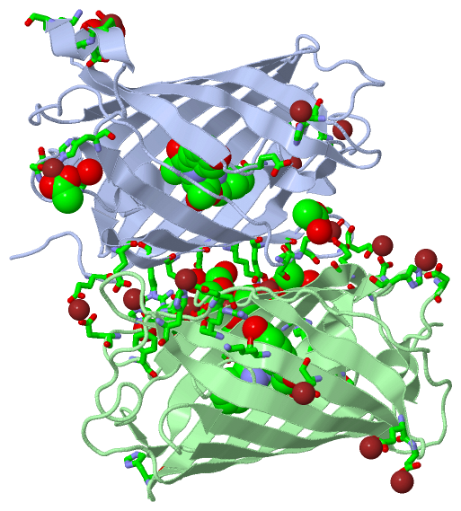
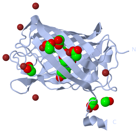
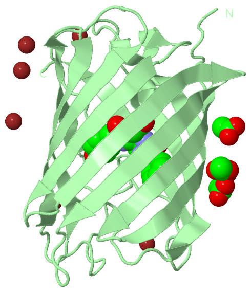
Asymmetric Unit (3, 28)
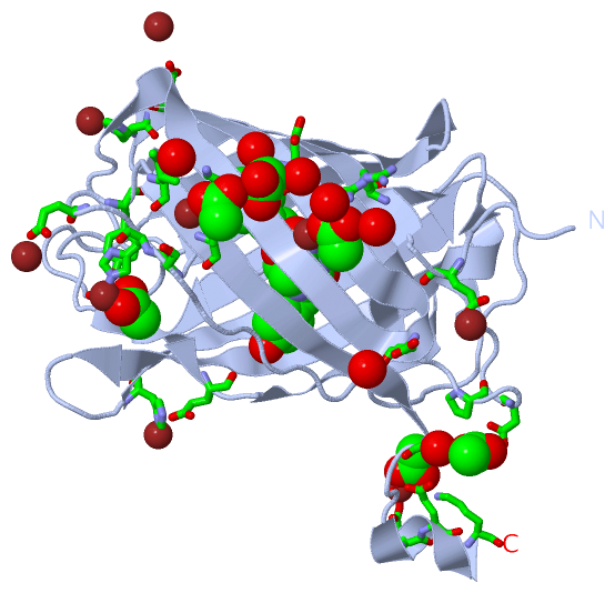
No.NameCountTypeFull Name
1ACT9Ligand/IonACETATE ION
2OFM2Mod. Amino Acid[(4Z)-2-{(2R,5R)-2-[(1S)-1-AMINO-2-PHENYLETHYL]-2-HYDROXY-5-METHYL-2,5-DIHYDRO-1,3-OXAZOL-4-YL}-4-(4-HYDROXYBENZYLIDENE)-5-OXO-4,5-DIHYDRO-1H-IMIDAZOL-1-YL]ACETIC ACID
3ZN17Ligand/IonZINC ION
Biological Unit 1 (2, 7)
No.NameCountTypeFull Name
1ACT6Ligand/IonACETATE ION
2OFM1Mod. Amino Acid[(4Z)-2-{(2R,5R)-2-[(1S)-1-AMINO-2-PHENYLETHYL]-2-HYDROXY-5-METHYL-2,5-DIHYDRO-1,3-OXAZOL-4-YL}-4-(4-HYDROXYBENZYLIDENE)-5-OXO-4,5-DIHYDRO-1H-IMIDAZOL-1-YL]ACETIC ACID
3ZN-1Ligand/IonZINC ION
Biological Unit 2 (2, 4)
No.NameCountTypeFull Name
1ACT3Ligand/IonACETATE ION
2OFM1Mod. Amino Acid[(4Z)-2-{(2R,5R)-2-[(1S)-1-AMINO-2-PHENYLETHYL]-2-HYDROXY-5-METHYL-2,5-DIHYDRO-1,3-OXAZOL-4-YL}-4-(4-HYDROXYBENZYLIDENE)-5-OXO-4,5-DIHYDRO-1H-IMIDAZOL-1-YL]ACETIC ACID
3ZN-1Ligand/IonZINC ION

(-) Sites  (26, 26)

Asymmetric Unit (26, 26)
No.NameEvidenceResiduesDescription
01AC1SOFTWAREGLU A:10 , HOH A:478 , GLU B:206 , HOH B:474BINDING SITE FOR RESIDUE ZN A 301
02AC2SOFTWAREHIS A:204 , TYR A:208 , ACT A:313 , HOH A:479 , GLU B:39BINDING SITE FOR RESIDUE ZN A 302
03AC3SOFTWAREGLU A:206 , HOH A:480 , GLU B:10 , HOH B:476BINDING SITE FOR RESIDUE ZN A 303
04AC4SOFTWAREASP A:227 , ACT A:315 , HOH A:481 , HOH A:482 , HIS B:25BINDING SITE FOR RESIDUE ZN A 304
05AC5SOFTWAREASP A:78 , HIS A:221 , ACT A:314BINDING SITE FOR RESIDUE ZN A 305
06AC6SOFTWAREGLU A:34 , ACT A:311 , GLU B:212 , ACT B:308BINDING SITE FOR RESIDUE ZN A 306
07AC7SOFTWAREGLU A:212 , ACT A:312 , GLU B:34 , ACT B:309BINDING SITE FOR RESIDUE ZN A 307
08AC8SOFTWAREGLU A:26 , HIS A:172 , HOH A:518BINDING SITE FOR RESIDUE ZN A 308
09AC9SOFTWAREGLU A:144 , HIS A:172BINDING SITE FOR RESIDUE ZN A 309
10BC1SOFTWAREHIS A:25 , HOH A:546 , HOH A:547 , HOH A:556BINDING SITE FOR RESIDUE ZN A 310
11BC2SOFTWAREARG A:13 , GLU A:34 , ZN A:306 , HOH A:423 , HOH A:483 , LYS B:45 , SER B:203 , GLU B:212 , ACT B:308BINDING SITE FOR RESIDUE ACT A 311
12BC3SOFTWARELYS A:45 , GLU A:212 , ZN A:307 , ACT A:316 , GLU B:34 , ARG B:164 , ZN B:306 , ACT B:309 , ACT B:310BINDING SITE FOR RESIDUE ACT A 312
13BC4SOFTWAREHIS A:204 , TYR A:208 , ZN A:302 , GLU B:39 , HOH B:427BINDING SITE FOR RESIDUE ACT A 313
14BC5SOFTWAREPRO A:76 , ASP A:78 , HIS A:221 , ZN A:305 , HOH A:460BINDING SITE FOR RESIDUE ACT A 314
15BC6SOFTWAREGLU A:228 , LYS A:231 , ZN A:304 , HOH A:481 , HOH A:482 , HIS B:25BINDING SITE FOR RESIDUE ACT A 315
16BC7SOFTWAREACT A:312 , HOH A:515 , HOH A:552 , ARG B:164 , ZN B:306 , ACT B:308BINDING SITE FOR RESIDUE ACT A 316
17BC8SOFTWAREGLU A:32 , GLU B:144 , HIS B:172 , HOH B:472BINDING SITE FOR RESIDUE ZN B 301
18BC9SOFTWAREGLU A:39 , HOH A:477 , HIS B:204 , TYR B:208 , HOH B:473BINDING SITE FOR RESIDUE ZN B 302
19CC1SOFTWAREASP B:78 , HIS B:221 , HOH B:477BINDING SITE FOR RESIDUE ZN B 303
20CC2SOFTWAREASP B:115 , GLU B:153 , HOH B:478BINDING SITE FOR RESIDUE ZN B 304
21CC3SOFTWARELYS B:15 , GLU B:117 , GLU B:160 , HOH B:479 , HOH B:500BINDING SITE FOR RESIDUE ZN B 305
22CC4SOFTWAREACT A:312 , ACT A:316 , ACT B:308 , ACT B:310BINDING SITE FOR RESIDUE ZN B 306
23CC5SOFTWAREASP B:154 , HOH B:522BINDING SITE FOR RESIDUE ZN B 307
24CC6SOFTWAREGLU A:34 , ZN A:306 , ACT A:311 , ACT A:316 , LYS B:45 , GLU B:212 , ZN B:306 , ACT B:310BINDING SITE FOR RESIDUE ACT B 308
25CC7SOFTWARELYS A:45 , SER A:203 , ILE A:210 , GLU A:212 , ZN A:307 , ACT A:312 , HOH A:493 , ARG B:13 , GLU B:34 , HOH B:533BINDING SITE FOR RESIDUE ACT B 309
26CC8SOFTWAREACT A:312 , GLU B:32 , LYS B:162 , ZN B:306 , ACT B:308BINDING SITE FOR RESIDUE ACT B 310

(-) SS Bonds  (0, 0)

(no "SS Bond" information available for 4Q7R)

(-) Cis Peptide Bonds  (4, 4)

Asymmetric Unit
No.Residues
1Gly A:52 -Pro A:53
2Phe A:87 -Pro A:88
3Gly B:52 -Pro B:53
4Phe B:87 -Pro B:88

 Sequence-Structure Mapping

(-) SAPs(SNPs)/Variants  (0, 0)

(no "SAP(SNP)/Variant" information available for 4Q7R)

(-) PROSITE Motifs  (0, 0)

(no "PROSITE Motif" information available for 4Q7R)

(-) Exons   (0, 0)

(no "Exon" information available for 4Q7R)

(-) Sequences/Alignments

Asymmetric Unit
   Reformat: Number of residues per line =  ('0' or empty: single-line sequence representation)
  Number of residues per labelling interval =   
  UniProt sequence: complete  aligned part    
   Show mapping: SCOP domains CATH domains Pfam domains Secondary structure (by author)
SAPs(SNPs) PROSITE motifs Exons
(details for a mapped element are shown in a popup box when the mouse pointer rests over it)
Chain A from PDB  Type:PROTEIN  Length:224
                                                                                                                                                                                                                                                                
               SCOP domains -------------------------------------------------------------------------------------------------------------------------------------------------------------------------------------------------------------------------------- SCOP domains
               CATH domains -------------------------------------------------------------------------------------------------------------------------------------------------------------------------------------------------------------------------------- CATH domains
               Pfam domains -------------------------------------------------------------------------------------------------------------------------------------------------------------------------------------------------------------------------------- Pfam domains
         Sec.struct. author .......eeeeeeeeeee..eeeeeeeeeeeeeeeeeeeeeeeeeeee.............hhhhh.......hhhhhh....eeeeeeeee....eeeeeeeeeee..eeeeeeeeeee............eeee..eeeeeeee..eeeeeeeeeeee....eeeeeeeeeeee..........................eeeeeeeeeee....hhhhhh. Sec.struct. author
                 SAPs(SNPs) -------------------------------------------------------------------------------------------------------------------------------------------------------------------------------------------------------------------------------- SAPs(SNPs)
                    PROSITE -------------------------------------------------------------------------------------------------------------------------------------------------------------------------------------------------------------------------------- PROSITE
                 Transcript -------------------------------------------------------------------------------------------------------------------------------------------------------------------------------------------------------------------------------- Transcript
                 4q7r A   5 MAIIKEFMRFKVHMEGSVNGHEFEIEGEGEGRPYEGFQTVKLKVTKGGPLPFAWDILSPQfSKAYVKHPADIPDYLKLSFPEGFKWERVMNFEDGGVVTVTQDSSLQDGEFIYKVKLRGTNFPSDGPVMQKKTMGMEASSERMYPEDGALKGEDKLRLKLKDGGHYTSEVKTTYKAKKPVQLPGAYIVDIKLDITSHNEDYTIVEQYERAEGRHSTGGMDELYK 231
                                    14        24        34        44        54        64||      77        87        97       107       117       127       137       147       157       167       177       187       197       207       217       227    
                                                                                      64||                                                                                                                                                                  
                                                                                       66-OFM                                                                                                                                                               
                                                                                        69                                                                                                                                                                  

Chain B from PDB  Type:PROTEIN  Length:215
                                                                                                                                                                                                                                                       
               SCOP domains ----------------------------------------------------------------------------------------------------------------------------------------------------------------------------------------------------------------------- SCOP domains
               CATH domains ----------------------------------------------------------------------------------------------------------------------------------------------------------------------------------------------------------------------- CATH domains
               Pfam domains ----------------------------------------------------------------------------------------------------------------------------------------------------------------------------------------------------------------------- Pfam domains
         Sec.struct. author .......eeeeeeeeeee..eeeeeeeeeeeeeeeeeeeeeeeeeeee.............hhhhh.......hhhhhh....eeeeeeeee....eeeeeeeeeee..eeeeeeeeeee..................eeeeeeee..eeeeeeeeeee.....eeeeeeeeeeee..........................eeeeeeeeeee.. Sec.struct. author
                 SAPs(SNPs) ----------------------------------------------------------------------------------------------------------------------------------------------------------------------------------------------------------------------- SAPs(SNPs)
                    PROSITE ----------------------------------------------------------------------------------------------------------------------------------------------------------------------------------------------------------------------- PROSITE
                 Transcript ----------------------------------------------------------------------------------------------------------------------------------------------------------------------------------------------------------------------- Transcript
                 4q7r B   5 MAIIKEFMRFKVHMEGSVNGHEFEIEGEGEGRPYEGFQTVKLKVTKGGPLPFAWDILSPQfSKAYVKHPADIPDYLKLSFPEGFKWERVMNFEDGGVVTVTQDSSLQDGEFIYKVKLRGTNFPSDGPVMQKKTMGMEASSERMYPEDGALKGEDKLRLKLKDGGHYTSEVKTTYKAKKPVQLPGAYIVDIKLDITSHNEDYTIVEQYERAEGRHS 222
                                    14        24        34        44        54        64||      77        87        97       107       117       127       137       147       157       167       177       187       197       207       217     
                                                                                      64||                                                                                                                                                         
                                                                                       66-OFM                                                                                                                                                      
                                                                                        69                                                                                                                                                         

   Legend:   → Mismatch (orange background)
  - → Gap (green background, '-', border residues have a numbering label)
    → Modified Residue (blue background, lower-case, 'x' indicates undefined single-letter code, labelled with number + name)
  x → Chemical Group (purple background, 'x', labelled with number + name, e.g. ACE or NH2)
  extra numbering lines below/above indicate numbering irregularities and modified residue names etc., number ends below/above '|'

 Classification and Annotation

(-) SCOP Domains  (0, 0)

(no "SCOP Domain" information available for 4Q7R)

(-) CATH Domains  (0, 0)

(no "CATH Domain" information available for 4Q7R)

(-) Pfam Domains  (0, 0)

(no "Pfam Domain" information available for 4Q7R)

(-) Gene Ontology  (2, 2)

Asymmetric Unit(hide GO term definitions)

 Visualization

(-) Interactive Views

Asymmetric Unit
  Complete Structure
    Jena3D(integrated viewing of ligand, site, SAP, PROSITE, SCOP information)
    WebMol | AstexViewer[tm]@PDBe
(Java Applets, require no local installation except for Java; loading may be slow)
    STRAP
(Java WebStart application, automatic local installation, requires Java; full application with system access!)
    RasMol
(require local installation)
    Molscript (VRML)
(requires installation of a VRML viewer; select preferred view via VRML and generate a mono or stereo PDF format file)
 
  Ligands, Modified Residues, Ions
    ACT  [ RasMol | Jena3D ]  +environment [ RasMol | Jena3D ]
    OFM  [ RasMol | Jena3D ]  +environment [ RasMol | Jena3D ]
    ZN  [ RasMol | Jena3D ]  +environment [ RasMol | Jena3D ]
 
  Sites
    AC1  [ RasMol ]  +environment [ RasMol ]
    AC2  [ RasMol ]  +environment [ RasMol ]
    AC3  [ RasMol ]  +environment [ RasMol ]
    AC4  [ RasMol ]  +environment [ RasMol ]
    AC5  [ RasMol ]  +environment [ RasMol ]
    AC6  [ RasMol ]  +environment [ RasMol ]
    AC7  [ RasMol ]  +environment [ RasMol ]
    AC8  [ RasMol ]  +environment [ RasMol ]
    AC9  [ RasMol ]  +environment [ RasMol ]
    BC1  [ RasMol ]  +environment [ RasMol ]
    BC2  [ RasMol ]  +environment [ RasMol ]
    BC3  [ RasMol ]  +environment [ RasMol ]
    BC4  [ RasMol ]  +environment [ RasMol ]
    BC5  [ RasMol ]  +environment [ RasMol ]
    BC6  [ RasMol ]  +environment [ RasMol ]
    BC7  [ RasMol ]  +environment [ RasMol ]
    BC8  [ RasMol ]  +environment [ RasMol ]
    BC9  [ RasMol ]  +environment [ RasMol ]
    CC1  [ RasMol ]  +environment [ RasMol ]
    CC2  [ RasMol ]  +environment [ RasMol ]
    CC3  [ RasMol ]  +environment [ RasMol ]
    CC4  [ RasMol ]  +environment [ RasMol ]
    CC5  [ RasMol ]  +environment [ RasMol ]
    CC6  [ RasMol ]  +environment [ RasMol ]
    CC7  [ RasMol ]  +environment [ RasMol ]
    CC8  [ RasMol ]  +environment [ RasMol ]
 
  Cis Peptide Bonds
    Gly A:52 - Pro A:53   [ RasMol ]  
    Gly B:52 - Pro B:53   [ RasMol ]  
    Phe A:87 - Pro A:88   [ RasMol ]  
    Phe B:87 - Pro B:88   [ RasMol ]  
 
Biological Units
  Complete Structure
    Biological Unit 1  [ Jena3D ]
    Biological Unit 2  [ Jena3D ]

(-) Still Images

Jmol
  protein: cartoon or spacefill or dots and stick; nucleic acid: cartoon and stick; ligands: spacefill; active site: stick
Molscript
  protein, nucleic acid: cartoon; ligands: spacefill; active site: ball and stick

 Databases and Analysis Tools

(-) Databases

Access by PDB/NDB ID
  4q7r
    Family and Domain InformationProDom | SYSTERS
    General Structural InformationGlycoscienceDB | MMDB | NDB | OCA | PDB | PDBe | PDBj | PDBsum | PDBWiki | PQS | PROTEOPEDIA
    Orientation in MembranesOPM
    Protein SurfaceSURFACE
    Secondary StructureDSSP (structure derived) | HSSP (homology derived)
    Structural GenomicsGeneCensus
    Structural NeighboursCE | VAST
    Structure ClassificationCATH | Dali | SCOP
    Validation and Original DataBMRB Data View | BMRB Restraints Grid | EDS | PROCHECK | RECOORD | WHAT_CHECK
 
Access by UniProt ID/Accession number
  D0VWW2_DISSP | D0VWW2
    Comparative Protein Structure ModelsModBase
    Genomic InformationEnsembl
    Protein-protein InteractionDIP
    Sequence, Family and Domain InformationInterPro | Pfam | SMART | UniProtKB/TrEMBL
 
Access by Enzyme Classificator   (EC Number)
  (no 'Enzyme Classificator' available)
    General Enzyme InformationBRENDA | EC-PDB | Enzyme | IntEnz
    PathwayKEGG | MetaCyc
 
Access by Disease Identifier   (MIM ID)
  (no 'MIM ID' available)
    Disease InformationOMIM
 
Access by GenAge ID
  (no 'GenAge ID' available)
    Age Related InformationGenAge

(-) Analysis Tools

Access by PDB/NDB ID
    Domain InformationXDom
    Interatomic Contacts of Structural UnitsCSU
    Ligand-protein ContactsLPC
    Protein CavitiescastP
    Sequence and Secondary StructurePDBCartoon
    Structure AlignmentSTRAP(Java WebStart application, automatic local installation, requires Java; full application with system access!)
    Structure and Sequence BrowserSTING
 
Access by UniProt ID/Accession number
  D0VWW2_DISSP | D0VWW2
    Protein Disorder PredictionDisEMBL | FoldIndex | GLOBPLOT (for more information see DisProt)

 Related Entries

(-) Entries Sharing at Least One Protein Chain (UniProt ID)

UniProtKB/TrEMBL
        D0VWW2_DISSP | D0VWW22h5o 4q7t 4q7u 5fhv 5h88

(-) Related Entries Specified in the PDB File

2h5o CRYSTAL STRUCTURE OF MORANGE
4q7t CRYSTAL STRUCTURE OF PSMORANGE
4q7u CRYSTAL STRUCTURE OF PSMORANGE2