Show PDB file:   
         Plain Text   HTML   (compressed file size)
QuickSearch:   
by PDB,NDB,UniProt,PROSITE Code or Search Term(s)  
(-)Asym./Biol. Unit
(-)Asym./Biol. Unit - sites
collapse expand < >
Image Asym./Biol. Unit
Asym./Biol. Unit  (Jmol Viewer)
Image Asym./Biol. Unit - sites
Asym./Biol. Unit - sites  (Jmol Viewer)

(-) Description

Title :  STRUCTURE OF PPSR N-Q-PAS1 FROM RB. SPHAEROIDES
 
Authors :  U. Heintz, A. Meinhart, I. Schlichting, A. Winkler
Date :  18 Jun 13  (Deposition) - 12 Feb 14  (Release) - 02 Apr 14  (Revision)
Method :  X-RAY DIFFRACTION
Resolution :  2.20
Chains :  Asym./Biol. Unit :  A,B
Keywords :  Pas Domain, Per-Arnt-Sim, Oligomerization, Appa, Transcription (Keyword Search: [Gene Ontology, PubMed, Web (Google))
 
Reference :  U. Heintz, A. Meinhart, A. Winkler
Multi-Pas Domain-Mediated Protein Oligomerization Of Ppsr From Rhodobacter Sphaeroides.
Acta Crystallogr. , Sect. D V. 70 863 2014
PubMed-ID: 24598755  |  Reference-DOI: 10.1107/S1399004713033634

(-) Compounds

Molecule 1 - TRANSCRIPTIONAL REGULATOR, PPSR
    ChainsA, B
    EngineeredYES
    Expression SystemESCHERICHIA COLI
    Expression System PlasmidPETM11
    Expression System StrainBL21 (DE3)
    Expression System Taxid469008
    Expression System Vector TypePLASMID
    FragmentUNP RESIDUES 2-257
    GenePPSR, RHOS4_18870, RSP_0282
    Organism ScientificRHODOBACTER SPHAEROIDES
    Organism Taxid272943
    StrainATCC 17023 / 2.4.1 / NCIB 8253 / DSM 158

 Structural Features

(-) Chains, Units

  12
Asymmetric/Biological Unit AB

Summary Information (see also Sequences/Alignments below)
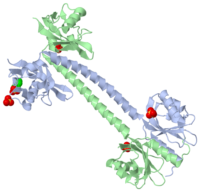
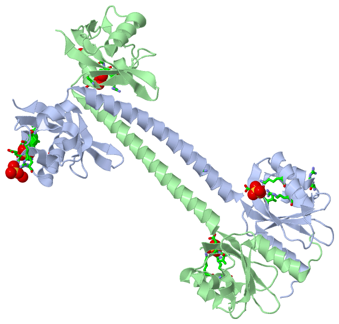
(-) Ligands, Modified Residues, Ions  (2, 5)

Asymmetric/Biological Unit (2, 5)
No.NameCountTypeFull Name
1MES1Ligand/Ion2-(N-MORPHOLINO)-ETHANESULFONIC ACID
2SO44Ligand/IonSULFATE ION

(-) Sites  (5, 5)

Asymmetric Unit (5, 5)
No.NameEvidenceResiduesDescription
1AC1SOFTWAREGLU A:200 , GLY A:201 , ARG A:228 , HOH A:472BINDING SITE FOR RESIDUE SO4 A 301
2AC2SOFTWARELYS A:69 , ARG A:73 , VAL A:86 , GLU A:87BINDING SITE FOR RESIDUE SO4 A 302
3AC3SOFTWAREARG A:202 , THR A:210 , ALA A:221 , PRO A:222 , VAL A:223 , GLY B:184 , GLY B:185 , VAL B:186 , HOH B:419BINDING SITE FOR RESIDUE MES A 303
4AC4SOFTWAREARG A:56 , SER B:177 , VAL B:186 , ARG B:187 , HOH B:432BINDING SITE FOR RESIDUE SO4 B 301
5AC5SOFTWAREGLN A:129 , LYS B:69 , ARG B:73 , VAL B:86 , GLU B:87BINDING SITE FOR RESIDUE SO4 B 302

(-) SS Bonds  (0, 0)

(no "SS Bond" information available for 4L9G)

(-) Cis Peptide Bonds  (0, 0)

(no "Cis Peptide Bond" information available for 4L9G)

 Sequence-Structure Mapping

(-) SAPs(SNPs)/Variants  (0, 0)

(no "SAP(SNP)/Variant" information available for 4L9G)

(-) PROSITE Motifs  (0, 0)

(no "PROSITE Motif" information available for 4L9G)

(-) Exons   (0, 0)

(no "Exon" information available for 4L9G)

(-) Sequences/Alignments

Asymmetric/Biological Unit
   Reformat: Number of residues per line =  ('0' or empty: single-line sequence representation)
  Number of residues per labelling interval =   
  UniProt sequence: complete  aligned part    
   Show mapping: SCOP domains CATH domains Pfam domains Secondary structure (by author)
SAPs(SNPs) PROSITE motifs Exons
(details for a mapped element are shown in a popup box when the mouse pointer rests over it)
Chain A from PDB  Type:PROTEIN  Length:250
                                                                                                                                                                                                                                                                                          
               SCOP domains ---------------------------------------------------------------------------------------------------------------------------------------------------------------------------------------------------------------------------------------------------------- SCOP domains
               CATH domains ---------------------------------------------------------------------------------------------------------------------------------------------------------------------------------------------------------------------------------------------------------- CATH domains
               Pfam domains ---------------------------------------------------------------------------------------------------------------------------------------------------------------------------------------------------------------------------------------------------------- Pfam domains
         Sec.struct. author ...hhhhhhhhhhhhh.eeeee.....eeeeee.........hhhhh..hhhhhhhhhhhhhhhhhhhh.......eeeeeeee.....eeeeeeeeee......eeeeeeehhhhhhhhhhhhhhhhhhhhhhhhhhhhhhhhhhhhhhh...eeeee.....eeeehhhhhhhhh.hhhhhh...hhhhh...hhhhhhhhhhhhhhhh....eeee......eeeeeeeeeee..eeeeeeeeee.. Sec.struct. author
                 SAPs(SNPs) ---------------------------------------------------------------------------------------------------------------------------------------------------------------------------------------------------------------------------------------------------------- SAPs(SNPs)
                    PROSITE ---------------------------------------------------------------------------------------------------------------------------------------------------------------------------------------------------------------------------------------------------------- PROSITE
                 Transcript ---------------------------------------------------------------------------------------------------------------------------------------------------------------------------------------------------------------------------------------------------------- Transcript
                 4l9g A   8 PSLAPDLVRDLIATAADISLLVSQEGVVREVMANPHHPSFGQLSEWEGRPLEEVLTAESVAKFRLRSEGLEPGRGSVAVELNHIDPRSFEFPIRYILHRLPADRSILMLGRDLRPIAEVQQQLVAAQLAMERDYETQREMETRYRVVLDVSRDPMVLVSMSTGRIVDLNSAAGLLLGGVRQDLLGAAIAQEFEGRRRGEFMETMTNLAATESAAPVEVLARRSQKRLLVVPRVFRAAGERLLLCQIDPAD 257
                                    17        27        37        47        57        67        77        87        97       107       117       127       137       147       157       167       177       187       197       207       217       227       237       247       257

Chain B from PDB  Type:PROTEIN  Length:246
                                                                                                                                                                                                                                                                                      
               SCOP domains ------------------------------------------------------------------------------------------------------------------------------------------------------------------------------------------------------------------------------------------------------ SCOP domains
               CATH domains ------------------------------------------------------------------------------------------------------------------------------------------------------------------------------------------------------------------------------------------------------ CATH domains
               Pfam domains ------------------------------------------------------------------------------------------------------------------------------------------------------------------------------------------------------------------------------------------------------ Pfam domains
         Sec.struct. author .....hhhhhhhhhhhhh.eeeee.....eeeeee........hhhhhh..hhhhhhhhhhhhhhhhhhhh.......eeeeee...eeeeeeee......eeeeeeehhhhhhhhhhhhhhhhhhhhhhhhhhhhhhhhhhhhhhh...eeeee.....eeeehhhhhhhhh.hhhhhh...hhhhh...hhhhhhhhhhhhhh......eeee......eeeeeeeeeee..eeeeeeeeee.. Sec.struct. author
                 SAPs(SNPs) ------------------------------------------------------------------------------------------------------------------------------------------------------------------------------------------------------------------------------------------------------ SAPs(SNPs)
                    PROSITE ------------------------------------------------------------------------------------------------------------------------------------------------------------------------------------------------------------------------------------------------------ PROSITE
                 Transcript ------------------------------------------------------------------------------------------------------------------------------------------------------------------------------------------------------------------------------------------------------ Transcript
                 4l9g B   6 SLPSLAPDLVRDLIATAADISLLVSQEGVVREVMANPHHPSFGQLSEWEGRPLEEVLTAESVAKFRLRSEGLEPGRGSVAVELNHIFPIRYILHRLPADRSILMLGRDLRPIAEVQQQLVAAQLAMERDYETQREMETRYRVVLDVSRDPMVLVSMSTGRIVDLNSAAGLLLGGVRQDLLGAAIAQEFEGRRRGEFMETMTNLAATESAAPVEVLARRSQKRLLVVPRVFRAAGERLLLCQIDPAD 257
                                    15        25        35        45        55        65        75        85     ||101       111       121       131       141       151       161       171       181       191       201       211       221       231       241       251      
                                                                                                                91|                                                                                                                                                               
                                                                                                                 98                                                                                                                                                               

   Legend:   → Mismatch (orange background)
  - → Gap (green background, '-', border residues have a numbering label)
    → Modified Residue (blue background, lower-case, 'x' indicates undefined single-letter code, labelled with number + name)
  x → Chemical Group (purple background, 'x', labelled with number + name, e.g. ACE or NH2)
  extra numbering lines below/above indicate numbering irregularities and modified residue names etc., number ends below/above '|'

 Classification and Annotation

(-) SCOP Domains  (0, 0)

(no "SCOP Domain" information available for 4L9G)

(-) CATH Domains  (0, 0)

(no "CATH Domain" information available for 4L9G)

(-) Pfam Domains  (0, 0)

(no "Pfam Domain" information available for 4L9G)

(-) Gene Ontology  (2, 2)

Asymmetric/Biological Unit(hide GO term definitions)

 Visualization

(-) Interactive Views

Asymmetric/Biological Unit
  Complete Structure
    Jena3D(integrated viewing of ligand, site, SAP, PROSITE, SCOP information)
    WebMol | AstexViewer[tm]@PDBe
(Java Applets, require no local installation except for Java; loading may be slow)
    STRAP
(Java WebStart application, automatic local installation, requires Java; full application with system access!)
    RasMol
(require local installation)
    Molscript (VRML)
(requires installation of a VRML viewer; select preferred view via VRML and generate a mono or stereo PDF format file)
 
  Ligands, Modified Residues, Ions
    MES  [ RasMol | Jena3D ]  +environment [ RasMol | Jena3D ]
    SO4  [ RasMol | Jena3D ]  +environment [ RasMol | Jena3D ]
 
  Sites
    AC1  [ RasMol ]  +environment [ RasMol ]
    AC2  [ RasMol ]  +environment [ RasMol ]
    AC3  [ RasMol ]  +environment [ RasMol ]
    AC4  [ RasMol ]  +environment [ RasMol ]
    AC5  [ RasMol ]  +environment [ RasMol ]
 
  Cis Peptide Bonds
(no "Cis Peptide Bonds" information available for 4l9g)
 

(-) Still Images

Jmol
  protein: cartoon or spacefill or dots and stick; nucleic acid: cartoon and stick; ligands: spacefill; active site: stick
Molscript
  protein, nucleic acid: cartoon; ligands: spacefill; active site: ball and stick

 Databases and Analysis Tools

(-) Databases

Access by PDB/NDB ID
  4l9g
    Family and Domain InformationProDom | SYSTERS
    General Structural InformationGlycoscienceDB | MMDB | NDB | OCA | PDB | PDBe | PDBj | PDBsum | PDBWiki | PQS | PROTEOPEDIA
    Orientation in MembranesOPM
    Protein SurfaceSURFACE
    Secondary StructureDSSP (structure derived) | HSSP (homology derived)
    Structural GenomicsGeneCensus
    Structural NeighboursCE | VAST
    Structure ClassificationCATH | Dali | SCOP
    Validation and Original DataBMRB Data View | BMRB Restraints Grid | EDS | PROCHECK | RECOORD | WHAT_CHECK
 
Access by UniProt ID/Accession number
  Q3J179_RHOS4 | Q3J179
    Comparative Protein Structure ModelsModBase
    Genomic InformationEnsembl
    Protein-protein InteractionDIP
    Sequence, Family and Domain InformationInterPro | Pfam | SMART | UniProtKB/TrEMBL
 
Access by Enzyme Classificator   (EC Number)
  (no 'Enzyme Classificator' available)
    General Enzyme InformationBRENDA | EC-PDB | Enzyme | IntEnz
    PathwayKEGG | MetaCyc
 
Access by Disease Identifier   (MIM ID)
  (no 'MIM ID' available)
    Disease InformationOMIM
 
Access by GenAge ID
  (no 'GenAge ID' available)
    Age Related InformationGenAge

(-) Analysis Tools

Access by PDB/NDB ID
    Domain InformationXDom
    Interatomic Contacts of Structural UnitsCSU
    Ligand-protein ContactsLPC
    Protein CavitiescastP
    Sequence and Secondary StructurePDBCartoon
    Structure AlignmentSTRAP(Java WebStart application, automatic local installation, requires Java; full application with system access!)
    Structure and Sequence BrowserSTING
 
Access by UniProt ID/Accession number
  Q3J179_RHOS4 | Q3J179
    Protein Disorder PredictionDisEMBL | FoldIndex | GLOBPLOT (for more information see DisProt)

 Related Entries

(-) Entries Sharing at Least One Protein Chain (UniProt ID)

UniProtKB/TrEMBL
        Q3J179_RHOS4 | Q3J1794hh2 4hh3 4l9e 4l9f

(-) Related Entries Specified in the PDB File

4hh0 4hh2 4hh3 4l9e 4l9f