Show PDB file:   
         Plain Text   HTML   (compressed file size)
QuickSearch:   
by PDB,NDB,UniProt,PROSITE Code or Search Term(s)  
(-)Asym./Biol. Unit
(-)Asym./Biol. Unit - sites
collapse expand < >
Image Asym./Biol. Unit
Asym./Biol. Unit  (Jmol Viewer)
Image Asym./Biol. Unit - sites
Asym./Biol. Unit - sites  (Jmol Viewer)

(-) Description

Title :  CRYSTAL STRUCTURE OF MOUSE RYANODINE RECEPTOR ISOFORM 2 (RYR2) 1-547
 
Authors :  L. Kimlicka, C. C. Tung, F. Van Petegem
Date :  07 Jun 13  (Deposition) - 24 Jul 13  (Release) - 24 Jul 13  (Revision)
Method :  X-RAY DIFFRACTION
Resolution :  2.00
Chains :  Asym./Biol. Unit :  A
Keywords :  Ca2+ Release, Ion Channel, Er/Sr Membrane, Metal Transport (Keyword Search: [Gene Ontology, PubMed, Web (Google))
 
Reference :  L. Kimlicka, C. C. Tung, A. C. Carlsson, P. A. Lobo, F. Van Petegem
Crystal Structure Of A Ryanodine Receptor Domain
To Be Published
PubMed: search

(-) Compounds

Molecule 1 - RYANODINE RECEPTOR 2
    ChainsA
    EngineeredYES
    Expression SystemESCHERICHIA COLI
    Expression System StrainROSETTA
    Expression System Taxid511693
    FragmentN-TERMINAL DOMAINS (UNP RESIDUES 1-547)
    GeneRYR2
    Organism CommonMOUSE
    Organism ScientificMUS MUSCULUS
    Organism Taxid10090
    SynonymRYR-2, RYR2, CARDIAC MUSCLE RYANODINE RECEPTOR, CARDIAC MUSCLE RYANODINE RECEPTOR-CALCIUM RELEASE CHANNEL, TYPE 2 RYANODINE RECEPTOR, CARDIAC CA2+ RELEASE CHANNEL

 Structural Features

(-) Chains, Units

  1
Asymmetric/Biological Unit A

Summary Information (see also Sequences/Alignments below)

(-) Ligands, Modified Residues, Ions  (3, 3)

Asymmetric/Biological Unit (3, 3)
No.NameCountTypeFull Name
1CL1Ligand/IonCHLORIDE ION
2PEG1Ligand/IonDI(HYDROXYETHYL)ETHER
3PGE1Ligand/IonTRIETHYLENE GLYCOL

(-) Sites  (3, 3)

Asymmetric Unit (3, 3)
No.NameEvidenceResiduesDescription
1AC1SOFTWAREARG A:276 , ARG A:420BINDING SITE FOR RESIDUE CL A 601
2AC2SOFTWAREGLY A:231 , ASP A:232 , VAL A:233 , ARG A:276 , ARG A:407 , SER A:408 , ARG A:417 , HOH A:718 , HOH A:763BINDING SITE FOR RESIDUE PGE A 602
3AC3SOFTWAREPHE A:42 , ARG A:414 , ASP A:458 , TYR A:462BINDING SITE FOR RESIDUE PEG A 603

(-) SS Bonds  (0, 0)

(no "SS Bond" information available for 4L4H)

(-) Cis Peptide Bonds  (0, 0)

(no "Cis Peptide Bond" information available for 4L4H)

 Sequence-Structure Mapping

(-) SAPs(SNPs)/Variants  (0, 0)

(no "SAP(SNP)/Variant" information available for 4L4H)

(-) PROSITE Motifs  (0, 0)

(no "PROSITE Motif" information available for 4L4H)

(-) Exons   (0, 0)

(no "Exon" information available for 4L4H)

(-) Sequences/Alignments

Asymmetric/Biological Unit
   Reformat: Number of residues per line =  ('0' or empty: single-line sequence representation)
  Number of residues per labelling interval =   
  UniProt sequence: complete  aligned part    
   Show mapping: SCOP domains CATH domains Pfam domains Secondary structure (by author)
SAPs(SNPs) PROSITE motifs Exons
(details for a mapped element are shown in a popup box when the mouse pointer rests over it)
Chain A from PDB  Type:PROTEIN  Length:466
                                                                                                                                                                                                                                                                                                                                                                                                                                                                                                                  
               SCOP domains ---------------------------------------------------------------------------------------------------------------------------------------------------------------------------------------------------------------------------------------------------------------------------------------------------------------------------------------------------------------------------------------------------------------------------------------------------------------------------------- SCOP domains
               CATH domains ---------------------------------------------------------------------------------------------------------------------------------------------------------------------------------------------------------------------------------------------------------------------------------------------------------------------------------------------------------------------------------------------------------------------------------------------------------------------------------- CATH domains
               Pfam domains ---------------------------------------------------------------------------------------------------------------------------------------------------------------------------------------------------------------------------------------------------------------------------------------------------------------------------------------------------------------------------------------------------------------------------------------------------------------------------------- Pfam domains
         Sec.struct. author .......eeeeeeeeeee..eeeeeeee.........eeee.........hhhhh.eeeeeee..........eeeeee.....eeee.......eeeeee........eeeeee...............eeeeee.....eeeeee....eeeeee.....eeeeeeee.hhh.........eeeeee....eeee........hhhhh.eeee.hhhhhhhh.eeeee..............eeeeee.....eeee...eeeehhhhhhhhhhheeee................eeeeee.....eeee.....eeeeee........eeeee.hhhhhhhhhhhhhhhhhhhhhhhhhhhhh....hhhhhhhhhhhhhhhh.......hhhhhhhhhhhhhhhhhhhhhhhhhhhhhhhhhhhh...hhhhhhhhhhhhhhhhhhhhhhhhhhhhhhhhh. Sec.struct. author
                 SAPs(SNPs) ---------------------------------------------------------------------------------------------------------------------------------------------------------------------------------------------------------------------------------------------------------------------------------------------------------------------------------------------------------------------------------------------------------------------------------------------------------------------------------- SAPs(SNPs)
                    PROSITE ---------------------------------------------------------------------------------------------------------------------------------------------------------------------------------------------------------------------------------------------------------------------------------------------------------------------------------------------------------------------------------------------------------------------------------------------------------------------------------- PROSITE
                 Transcript ---------------------------------------------------------------------------------------------------------------------------------------------------------------------------------------------------------------------------------------------------------------------------------------------------------------------------------------------------------------------------------------------------------------------------------------------------------------------------------- Transcript
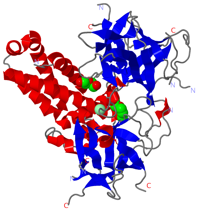
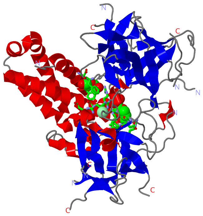
                 4l4h A  11 IQFLRTDDEVVLQCTATIHKEQQKLCLAAEGFGNRLCFLESTSNSKNVPPDLSICTFVLEQSLSVHRTLLYGHAILLRHSYSGMYLCCLSTSKLAFDVGLQEDTTGEACWWTIHPASKQRSEGEKVRVGDDLILVSVSSERYLHLSYGNSSWHVDAAFQQTLWSVAPISSGSEAAQGYLIGGDVLRLLHGHMDECLTVPSGEHGEEQRRTVHYEGGAVSVHARSLWRLETLRVAWSGSHIRWGQPFRLRHVTTGKYLSLMKNLLLMDKEKADVKSTAFAFRSSKEGMGTSEIKYGDSICYIQHVDTGLWLTYQAVDQRKAIMHHEGHMDDGLNLSRSQHEESRTARVIRSTVFLFNRFIRGLDALSKIDLPIESVSLSLQDLIGYFHPPDEHLEHEDKQNRLRALKNRQNLFQEEGMINLVLECIDRLHVYSSAAHFADVAGREAGESWKSILNSLYELLAALIRG 543
                                    20        30        40        50        60        70    || 114       124       134 ||    149       159       169       179       189       199       209       219       229       239       249       259       269       279       289       299       309|      321       331    || 352       362       372     ||391       401       411       421       431      |447       457       467       477       487       497       507       517       527       537      
                                                                                           75|                       136|                                                                                                                                                                    309|                     336|                           378|                                               438|                                                                                                  
                                                                                           110                        142                                                                                                                                                                     312                      348                            388                                                445                                                                                                  

   Legend:   → Mismatch (orange background)
  - → Gap (green background, '-', border residues have a numbering label)
    → Modified Residue (blue background, lower-case, 'x' indicates undefined single-letter code, labelled with number + name)
  x → Chemical Group (purple background, 'x', labelled with number + name, e.g. ACE or NH2)
  extra numbering lines below/above indicate numbering irregularities and modified residue names etc., number ends below/above '|'

 Classification and Annotation

(-) SCOP Domains  (0, 0)

(no "SCOP Domain" information available for 4L4H)

(-) CATH Domains  (0, 0)

(no "CATH Domain" information available for 4L4H)

(-) Pfam Domains  (0, 0)

(no "Pfam Domain" information available for 4L4H)

(-) Gene Ontology  (69, 69)

Asymmetric/Biological Unit(hide GO term definitions)

 Visualization

(-) Interactive Views

Asymmetric/Biological Unit
  Complete Structure
    Jena3D(integrated viewing of ligand, site, SAP, PROSITE, SCOP information)
    WebMol | AstexViewer[tm]@PDBe
(Java Applets, require no local installation except for Java; loading may be slow)
    STRAP
(Java WebStart application, automatic local installation, requires Java; full application with system access!)
    RasMol
(require local installation)
    Molscript (VRML)
(requires installation of a VRML viewer; select preferred view via VRML and generate a mono or stereo PDF format file)
 
  Ligands, Modified Residues, Ions
    CL  [ RasMol | Jena3D ]  +environment [ RasMol | Jena3D ]
    PEG  [ RasMol | Jena3D ]  +environment [ RasMol | Jena3D ]
    PGE  [ RasMol | Jena3D ]  +environment [ RasMol | Jena3D ]
 
  Sites
    AC1  [ RasMol ]  +environment [ RasMol ]
    AC2  [ RasMol ]  +environment [ RasMol ]
    AC3  [ RasMol ]  +environment [ RasMol ]
 
  Cis Peptide Bonds
(no "Cis Peptide Bonds" information available for 4l4h)
 

(-) Still Images

Jmol
  protein: cartoon or spacefill or dots and stick; nucleic acid: cartoon and stick; ligands: spacefill; active site: stick
Molscript
  protein, nucleic acid: cartoon; ligands: spacefill; active site: ball and stick

 Databases and Analysis Tools

(-) Databases

Access by PDB/NDB ID
  4l4h
    Family and Domain InformationProDom | SYSTERS
    General Structural InformationGlycoscienceDB | MMDB | NDB | OCA | PDB | PDBe | PDBj | PDBsum | PDBWiki | PQS | PROTEOPEDIA
    Orientation in MembranesOPM
    Protein SurfaceSURFACE
    Secondary StructureDSSP (structure derived) | HSSP (homology derived)
    Structural GenomicsGeneCensus
    Structural NeighboursCE | VAST
    Structure ClassificationCATH | Dali | SCOP
    Validation and Original DataBMRB Data View | BMRB Restraints Grid | EDS | PROCHECK | RECOORD | WHAT_CHECK
 
Access by UniProt ID/Accession number
  RYR2_MOUSE | E9Q401
    Comparative Protein Structure ModelsModBase
    Genomic InformationEnsembl
    Protein-protein InteractionDIP
    Sequence, Family and Domain InformationInterPro | Pfam | SMART | UniProtKB/SwissProt
 
Access by Enzyme Classificator   (EC Number)
  (no 'Enzyme Classificator' available)
    General Enzyme InformationBRENDA | EC-PDB | Enzyme | IntEnz
    PathwayKEGG | MetaCyc
 
Access by Disease Identifier   (MIM ID)
  (no 'MIM ID' available)
    Disease InformationOMIM
 
Access by GenAge ID
  (no 'GenAge ID' available)
    Age Related InformationGenAge

(-) Analysis Tools

Access by PDB/NDB ID
    Domain InformationXDom
    Interatomic Contacts of Structural UnitsCSU
    Ligand-protein ContactsLPC
    Protein CavitiescastP
    Sequence and Secondary StructurePDBCartoon
    Structure AlignmentSTRAP(Java WebStart application, automatic local installation, requires Java; full application with system access!)
    Structure and Sequence BrowserSTING
 
Access by UniProt ID/Accession number
  RYR2_MOUSE | E9Q401
    Protein Disorder PredictionDisEMBL | FoldIndex | GLOBPLOT (for more information see DisProt)

 Related Entries

(-) Entries Sharing at Least One Protein Chain (UniProt ID)

UniProtKB/Swiss-Prot
        RYR2_MOUSE | E9Q4012mc2 3im5 3im6 3im7 3qr5 4etv 4kei 4kej 4kek 4l4i 4p9i 4p9l 5c33

(-) Related Entries Specified in the PDB File

4l4i