Show PDB file:   
         Plain Text   HTML   (compressed file size)
QuickSearch:   
by PDB,NDB,UniProt,PROSITE Code or Search Term(s)  
(-)Asymmetric Unit
(-)Asym. Unit - sites
(-)Biological Unit 1
(-)Biol. Unit 1 - sites
(-)Biological Unit 2
(-)Biological Unit 3
collapse expand < >
Image Asymmetric Unit
Asymmetric Unit  (Jmol Viewer)
Image Asym. Unit - sites
Asym. Unit - sites  (Jmol Viewer)
Image Biological Unit 1
Biological Unit 1  (Jmol Viewer)
Image Biol. Unit 1 - sites
Biol. Unit 1 - sites  (Jmol Viewer)
Image Biological Unit 2
Biological Unit 2  (Jmol Viewer)
Image Biological Unit 3
Biological Unit 3  (Jmol Viewer)

(-) Description

Title :  CRYSTAL STRUCTURE OF FGF RECEPTOR 2 (FGFR2) KINASE DOMAIN HARBORING THE GAIN-OF-FUNCTION K659Q MUTATION.
 
Authors :  Z. Huang, H. Chen, M. Mohammadi
Date :  15 Feb 13  (Deposition) - 07 Aug 13  (Release) - 28 Dec 16  (Revision)
Method :  X-RAY DIFFRACTION
Resolution :  2.31
Chains :  Asym. Unit :  A,B
Biol. Unit 1:  A  (1x)
Biol. Unit 2:  B  (1x)
Biol. Unit 3:  A,B  (1x)
Keywords :  Fgf Receptor, Receptor Tyrosine Kinase, Gain-Of-Function Mutation, Atp Binding, Transferase (Keyword Search: [Gene Ontology, PubMed, Web (Google))
 
Reference :  H. Chen, Z. Huang, K. Dutta, S. Blais, T. A. Neubert, X. Li, D. Cowburn, N. J. Traaseth, M. Mohammadi
Cracking The Molecular Origin Of Intrinsic Tyrosine Kinase Activity Through Analysis Of Pathogenic Gain-Of-Function Mutations.
Cell Rep V. 4 376 2013
PubMed-ID: 23871672  |  Reference-DOI: 10.1016/J.CELREP.2013.06.025

(-) Compounds

Molecule 1 - FIBROBLAST GROWTH FACTOR RECEPTOR 2
    ChainsA, B
    EC Number2.7.10.1
    EngineeredYES
    Expression SystemESCHERICHIA COLI
    Expression System StrainBL21(DE3)PLYS-S
    Expression System Taxid469008
    FragmentHUMAN FGF RECEPTOR 2 KINASE DOMAIN (UNP RESIDUES 458-768)
    GeneBEK, FGFR2, KGFR, KSAM
    MutationYES
    Organism CommonHUMAN
    Organism ScientificHOMO SAPIENS
    Organism Taxid9606
    SynonymFGFR-2, K-SAM, KGFR, KERATINOCYTE GROWTH FACTOR RECEPTOR

 Structural Features

(-) Chains, Units

  12
Asymmetric Unit AB
Biological Unit 1 (1x)A 
Biological Unit 2 (1x) B
Biological Unit 3 (1x)AB

Summary Information (see also Sequences/Alignments below)

(-) Ligands, Modified Residues, Ions  (2, 6)

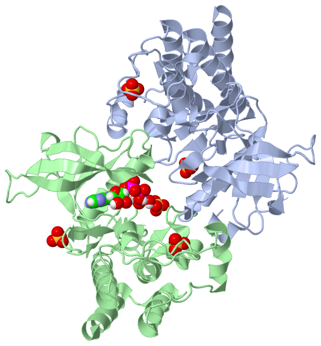
Asymmetric Unit (2, 6)
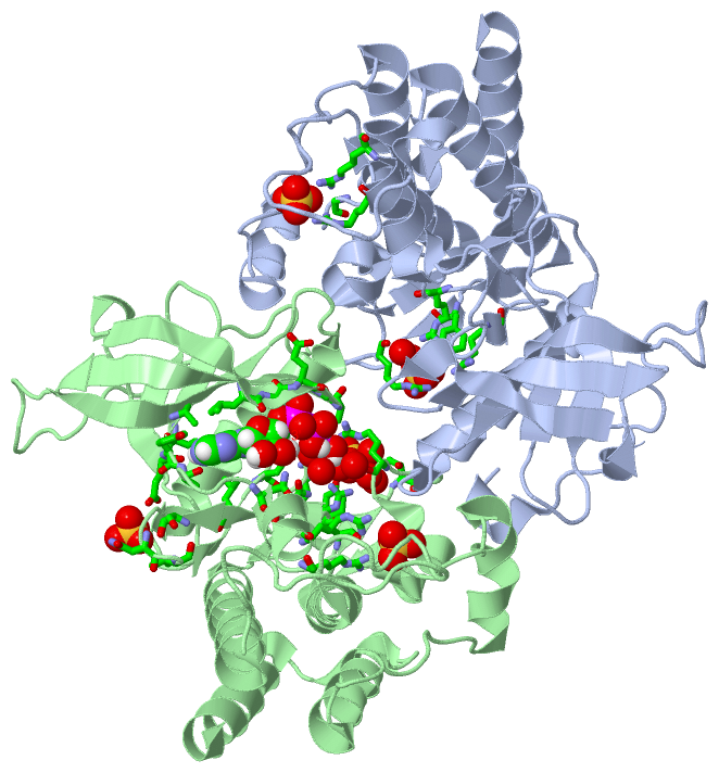
No.NameCountTypeFull Name
1ACP1Ligand/IonPHOSPHOMETHYLPHOSPHONIC ACID ADENYLATE ESTER
2SO45Ligand/IonSULFATE ION
Biological Unit 1 (1, 2)
No.NameCountTypeFull Name
1ACP-1Ligand/IonPHOSPHOMETHYLPHOSPHONIC ACID ADENYLATE ESTER
2SO42Ligand/IonSULFATE ION
Biological Unit 2 (2, 4)
No.NameCountTypeFull Name
1ACP1Ligand/IonPHOSPHOMETHYLPHOSPHONIC ACID ADENYLATE ESTER
2SO43Ligand/IonSULFATE ION
Biological Unit 3 (2, 6)
No.NameCountTypeFull Name
1ACP1Ligand/IonPHOSPHOMETHYLPHOSPHONIC ACID ADENYLATE ESTER
2SO45Ligand/IonSULFATE ION

(-) Sites  (6, 6)

Asymmetric Unit (6, 6)
No.NameEvidenceResiduesDescription
1AC1SOFTWAREARG A:625 , ARG A:649 , TYR A:657 , ARG A:664 , HOH A:1004 , HOH A:1005BINDING SITE FOR RESIDUE SO4 A 801
2AC2SOFTWAREARG A:577 , ARG A:580 , GLY A:701BINDING SITE FOR RESIDUE SO4 A 802
3AC3SOFTWAREARG B:625 , ARG B:649 , TYR B:657 , THR B:660 , ARG B:664 , HOH B:904 , HOH B:1003 , HOH B:1004BINDING SITE FOR RESIDUE SO4 B 801
4AC4SOFTWAREARG B:577 , ARG B:580 , SER B:702BINDING SITE FOR RESIDUE SO4 B 802
5AC5SOFTWARETHR B:635 , ASN B:637 , VAL B:639BINDING SITE FOR RESIDUE SO4 B 803
6AC6SOFTWARELEU B:487 , GLY B:488 , GLU B:489 , GLY B:490 , ALA B:491 , ALA B:515 , VAL B:564 , GLU B:565 , TYR B:566 , ALA B:567 , ASN B:571 , ASN B:631 , LEU B:633 , ASP B:644 , HOH B:901 , HOH B:936 , HOH B:964BINDING SITE FOR RESIDUE ACP B 804

(-) SS Bonds  (0, 0)

(no "SS Bond" information available for 4J98)

(-) Cis Peptide Bonds  (1, 1)

Asymmetric Unit
No.Residues
1Tyr A:586 -Ser A:587

 Sequence-Structure Mapping

(-) SAPs(SNPs)/Variants  (0, 0)

(no "SAP(SNP)/Variant" information available for 4J98)

(-) PROSITE Motifs  (0, 0)

(no "PROSITE Motif" information available for 4J98)

(-) Exons   (0, 0)

(no "Exon" information available for 4J98)

(-) Sequences/Alignments

Asymmetric Unit
   Reformat: Number of residues per line =  ('0' or empty: single-line sequence representation)
  Number of residues per labelling interval =   
  UniProt sequence: complete  aligned part    
   Show mapping: SCOP domains CATH domains Pfam domains Secondary structure (by author)
SAPs(SNPs) PROSITE motifs Exons
(details for a mapped element are shown in a popup box when the mouse pointer rests over it)
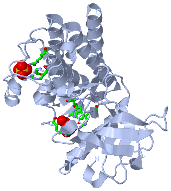
Chain A from PDB  Type:PROTEIN  Length:304
                                                                                                                                                                                                                                                                                                                                                
               SCOP domains d4j98a_ A: Fibroblast growth factor receptor 2                                                                                                                                                                                                                                                                   SCOP domains
               CATH domains ---------------------------------------------------------------------------------------------------------------------------------------------------------------------------------------------------------------------------------------------------------------------------------------------------------------- CATH domains
               Pfam domains ---------------------------------------------------------------------------------------------------------------------------------------------------------------------------------------------------------------------------------------------------------------------------------------------------------------- Pfam domains
         Sec.struct. author .hhhhh..........hhh.eeeeeeeee....eeeeeeee.........eeeeeeee.....hhhhhhhhhhhhhhhhhhh.......eeeee......eeeee.....hhhhhhhhh.............hhhhh.hhhhhhhhhhhhhhhhhhhhhh.ee....hhh.eee.....eee......ee......ee.......hhhhhhhhhhhhheehhhhhhhhhhhhhhhhhh.........hhhhhhhhhhh..........hhhhhhhhhhhh..hhhhh.hhhhhhhhhhhhhhhh Sec.struct. author
                 SAPs(SNPs) ---------------------------------------------------------------------------------------------------------------------------------------------------------------------------------------------------------------------------------------------------------------------------------------------------------------- SAPs(SNPs)
                    PROSITE ---------------------------------------------------------------------------------------------------------------------------------------------------------------------------------------------------------------------------------------------------------------------------------------------------------------- PROSITE
                 Transcript ---------------------------------------------------------------------------------------------------------------------------------------------------------------------------------------------------------------------------------------------------------------------------------------------------------------- Transcript
                 4j98 A 461 AGVSEYELPEDPKWEFPRDKLTLGKPLGEGAFGQVVMAEAVGIDKDKPKEAVTVAVKMLKDDATEKDLSDLVSEMEMMKMIGKHKNIINLLGACTQDGPLYVIVEYASKGNLREYLRARRPPGMEYSYDINRVPEEQMTFKDLVSCTYQLARGMEYLASQKCIHRDLAARNVLVTENNVMKIADFGLARDINNIDYYKQTTNGRLPVKWMAPEALFDRVYTHQSDVWSFGVLMWEIFTLGGSPYPGIPVEELFKLLKEGHRMDKPANCTNELYMMMRDCWHAVPSQRPTFKQLVEDLDRILTLT 764
                                   470       480       490       500       510       520       530       540       550       560       570       580       590       600       610       620       630       640       650       660       670       680       690       700       710       720       730       740       750       760    

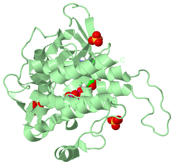
Chain B from PDB  Type:PROTEIN  Length:304
                                                                                                                                                                                                                                                                                                                                                
               SCOP domains d4j98b_ B: Fibroblast growth factor receptor 2                                                                                                                                                                                                                                                                   SCOP domains
               CATH domains ---------------------------------------------------------------------------------------------------------------------------------------------------------------------------------------------------------------------------------------------------------------------------------------------------------------- CATH domains
               Pfam domains ---------------------------------------------------------------------------------------------------------------------------------------------------------------------------------------------------------------------------------------------------------------------------------------------------------------- Pfam domains
         Sec.struct. author ................hhh.eeeeeeeeee..eeeeeeeee.........eeeeeeee.....hhhhhhhhhhhhhhhhhhh.......eeeee......eeeee.....hhhhhhhh....................hhhhhhhhhhhhhhhhhhhhhh.ee....hhh.eee.....eee......ee......ee.......hhhhhhhhhhhhheehhhhhhhhhhhhhhhhhh.........hhhhhhhhhhh..........hhhhhhhhhhhh..hhhhh.hhhhhhhhhhhhhhhh Sec.struct. author
                 SAPs(SNPs) ---------------------------------------------------------------------------------------------------------------------------------------------------------------------------------------------------------------------------------------------------------------------------------------------------------------- SAPs(SNPs)
                    PROSITE ---------------------------------------------------------------------------------------------------------------------------------------------------------------------------------------------------------------------------------------------------------------------------------------------------------------- PROSITE
                 Transcript ---------------------------------------------------------------------------------------------------------------------------------------------------------------------------------------------------------------------------------------------------------------------------------------------------------------- Transcript
                 4j98 B 461 AGVSEYELPEDPKWEFPRDKLTLGKPLGEGAFGQVVMAEAVGIDKDKPKEAVTVAVKMLKDDATEKDLSDLVSEMEMMKMIGKHKNIINLLGACTQDGPLYVIVEYASKGNLREYLRARRPPGMEYSYDINRVPEEQMTFKDLVSCTYQLARGMEYLASQKCIHRDLAARNVLVTENNVMKIADFGLARDINNIDYYKQTTNGRLPVKWMAPEALFDRVYTHQSDVWSFGVLMWEIFTLGGSPYPGIPVEELFKLLKEGHRMDKPANCTNELYMMMRDCWHAVPSQRPTFKQLVEDLDRILTLT 764
                                   470       480       490       500       510       520       530       540       550       560       570       580       590       600       610       620       630       640       650       660       670       680       690       700       710       720       730       740       750       760    

   Legend:   → Mismatch (orange background)
  - → Gap (green background, '-', border residues have a numbering label)
    → Modified Residue (blue background, lower-case, 'x' indicates undefined single-letter code, labelled with number + name)
  x → Chemical Group (purple background, 'x', labelled with number + name, e.g. ACE or NH2)
  extra numbering lines below/above indicate numbering irregularities and modified residue names etc., number ends below/above '|'

 Classification and Annotation

(-) SCOP Domains  (1, 2)

Asymmetric Unit

(-) CATH Domains  (0, 0)

(no "CATH Domain" information available for 4J98)

(-) Pfam Domains  (0, 0)

(no "Pfam Domain" information available for 4J98)

(-) Gene Ontology  (133, 133)

Asymmetric Unit(hide GO term definitions)

 Visualization

(-) Interactive Views

Asymmetric Unit
  Complete Structure
    Jena3D(integrated viewing of ligand, site, SAP, PROSITE, SCOP information)
    WebMol | AstexViewer[tm]@PDBe
(Java Applets, require no local installation except for Java; loading may be slow)
    STRAP
(Java WebStart application, automatic local installation, requires Java; full application with system access!)
    RasMol
(require local installation)
    Molscript (VRML)
(requires installation of a VRML viewer; select preferred view via VRML and generate a mono or stereo PDF format file)
 
  Ligands, Modified Residues, Ions
    ACP  [ RasMol | Jena3D ]  +environment [ RasMol | Jena3D ]
    SO4  [ RasMol | Jena3D ]  +environment [ RasMol | Jena3D ]
 
  Sites
    AC1  [ RasMol ]  +environment [ RasMol ]
    AC2  [ RasMol ]  +environment [ RasMol ]
    AC3  [ RasMol ]  +environment [ RasMol ]
    AC4  [ RasMol ]  +environment [ RasMol ]
    AC5  [ RasMol ]  +environment [ RasMol ]
    AC6  [ RasMol ]  +environment [ RasMol ]
 
  Cis Peptide Bonds
    Tyr A:586 - Ser A:587   [ RasMol ]  
 
Biological Units
  Complete Structure
    Biological Unit 1  [ Jena3D ]
    Biological Unit 2  [ Jena3D ]
    Biological Unit 3  [ Jena3D ]

(-) Still Images

Jmol
  protein: cartoon or spacefill or dots and stick; nucleic acid: cartoon and stick; ligands: spacefill; active site: stick
Molscript
  protein, nucleic acid: cartoon; ligands: spacefill; active site: ball and stick

 Databases and Analysis Tools

(-) Databases

Access by PDB/NDB ID
  4j98
    Family and Domain InformationProDom | SYSTERS
    General Structural InformationGlycoscienceDB | MMDB | NDB | OCA | PDB | PDBe | PDBj | PDBsum | PDBWiki | PQS | PROTEOPEDIA
    Orientation in MembranesOPM
    Protein SurfaceSURFACE
    Secondary StructureDSSP (structure derived) | HSSP (homology derived)
    Structural GenomicsGeneCensus
    Structural NeighboursCE | VAST
    Structure ClassificationCATH | Dali | SCOP
    Validation and Original DataBMRB Data View | BMRB Restraints Grid | EDS | PROCHECK | RECOORD | WHAT_CHECK
 
Access by UniProt ID/Accession number
  FGFR2_HUMAN | P21802
    Comparative Protein Structure ModelsModBase
    Genomic InformationEnsembl
    Protein-protein InteractionDIP
    Sequence, Family and Domain InformationInterPro | Pfam | SMART | UniProtKB/SwissProt
 
Access by Enzyme Classificator   (EC Number)
  2.7.10.1
    General Enzyme InformationBRENDA | EC-PDB | Enzyme | IntEnz
    PathwayKEGG | MetaCyc
 
Access by Disease Identifier   (MIM ID)
  (no 'MIM ID' available)
    Disease InformationOMIM
 
Access by GenAge ID
  (no 'GenAge ID' available)
    Age Related InformationGenAge

(-) Analysis Tools

Access by PDB/NDB ID
    Domain InformationXDom
    Interatomic Contacts of Structural UnitsCSU
    Ligand-protein ContactsLPC
    Protein CavitiescastP
    Sequence and Secondary StructurePDBCartoon
    Structure AlignmentSTRAP(Java WebStart application, automatic local installation, requires Java; full application with system access!)
    Structure and Sequence BrowserSTING
 
Access by UniProt ID/Accession number
  FGFR2_HUMAN | P21802
    Protein Disorder PredictionDisEMBL | FoldIndex | GLOBPLOT (for more information see DisProt)

 Related Entries

(-) Entries Sharing at Least One Protein Chain (UniProt ID)

UniProtKB/Swiss-Prot
        FGFR2_HUMAN | P218021djs 1e0o 1ev2 1gjo 1ii4 1iil 1nun 1oec 1wvz 2fdb 2psq 2pvf 2pvy 2pwl 2py3 2pz5 2pzp 2pzr 2q0b 3b2t 3caf 3cly 3cu1 3dar 3euu 3oj2 3ojm 3ri1 4j23 4j95 4j96 4j97 4j99 4wv1 5eg3 5ugl 5ugx 5uhn 5ui0

(-) Related Entries Specified in the PDB File

2psq UNPHOSPHORYLATED UNACTIVATED WILD TYPE FGFR2 KINASE DOMAIN
2pvf PHOSPHORYLATED ACTIVATED FGFR2 KINASE DOMAIN
4j95 FGFR2 KINASE DOMAIN HARBORING THE PATHOGENIC K659N MUTATION IN SPACE GROUP C2
4j96 FGFR2 KINASE DOMAIN HARBORING THE PATHOGENIC K659M MUTATION
4j97 FGFR2 KINASE DOMAIN HARBORING THE PATHOGENIC K659E MUTATION
4j99 FGFR2 KINASE DOMAIN HARBORING THE PATHOGENIC K659T MUTATION