Show PDB file:   
         Plain Text   HTML   (compressed file size)
QuickSearch:   
by PDB,NDB,UniProt,PROSITE Code or Search Term(s)  
(-)Asym./Biol. Unit
(-)Asym./Biol. Unit - sites
collapse expand < >
Image Asym./Biol. Unit
Asym./Biol. Unit  (Jmol Viewer)
Image Asym./Biol. Unit - sites
Asym./Biol. Unit - sites  (Jmol Viewer)

(-) Description

Title :  HUMAN HISTIDINE TRIAD NUCLEOTIDE BINDING PROTEIN 2
 
Authors :  K. M. Maize, C. R. Wagner, B. C. Finzel
Date :  04 Jan 13  (Deposition) - 22 May 13  (Release) - 17 Jul 13  (Revision)
Method :  X-RAY DIFFRACTION
Resolution :  1.19
Chains :  Asym./Biol. Unit :  A,B
Keywords :  Hint, Histidine Triad, Hit, Phosphoramidase, Hydrolase (Keyword Search: [Gene Ontology, PubMed, Web (Google))
 
Reference :  K. M. Maize, C. R. Wagner, B. C. Finzel
Structural Characterization Of Human Histidine Triad Nucleotide-Binding Protein 2, A Member Of The Histidine Triad Superfamily.
Febs J. V. 280 3389 2013
PubMed-ID: 23659632  |  Reference-DOI: 10.1111/FEBS.12330

(-) Compounds

Molecule 1 - HISTIDINE TRIAD NUCLEOTIDE-BINDING PROTEIN 2, MITOCHONDRIAL
    ChainsA, B
    EC Number3.-.-.-
    EngineeredYES
    Expression SystemESCHERICHIA COLI
    Expression System PlasmidPMCSG9
    Expression System StrainROSETTA 2 PLYSS
    Expression System Taxid562
    Expression System Vector TypePLASMID
    GeneHINT2
    Organism CommonHUMAN
    Organism ScientificHOMO SAPIENS
    Organism Taxid9606
    SynonymHINT-2, HINT-3, HIT-17KDA, PKCI-1-RELATED HIT PROTEIN

 Structural Features

(-) Chains, Units

  12
Asymmetric/Biological Unit AB

Summary Information (see also Sequences/Alignments below)

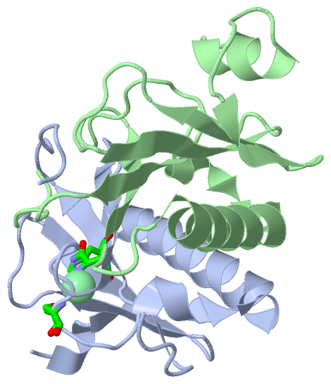
(-) Ligands, Modified Residues, Ions  (1, 1)

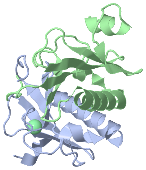
Asymmetric/Biological Unit (1, 1)
No.NameCountTypeFull Name
1CL1Ligand/IonCHLORIDE ION

(-) Sites  (1, 1)

Asymmetric Unit (1, 1)
No.NameEvidenceResiduesDescription
1AC1SOFTWAREASN A:136 , SER A:144 , HIS A:151BINDING SITE FOR RESIDUE CL A 201

(-) SS Bonds  (0, 0)

(no "SS Bond" information available for 4INC)

(-) Cis Peptide Bonds  (2, 2)

Asymmetric/Biological Unit
No.Residues
1Trp A:160 -Pro A:161
2Trp B:160 -Pro B:161

 Sequence-Structure Mapping

(-) SAPs(SNPs)/Variants  (0, 0)

(no "SAP(SNP)/Variant" information available for 4INC)

(-) PROSITE Motifs  (0, 0)

(no "PROSITE Motif" information available for 4INC)

(-) Exons   (0, 0)

(no "Exon" information available for 4INC)

(-) Sequences/Alignments

Asymmetric/Biological Unit
   Reformat: Number of residues per line =  ('0' or empty: single-line sequence representation)
  Number of residues per labelling interval =   
  UniProt sequence: complete  aligned part    
   Show mapping: SCOP domains CATH domains Pfam domains Secondary structure (by author)
SAPs(SNPs) PROSITE motifs Exons
(details for a mapped element are shown in a popup box when the mouse pointer rests over it)
Chain A from PDB  Type:PROTEIN  Length:101
                                                                                                                                     
               SCOP domains d4inca_ A: automated matches                                                                          SCOP domains
               CATH domains ----------------------------------------------------------------------------------------------------- CATH domains
               Pfam domains ----------------------------------------------------------------------------------------------------- Pfam domains
         Sec.struct. author .hhh.eeee...eeeee.......eeeeeee.....hhhhhhhhhhhhhhhhhhhhhhhhhhh.....eeeeeee............eeeee......... Sec.struct. author
                 SAPs(SNPs) ----------------------------------------------------------------------------------------------------- SAPs(SNPs)
                    PROSITE ----------------------------------------------------------------------------------------------------- PROSITE
                 Transcript ----------------------------------------------------------------------------------------------------- Transcript
                 4inc A  63 SLPADILYEDQQCLVFRDVAPQAPVHFLVIPKKPIPRISQAEEEDQQLLGHLLLVAKQTAKAEGLGDGYRLVINDGKLGAQSVYHLHIHVLGGRQLQWPPG 163
                                    72        82        92       102       112       122       132       142       152       162 

Chain B from PDB  Type:PROTEIN  Length:112
                                                                                                                                                
               SCOP domains d4incb_ B: automated matches                                                                                     SCOP domains
               CATH domains ---------------------------------------------------------------------------------------------------------------- CATH domains
               Pfam domains ---------------------------------------------------------------------------------------------------------------- Pfam domains
         Sec.struct. author ...hhhhhhh......eeee...eeeee.......eeeeeee.....hhhhhhhhhhhhhhhhhhhhhhhhhhh.....eeeeeee............eeeee......... Sec.struct. author
                 SAPs(SNPs) ---------------------------------------------------------------------------------------------------------------- SAPs(SNPs)
                    PROSITE ---------------------------------------------------------------------------------------------------------------- PROSITE
                 Transcript ---------------------------------------------------------------------------------------------------------------- Transcript
                 4inc B  52 APTIFSRILDKSLPADILYEDQQCLVFRDVAPQAPVHFLVIPKKPIPRISQAEEEDQQLLGHLLLVAKQTAKAEGLGDGYRLVINDGKLGAQSVYHLHIHVLGGRQLQWPPG 163
                                    61        71        81        91       101       111       121       131       141       151       161  

   Legend:   → Mismatch (orange background)
  - → Gap (green background, '-', border residues have a numbering label)
    → Modified Residue (blue background, lower-case, 'x' indicates undefined single-letter code, labelled with number + name)
  x → Chemical Group (purple background, 'x', labelled with number + name, e.g. ACE or NH2)
  extra numbering lines below/above indicate numbering irregularities and modified residue names etc., number ends below/above '|'

 Classification and Annotation

(-) SCOP Domains  (1, 2)

Asymmetric/Biological Unit

(-) CATH Domains  (0, 0)

(no "CATH Domain" information available for 4INC)

(-) Pfam Domains  (0, 0)

(no "Pfam Domain" information available for 4INC)

(-) Gene Ontology  (8, 8)

Asymmetric/Biological Unit(hide GO term definitions)

 Visualization

(-) Interactive Views

Asymmetric/Biological Unit
  Complete Structure
    Jena3D(integrated viewing of ligand, site, SAP, PROSITE, SCOP information)
    WebMol | AstexViewer[tm]@PDBe
(Java Applets, require no local installation except for Java; loading may be slow)
    STRAP
(Java WebStart application, automatic local installation, requires Java; full application with system access!)
    RasMol
(require local installation)
    Molscript (VRML)
(requires installation of a VRML viewer; select preferred view via VRML and generate a mono or stereo PDF format file)
 
  Ligands, Modified Residues, Ions
    CL  [ RasMol | Jena3D ]  +environment [ RasMol | Jena3D ]
 
  Sites
    AC1  [ RasMol ]  +environment [ RasMol ]
 
  Cis Peptide Bonds
    Trp A:160 - Pro A:161   [ RasMol ]  
    Trp B:160 - Pro B:161   [ RasMol ]  
 

(-) Still Images

Jmol
  protein: cartoon or spacefill or dots and stick; nucleic acid: cartoon and stick; ligands: spacefill; active site: stick
Molscript
  protein, nucleic acid: cartoon; ligands: spacefill; active site: ball and stick

 Databases and Analysis Tools

(-) Databases

Access by PDB/NDB ID
  4inc
    Family and Domain InformationProDom | SYSTERS
    General Structural InformationGlycoscienceDB | MMDB | NDB | OCA | PDB | PDBe | PDBj | PDBsum | PDBWiki | PQS | PROTEOPEDIA
    Orientation in MembranesOPM
    Protein SurfaceSURFACE
    Secondary StructureDSSP (structure derived) | HSSP (homology derived)
    Structural GenomicsGeneCensus
    Structural NeighboursCE | VAST
    Structure ClassificationCATH | Dali | SCOP
    Validation and Original DataBMRB Data View | BMRB Restraints Grid | EDS | PROCHECK | RECOORD | WHAT_CHECK
 
Access by UniProt ID/Accession number
  HINT2_HUMAN | Q9BX68
    Comparative Protein Structure ModelsModBase
    Genomic InformationEnsembl
    Protein-protein InteractionDIP
    Sequence, Family and Domain InformationInterPro | Pfam | SMART | UniProtKB/SwissProt
 
Access by Enzyme Classificator   (EC Number)
  3.-.-.-
    General Enzyme InformationBRENDA | EC-PDB | Enzyme | IntEnz
    PathwayKEGG | MetaCyc
 
Access by Disease Identifier   (MIM ID)
  (no 'MIM ID' available)
    Disease InformationOMIM
 
Access by GenAge ID
  (no 'GenAge ID' available)
    Age Related InformationGenAge

(-) Analysis Tools

Access by PDB/NDB ID
    Domain InformationXDom
    Interatomic Contacts of Structural UnitsCSU
    Ligand-protein ContactsLPC
    Protein CavitiescastP
    Sequence and Secondary StructurePDBCartoon
    Structure AlignmentSTRAP(Java WebStart application, automatic local installation, requires Java; full application with system access!)
    Structure and Sequence BrowserSTING
 
Access by UniProt ID/Accession number
  HINT2_HUMAN | Q9BX68
    Protein Disorder PredictionDisEMBL | FoldIndex | GLOBPLOT (for more information see DisProt)

 Related Entries

(-) Entries Sharing at Least One Protein Chain (UniProt ID)

UniProtKB/Swiss-Prot
        HINT2_HUMAN | Q9BX684ini 4njx 4njy 4njz 4nk0 5km5 5km8 5km9

(-) Related Entries Specified in the PDB File

4ini