|
|
|
|
 Description
|
|
Compounds
|
||||||||||||||||||||||||||||||||||||||||||||||||||||||||
Chains, Units
Summary Information (see also Sequences/Alignments below) |
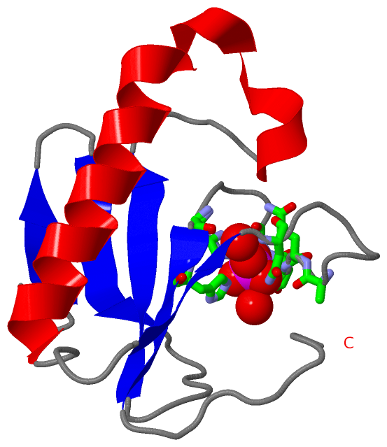
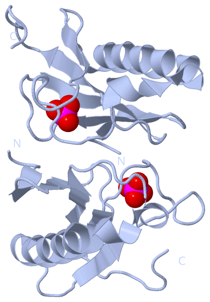
Ligands, Modified Residues, Ions (1, 1)
Asymmetric Unit (1, 1)
|
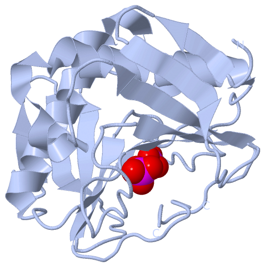
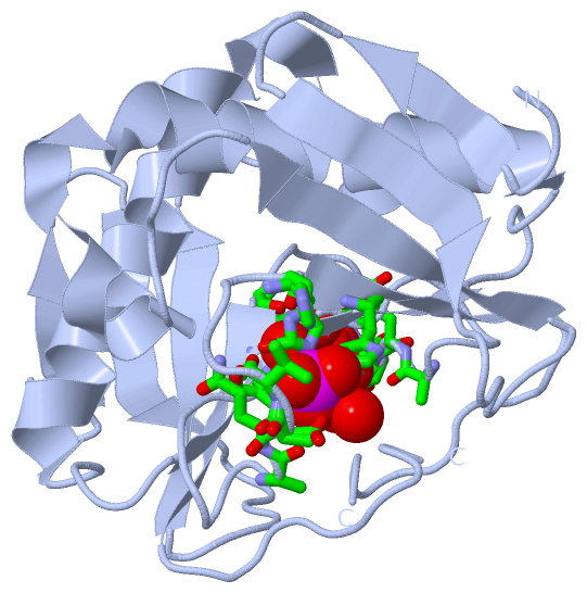
Sites (1, 1)
Asymmetric Unit (1, 1)
|
SS Bonds (0, 0)| (no "SS Bond" information available for 4NJY) |
Cis Peptide Bonds (1, 1)
Asymmetric Unit
|
||||||||
SAPs(SNPs)/Variants (0, 0)| (no "SAP(SNP)/Variant" information available for 4NJY) |
PROSITE Motifs (0, 0)| (no "PROSITE Motif" information available for 4NJY) |
Exons (0, 0)| (no "Exon" information available for 4NJY) |
Sequences/Alignments
Asymmetric Unit
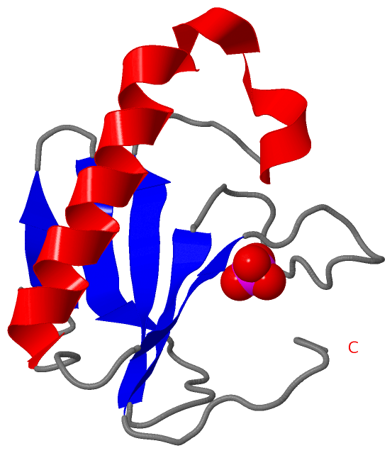
Chain A from PDB Type:PROTEIN Length:99
SCOP domains d4njya_ A: automated matches SCOP domains
CATH domains --------------------------------------------------------------------------------------------------- CATH domains
Pfam domains --------------------------------------------------------------------------------------------------- Pfam domains
SAPs(SNPs) --------------------------------------------------------------------------------------------------- SAPs(SNPs)
PROSITE --------------------------------------------------------------------------------------------------- PROSITE
Transcript --------------------------------------------------------------------------------------------------- Transcript
4njy A 65 PADILYEDQQCLVFRDVAPQAPVHFLVIPKKPIPRISQAEEEDQQLLGHLLLVAKQTAKAEGLGDGYRLVINDGKLGAQSVYHLHIHVLGGRQLQWPPG 163
74 84 94 104 114 124 134 144 154
|
||||||||||||||||||||
SCOP Domains (1, 1)
Asymmetric Unit
 |
CATH Domains (0, 0)| (no "CATH Domain" information available for 4NJY) |
Pfam Domains (0, 0)| (no "Pfam Domain" information available for 4NJY) |
Gene Ontology (8, 8)|
Asymmetric Unit(hide GO term definitions) |
Interactive Views
|
||||||||||||||||||||||||||||||||||||||||||||||||||||||||||||||||||||||||||||||||||||||||||||||||||||||||||||||||||||||||||||||||||||||||||||||
Still Images
|
||||||||||||||||
Databases
|
||||||||||||||||||||||||||||||||||||||||||||||||||||||||||||||||||||||||||||||||||||||||||||||||||||||||||||||||||||||||||||||||||||||||||||||||||||||||||||||||
Analysis Tools
|
|||||||||||||||||||||||||||||||||||||||||||||||||||||||||||||
Entries Sharing at Least One Protein Chain (UniProt ID)
Related Entries Specified in the PDB File
|
|