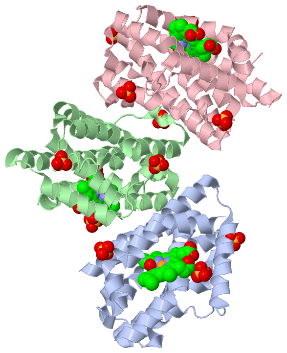
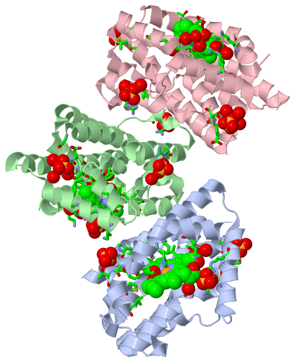
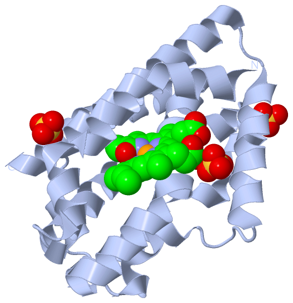
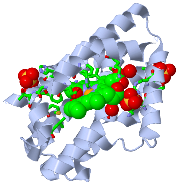
Show PDB file:   
         Plain Text   HTML   (compressed file size)
QuickSearch:   
by PDB,NDB,UniProt,PROSITE Code or Search Term(s)  
(-)Asymmetric Unit
(-)Asym. Unit - sites
(-)Biological Unit 1
(-)Biol. Unit 1 - sites
(-)Biological Unit 2
(-)Biological Unit 3
collapse expand < >
Image Asymmetric Unit
Asymmetric Unit  (Jmol Viewer)
Image Asym. Unit - sites
Asym. Unit - sites  (Jmol Viewer)
Image Biological Unit 1
Biological Unit 1  (Jmol Viewer)
Image Biol. Unit 1 - sites
Biol. Unit 1 - sites  (Jmol Viewer)
Image Biological Unit 2
Biological Unit 2  (Jmol Viewer)
Image Biological Unit 3
Biological Unit 3  (Jmol Viewer)

(-) Description

Title :  STRUCTURE OF THE FE3+-BILIVERDIN-HMUO, HEME OXYGENASE FROM CORYNEBACTERIUM DIPHTHERIAE (DATA SET III)
 
Authors :  M. Unno, M. Ikeda-Saito
Date :  20 Aug 12  (Deposition) - 21 Aug 13  (Release) - 21 Aug 13  (Revision)
Method :  X-RAY DIFFRACTION
Resolution :  1.90
Chains :  Asym. Unit :  A,B,C
Biol. Unit 1:  A  (1x)
Biol. Unit 2:  B  (1x)
Biol. Unit 3:  C  (1x)
Keywords :  Helix, Heme Degradation, Heme, Oxidoreductase (Keyword Search: [Gene Ontology, PubMed, Web (Google))
 
Reference :  M. Unno, A. Ardevol, C. Rovira, M. Ikeda-Saito
Crystal Structure Of The Substrate-Free And The Product-Bound Forms Of Hmuo, A Heme Oxygenase From Corynebacterium Diphtheriae
To Be Published
PubMed: search

(-) Compounds

Molecule 1 - HEME OXYGENASE
    ChainsA, B, C
    EC Number1.14.99.3
    EngineeredYES
    Expression SystemESCHERICHIA COLI
    Expression System Taxid562
    Organism ScientificCORYNEBACTERIUM DIPHTHERIAE
    Organism Taxid1717

 Structural Features

(-) Chains, Units

  123
Asymmetric Unit ABC
Biological Unit 1 (1x)A  
Biological Unit 2 (1x) B 
Biological Unit 3 (1x)  C

Summary Information (see also Sequences/Alignments below)

(-) Ligands, Modified Residues, Ions  (3, 16)

Asymmetric Unit (3, 16)
No.NameCountTypeFull Name
1BLA3Ligand/IonBILIVERDINE IX ALPHA
2FE1Ligand/IonFE (III) ION
3SO412Ligand/IonSULFATE ION
Biological Unit 1 (2, 4)
No.NameCountTypeFull Name
1BLA1Ligand/IonBILIVERDINE IX ALPHA
2FE-1Ligand/IonFE (III) ION
3SO43Ligand/IonSULFATE ION
Biological Unit 2 (2, 6)
No.NameCountTypeFull Name
1BLA1Ligand/IonBILIVERDINE IX ALPHA
2FE-1Ligand/IonFE (III) ION
3SO45Ligand/IonSULFATE ION
Biological Unit 3 (2, 5)
No.NameCountTypeFull Name
1BLA1Ligand/IonBILIVERDINE IX ALPHA
2FE-1Ligand/IonFE (III) ION
3SO44Ligand/IonSULFATE ION

(-) Sites  (16, 16)

Asymmetric Unit (16, 16)
No.NameEvidenceResiduesDescription
01AC1SOFTWARELYS A:13 , HIS A:20 , GLU A:24 , MET A:29 , LEU A:33 , TYR A:130 , VAL A:131 , GLY A:135 , SER A:138 , ARG A:177 , PHE A:201 , ASN A:204 , PHE A:208 , FE A:902 , SO4 A:905 , HOH A:1039 , HOH A:1062 , HOH A:1069 , HOH A:1070BINDING SITE FOR RESIDUE BLA A 901
02AC2SOFTWAREHIS A:20 , BLA A:901BINDING SITE FOR RESIDUE FE A 902
03AC3SOFTWAREGLY A:67 , ALA A:69 , GLU A:70 , SER A:71 , HOH A:1021 , HOH A:1036BINDING SITE FOR RESIDUE SO4 A 903
04AC4SOFTWARESER A:26 , THR A:27 , HIS A:205 , HOH A:1038BINDING SITE FOR RESIDUE SO4 A 904
05AC5SOFTWARELYS A:173 , ARG A:177 , BLA A:901BINDING SITE FOR RESIDUE SO4 A 905
06AC6SOFTWARESER B:326 , THR B:327 , HIS B:505 , HOH B:785BINDING SITE FOR RESIDUE SO4 B 601
07AC7SOFTWARELYS B:470 , LYS B:473 , ARG B:477 , HOH B:792BINDING SITE FOR RESIDUE SO4 B 602
08AC8SOFTWAREGLY B:367 , ALA B:369 , GLU B:370 , SER B:371 , HOH B:747 , HOH B:761 , HOH B:787BINDING SITE FOR RESIDUE SO4 B 603
09AC9SOFTWAREARG B:385 , HOH B:706BINDING SITE FOR RESIDUE SO4 B 604
10BC1SOFTWAREARG B:399BINDING SITE FOR RESIDUE SO4 B 605
11BC2SOFTWARELYS B:313 , HIS B:320 , GLU B:324 , MET B:329 , LEU B:333 , TYR B:430 , VAL B:431 , GLY B:435 , SER B:438 , GLY B:439 , ILE B:443 , ARG B:477 , PHE B:501 , PHE B:508 , HOH B:726 , HOH B:814BINDING SITE FOR RESIDUE BLA B 606
12BC3SOFTWARESER C:626 , THR C:627 , HIS C:805 , HOH C:1129BINDING SITE FOR RESIDUE SO4 C 901
13BC4SOFTWAREGLY C:638 , VAL C:639 , ALA C:640BINDING SITE FOR RESIDUE SO4 C 902
14BC5SOFTWAREALA C:669 , GLU C:670 , SER C:671 , HOH C:1021 , HOH C:1069 , HOH C:1075BINDING SITE FOR RESIDUE SO4 C 903
15BC6SOFTWAREVAL C:682 , ARG C:685 , HOH C:1018 , HOH C:1026BINDING SITE FOR RESIDUE SO4 C 904
16BC7SOFTWARELYS C:613 , HIS C:620 , ALA C:623 , GLU C:624 , TYR C:730 , VAL C:731 , GLY C:735 , SER C:738 , ILE C:743 , ARG C:777 , PHE C:808 , HOH C:1023 , HOH C:1029 , HOH C:1057 , HOH C:1066 , HOH C:1105 , HOH C:1106BINDING SITE FOR RESIDUE BLA C 905

(-) SS Bonds  (0, 0)

(no "SS Bond" information available for 4GPF)

(-) Cis Peptide Bonds  (0, 0)

(no "Cis Peptide Bond" information available for 4GPF)

 Sequence-Structure Mapping

(-) SAPs(SNPs)/Variants  (0, 0)

(no "SAP(SNP)/Variant" information available for 4GPF)

(-) PROSITE Motifs  (0, 0)

(no "PROSITE Motif" information available for 4GPF)

(-) Exons   (0, 0)

(no "Exon" information available for 4GPF)

(-) Sequences/Alignments

Asymmetric Unit
   Reformat: Number of residues per line =  ('0' or empty: single-line sequence representation)
  Number of residues per labelling interval =   
  UniProt sequence: complete  aligned part    
   Show mapping: SCOP domains CATH domains Pfam domains Secondary structure (by author)
SAPs(SNPs) PROSITE motifs Exons
(details for a mapped element are shown in a popup box when the mouse pointer rests over it)
Chain A from PDB  Type:PROTEIN  Length:207
                                                                                                                                                                                                                                               
               SCOP domains d4gpfa_ A: Heme oxygenase HmuO                                                                                                                                                                                  SCOP domains
               CATH domains --------------------------------------------------------------------------------------------------------------------------------------------------------------------------------------------------------------- CATH domains
               Pfam domains --------------------------------------------------------------------------------------------------------------------------------------------------------------------------------------------------------------- Pfam domains
         Sec.struct. author hhhhhhhhhhhhhhhhhhhhhhhhhhhh...hhhhhhhhhhhhhhhhhhhhhhhhhhhhh..hhhhhhhhhhhhhhhhhhhhhhhh..hhhhhh..hhhhhhhhhhhhhhhhhhhhhhhhhhhhhhhhhhhhhhhhhhhhhhhhhh.hhhhhhhhh.....hhhhhhhhhhhhhhhh..hhhhhhhhhhhhhhhhhhhhhhhhhhhh Sec.struct. author
                 SAPs(SNPs) --------------------------------------------------------------------------------------------------------------------------------------------------------------------------------------------------------------- SAPs(SNPs)
                    PROSITE --------------------------------------------------------------------------------------------------------------------------------------------------------------------------------------------------------------- PROSITE
                 Transcript --------------------------------------------------------------------------------------------------------------------------------------------------------------------------------------------------------------- Transcript
                 4gpf A   7 GLAVELKQSTAQAHEKAEHSTFMSDLLKGRLGVAEFTRLQEQAWLFYTALEQAVDAVRASGFAESLLDPALNRAEVLARDLDKLNGSSEWRSRITASPAVIDYVNRLEEIRDNVDGPALVAHHYVRYLGDLSGGQVIARMMQRHYGVDPEALGFYHFEGIAKLKVYKDEYREKLNNLELSDEQREHLLKEATDAFVFNHQVFADLGK 213
                                    16        26        36        46        56        66        76        86        96       106       116       126       136       146       156       166       176       186       196       206       

Chain B from PDB  Type:PROTEIN  Length:209
                                                                                                                                                                                                                                                 
               SCOP domains d4gpfb_ B: Heme oxygenase HmuO                                                                                                                                                                                    SCOP domains
               CATH domains ----------------------------------------------------------------------------------------------------------------------------------------------------------------------------------------------------------------- CATH domains
               Pfam domains ----------------------------------------------------------------------------------------------------------------------------------------------------------------------------------------------------------------- Pfam domains
         Sec.struct. author .hhhhhhhhhhhhhhhhhhhhhhhhhhh...hhhhhhhhhhhhhhhhhhhhhhhhhhhhh.......hhhhhhhhhhhhhhhhhhh.hhhhhh...hhhhhhhhhhhhhhhhhhhhhhhhhhhhhhhhhhhhhhhhhhhhhhhhhh.hhhhhhhhh.....hhhhhhhhhhhhhhhh..hhhhhhhhhhhhhhhhhhhhhhhhhhhhh. Sec.struct. author
                 SAPs(SNPs) ----------------------------------------------------------------------------------------------------------------------------------------------------------------------------------------------------------------- SAPs(SNPs)
                    PROSITE ----------------------------------------------------------------------------------------------------------------------------------------------------------------------------------------------------------------- PROSITE
                 Transcript ----------------------------------------------------------------------------------------------------------------------------------------------------------------------------------------------------------------- Transcript
                 4gpf B 307 GLAVELKQSTAQAHEKAEHSTFMSDLLKGRLGVAEFTRLQEQAWLFYTALEQAVDAVRASGFAESLLDPALNRAEVLARDLDKLNGSSEWRSRITASPAVIDYVNRLEEIRDNVDGPALVAHHYVRYLGDLSGGQVIARMMQRHYGVDPEALGFYHFEGIAKLKVYKDEYREKLNNLELSDEQREHLLKEATDAFVFNHQVFADLGKGL 515
                                   316       326       336       346       356       366       376       386       396       406       416       426       436       446       456       466       476       486       496       506         

Chain C from PDB  Type:PROTEIN  Length:208
                                                                                                                                                                                                                                                
               SCOP domains d4gpfc_ C: Heme oxygenase HmuO                                                                                                                                                                                   SCOP domains
               CATH domains ---------------------------------------------------------------------------------------------------------------------------------------------------------------------------------------------------------------- CATH domains
               Pfam domains ---------------------------------------------------------------------------------------------------------------------------------------------------------------------------------------------------------------- Pfam domains
         Sec.struct. author .hhhhhhhhhhhhhhhhhhhhhhhhhhhh...hhhhhhhhhhhhhhhhhhhhhhhhhhhhh.......hhhhhhhhhhhhhhhhhhh.hhhhhh...hhhhhhhhhhhhhhhhhhhhhhhhhhhhhhhhhhhhhhhhhhhhhhhhhh.hhhhhhhhh.....hhhhhhhhhhhhhhhh..hhhhhhhhhhhhhhhhhhhhhhhhhhh. Sec.struct. author
                 SAPs(SNPs) ---------------------------------------------------------------------------------------------------------------------------------------------------------------------------------------------------------------- SAPs(SNPs)
                    PROSITE ---------------------------------------------------------------------------------------------------------------------------------------------------------------------------------------------------------------- PROSITE
                 Transcript ---------------------------------------------------------------------------------------------------------------------------------------------------------------------------------------------------------------- Transcript
                 4gpf C 606 AGLAVELKQSTAQAHEKAEHSTFMSDLLKGRLGVAEFTRLQEQAWLFYTALEQAVDAVRASGFAESLLDPALNRAEVLARDLDKLNGSSEWRSRITASPAVIDYVNRLEEIRDNVDGPALVAHHYVRYLGDLSGGQVIARMMQRHYGVDPEALGFYHFEGIAKLKVYKDEYREKLNNLELSDEQREHLLKEATDAFVFNHQVFADLGK 813
                                   615       625       635       645       655       665       675       685       695       705       715       725       735       745       755       765       775       785       795       805        

   Legend:   → Mismatch (orange background)
  - → Gap (green background, '-', border residues have a numbering label)
    → Modified Residue (blue background, lower-case, 'x' indicates undefined single-letter code, labelled with number + name)
  x → Chemical Group (purple background, 'x', labelled with number + name, e.g. ACE or NH2)
  extra numbering lines below/above indicate numbering irregularities and modified residue names etc., number ends below/above '|'

 Classification and Annotation

(-) SCOP Domains  (1, 3)

Asymmetric Unit

(-) CATH Domains  (0, 0)

(no "CATH Domain" information available for 4GPF)

(-) Pfam Domains  (0, 0)

(no "Pfam Domain" information available for 4GPF)

(-) Gene Ontology  (5, 5)

Asymmetric Unit(hide GO term definitions)

 Visualization

(-) Interactive Views

Asymmetric Unit
  Complete Structure
    Jena3D(integrated viewing of ligand, site, SAP, PROSITE, SCOP information)
    WebMol | AstexViewer[tm]@PDBe
(Java Applets, require no local installation except for Java; loading may be slow)
    STRAP
(Java WebStart application, automatic local installation, requires Java; full application with system access!)
    RasMol
(require local installation)
    Molscript (VRML)
(requires installation of a VRML viewer; select preferred view via VRML and generate a mono or stereo PDF format file)
 
  Ligands, Modified Residues, Ions
    BLA  [ RasMol | Jena3D ]  +environment [ RasMol | Jena3D ]
    FE  [ RasMol | Jena3D ]  +environment [ RasMol | Jena3D ]
    SO4  [ RasMol | Jena3D ]  +environment [ RasMol | Jena3D ]
 
  Sites
    AC1  [ RasMol ]  +environment [ RasMol ]
    AC2  [ RasMol ]  +environment [ RasMol ]
    AC3  [ RasMol ]  +environment [ RasMol ]
    AC4  [ RasMol ]  +environment [ RasMol ]
    AC5  [ RasMol ]  +environment [ RasMol ]
    AC6  [ RasMol ]  +environment [ RasMol ]
    AC7  [ RasMol ]  +environment [ RasMol ]
    AC8  [ RasMol ]  +environment [ RasMol ]
    AC9  [ RasMol ]  +environment [ RasMol ]
    BC1  [ RasMol ]  +environment [ RasMol ]
    BC2  [ RasMol ]  +environment [ RasMol ]
    BC3  [ RasMol ]  +environment [ RasMol ]
    BC4  [ RasMol ]  +environment [ RasMol ]
    BC5  [ RasMol ]  +environment [ RasMol ]
    BC6  [ RasMol ]  +environment [ RasMol ]
    BC7  [ RasMol ]  +environment [ RasMol ]
 
  Cis Peptide Bonds
(no "Cis Peptide Bonds" information available for 4gpf)
 
Biological Units
  Complete Structure
    Biological Unit 1  [ Jena3D ]
    Biological Unit 2  [ Jena3D ]
    Biological Unit 3  [ Jena3D ]

(-) Still Images

Jmol
  protein: cartoon or spacefill or dots and stick; nucleic acid: cartoon and stick; ligands: spacefill; active site: stick
Molscript
  protein, nucleic acid: cartoon; ligands: spacefill; active site: ball and stick

 Databases and Analysis Tools

(-) Databases

Access by PDB/NDB ID
  4gpf
    Family and Domain InformationProDom | SYSTERS
    General Structural InformationGlycoscienceDB | MMDB | NDB | OCA | PDB | PDBe | PDBj | PDBsum | PDBWiki | PQS | PROTEOPEDIA
    Orientation in MembranesOPM
    Protein SurfaceSURFACE
    Secondary StructureDSSP (structure derived) | HSSP (homology derived)
    Structural GenomicsGeneCensus
    Structural NeighboursCE | VAST
    Structure ClassificationCATH | Dali | SCOP
    Validation and Original DataBMRB Data View | BMRB Restraints Grid | EDS | PROCHECK | RECOORD | WHAT_CHECK
 
Access by UniProt ID/Accession number
  Q54AI1_CORDP | Q54AI1
    Comparative Protein Structure ModelsModBase
    Genomic InformationEnsembl
    Protein-protein InteractionDIP
    Sequence, Family and Domain InformationInterPro | Pfam | SMART | UniProtKB/TrEMBL
 
Access by Enzyme Classificator   (EC Number)
  1.14.99.3
    General Enzyme InformationBRENDA | EC-PDB | Enzyme | IntEnz
    PathwayKEGG | MetaCyc
 
Access by Disease Identifier   (MIM ID)
  (no 'MIM ID' available)
    Disease InformationOMIM
 
Access by GenAge ID
  (no 'GenAge ID' available)
    Age Related InformationGenAge

(-) Analysis Tools

Access by PDB/NDB ID
    Domain InformationXDom
    Interatomic Contacts of Structural UnitsCSU
    Ligand-protein ContactsLPC
    Protein CavitiescastP
    Sequence and Secondary StructurePDBCartoon
    Structure AlignmentSTRAP(Java WebStart application, automatic local installation, requires Java; full application with system access!)
    Structure and Sequence BrowserSTING
 
Access by UniProt ID/Accession number
  Q54AI1_CORDP | Q54AI1
    Protein Disorder PredictionDisEMBL | FoldIndex | GLOBPLOT (for more information see DisProt)

 Related Entries

(-) Entries Sharing at Least One Protein Chain (UniProt ID)

UniProtKB/TrEMBL
        Q54AI1_CORDP | Q54AI11wzd 2z68 3i8r 3moo 4goh 4gpc 4gph

(-) Related Entries Specified in the PDB File

1iw0 1iw1 4goh 4gpc 4gph