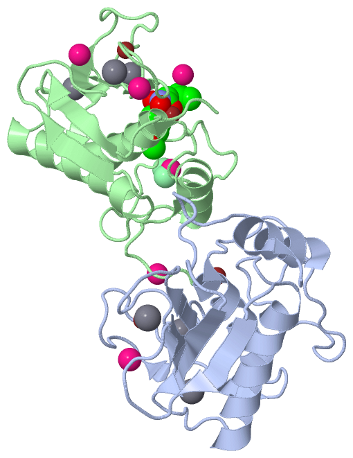
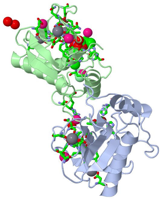
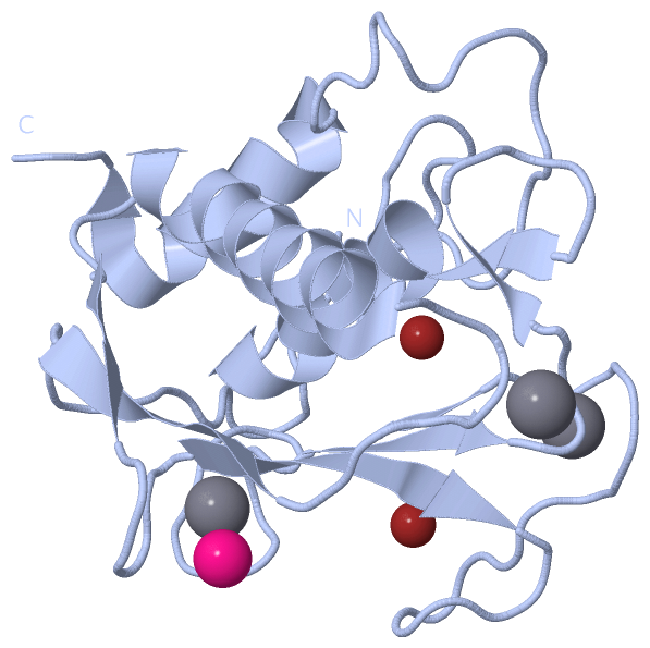
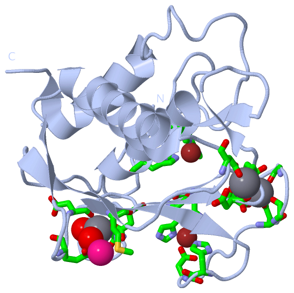
Show PDB file:   
         Plain Text   HTML   (compressed file size)
QuickSearch:   
by PDB,NDB,UniProt,PROSITE Code or Search Term(s)  
(-)Asymmetric Unit
(-)Asym. Unit - sites
(-)Biological Unit 1
(-)Biol. Unit 1 - sites
(-)Biological Unit 2
collapse expand < >
Image Asymmetric Unit
Asymmetric Unit  (Jmol Viewer)
Image Asym. Unit - sites
Asym. Unit - sites  (Jmol Viewer)
Image Biological Unit 1
Biological Unit 1  (Jmol Viewer)
Image Biol. Unit 1 - sites
Biol. Unit 1 - sites  (Jmol Viewer)
Image Biological Unit 2
Biological Unit 2  (Jmol Viewer)

(-) Description

Title :  STRUCTURE OF MMP3 COMPLEXED WITH A PLATINUM-BASED INHIBITOR.
 
Authors :  B. D. Belviso, F. Arnesano, V. Calderone, R. Caliandro, G. Natile, D. Si
Date :  13 Feb 12  (Deposition) - 20 Feb 13  (Release) - 10 Jul 13  (Revision)
Method :  X-RAY DIFFRACTION
Resolution :  1.96
Chains :  Asym. Unit :  A,B
Biol. Unit 1:  A  (1x)
Biol. Unit 2:  B  (1x)
Keywords :  Matrix Metalloprotease, Hydrolase-Hydrolase Inhibitor Complex (Keyword Search: [Gene Ontology, PubMed, Web (Google))
 
Reference :  B. D. Belviso, R. Caliandro, D. Siliqi, V. Calderone, F. Arnesano, G. Natile
Structure Of Matrix Metalloproteinase-3 With A Platinum-Based Inhibitor.
Chem. Commun. (Camb. ) V. 49 5492 2013
PubMed-ID: 23660647  |  Reference-DOI: 10.1039/C3CC41278D

(-) Compounds

Molecule 1 - STROMELYSIN-1
    ChainsA, B
    EC Number3.4.24.17
    EngineeredYES
    Expression SystemESCHERICHIA COLI
    Expression System Taxid562
    FragmentUNP RESIDUES 100-272
    GeneMMP3, STMY1
    Organism CommonHUMAN
    Organism ScientificHOMO SAPIENS
    Organism Taxid9606
    SynonymSL-1, MATRIX METALLOPROTEINASE-3, MMP-3, TRANSIN-1

 Structural Features

(-) Chains, Units

  12
Asymmetric Unit AB
Biological Unit 1 (1x)A 
Biological Unit 2 (1x) B

Summary Information (see also Sequences/Alignments below)

(-) Ligands, Modified Residues, Ions  (5, 19)

Asymmetric Unit (5, 19)
No.NameCountTypeFull Name
1CA6Ligand/IonCALCIUM ION
2CL2Ligand/IonCHLORIDE ION
3NGH1Ligand/IonN-ISOBUTYL-N-[4-METHOXYPHENYLSULFONYL]GLYCYL HYDROXAMICACID
4PT6Ligand/IonPLATINUM (II) ION
5ZN4Ligand/IonZINC ION
Biological Unit 1 (0, 0)
No.NameCountTypeFull Name
1CA-1Ligand/IonCALCIUM ION
2CL-1Ligand/IonCHLORIDE ION
3NGH-1Ligand/IonN-ISOBUTYL-N-[4-METHOXYPHENYLSULFONYL]GLYCYL HYDROXAMICACID
4PT-1Ligand/IonPLATINUM (II) ION
5ZN-1Ligand/IonZINC ION
Biological Unit 2 (1, 1)
No.NameCountTypeFull Name
1CA-1Ligand/IonCALCIUM ION
2CL-1Ligand/IonCHLORIDE ION
3NGH1Ligand/IonN-ISOBUTYL-N-[4-METHOXYPHENYLSULFONYL]GLYCYL HYDROXAMICACID
4PT-1Ligand/IonPLATINUM (II) ION
5ZN-1Ligand/IonZINC ION

(-) Sites  (19, 19)

Asymmetric Unit (19, 19)
No.NameEvidenceResiduesDescription
01AC1SOFTWAREHIS A:201 , HIS A:205 , HIS A:211 , THR B:255 , HOH B:483BINDING SITE FOR RESIDUE ZN A 301
02AC2SOFTWAREHIS A:151 , ASP A:153 , HIS A:166 , HIS A:179BINDING SITE FOR RESIDUE ZN A 302
03AC3SOFTWAREASP A:158 , GLY A:159 , GLY A:161 , VAL A:163 , ASP A:181 , GLU A:184BINDING SITE FOR RESIDUE CA A 303
04AC4SOFTWAREASP A:141 , GLY A:173 , ASN A:175 , ASP A:177 , HOH A:402 , HOH A:411BINDING SITE FOR RESIDUE CA A 304
05AC5SOFTWAREASP A:107 , ASP A:182 , GLU A:184 , HOH B:410 , HOH B:411BINDING SITE FOR RESIDUE CA A 305
06AC6SOFTWAREGLU A:139 , MET A:143 , HOH A:460BINDING SITE FOR RESIDUE PT A 306
07AC7SOFTWAREHIS B:201 , HIS B:205 , HIS B:211 , NGH B:313BINDING SITE FOR RESIDUE ZN B 301
08AC8SOFTWAREHIS B:151 , ASP B:153 , HIS B:166 , HIS B:179BINDING SITE FOR RESIDUE ZN B 302
09AC9SOFTWAREASP B:158 , GLY B:159 , GLY B:161 , VAL B:163 , ASP B:181 , GLU B:184BINDING SITE FOR RESIDUE CA B 303
10BC1SOFTWAREASP B:141 , GLY B:173 , ASN B:175 , ASP B:177 , HOH B:408 , HOH B:416BINDING SITE FOR RESIDUE CA B 304
11BC2SOFTWAREASP B:107 , ASP B:182 , GLU B:184 , HOH B:422BINDING SITE FOR RESIDUE CA B 305
12BC3SOFTWAREGLU B:139 , MET B:143 , HOH B:500 , HOH B:501BINDING SITE FOR RESIDUE PT B 306
13BC4SOFTWAREHIS B:224 , CL B:311 , CL B:312 , HOH B:491BINDING SITE FOR RESIDUE PT B 307
14BC5SOFTWAREHOH B:494 , HOH B:495BINDING SITE FOR RESIDUE PT B 308
15BC6SOFTWAREARG B:231 , ARG B:233 , HOH B:401 , HOH B:456 , HOH B:490BINDING SITE FOR RESIDUE PT B 309
16BC7SOFTWARELYS B:94 , HOH B:402 , HOH B:403 , HOH B:404BINDING SITE FOR RESIDUE PT B 310
17BC8SOFTWAREHIS B:224 , PT B:307 , CL B:312 , HOH B:491BINDING SITE FOR RESIDUE CL B 311
18BC9SOFTWAREPT B:307 , CL B:311 , HOH B:491BINDING SITE FOR RESIDUE CL B 312
19CC1SOFTWAREVAL B:163 , LEU B:164 , ALA B:165 , HIS B:201 , GLU B:202 , HIS B:205 , HIS B:211 , PRO B:221 , TYR B:223 , ZN B:301 , HOH B:426BINDING SITE FOR RESIDUE NGH B 313

(-) SS Bonds  (0, 0)

(no "SS Bond" information available for 4DPE)

(-) Cis Peptide Bonds  (0, 0)

(no "Cis Peptide Bond" information available for 4DPE)

 Sequence-Structure Mapping

(-) SAPs(SNPs)/Variants  (0, 0)

(no "SAP(SNP)/Variant" information available for 4DPE)

(-) PROSITE Motifs  (1, 2)

Asymmetric Unit (1, 2)
 PROSITEUniProtKBPDB
No.IDACDescriptionIDLocationCountLocation
1ZINC_PROTEASEPS00142 Neutral zinc metallopeptidases, zinc-binding region signature.MMP3_HUMAN215-224
 
  2A:198-207
B:198-207
Biological Unit 1 (1, 1)
 PROSITEUniProtKBPDB
No.IDACDescriptionIDLocationCountLocation
1ZINC_PROTEASEPS00142 Neutral zinc metallopeptidases, zinc-binding region signature.MMP3_HUMAN215-224
 
  1A:198-207
-
Biological Unit 2 (1, 1)
 PROSITEUniProtKBPDB
No.IDACDescriptionIDLocationCountLocation
1ZINC_PROTEASEPS00142 Neutral zinc metallopeptidases, zinc-binding region signature.MMP3_HUMAN215-224
 
  1-
B:198-207

(-) Exons   (5, 10)

Asymmetric Unit (5, 10)
 ENSEMBLUniProtKBPDB
No.Transcript IDExonExon IDGenome LocationLengthIDLocationLengthCountLocationLength
1.1aENST000002998551aENSE00002171374chr11:102714534-102714173362MMP3_HUMAN1-35350--
1.2ENST000002998552ENSE00001105428chr11:102713647-102713403245MMP3_HUMAN36-117822A:83-100
B:89-100
18
12
1.3ENST000002998553ENSE00001642002chr11:102713310-102713162149MMP3_HUMAN117-167512A:100-150
B:100-150
51
51
1.4bENST000002998554bENSE00001105420chr11:102713010-102712885126MMP3_HUMAN167-209432A:150-192
B:150-192
43
43
1.5ENST000002998555ENSE00001657829chr11:102711324-102711160165MMP3_HUMAN209-264562A:192-247
B:192-247
56
56
1.6ENST000002998556ENSE00001105427chr11:102710983-102710839145MMP3_HUMAN264-312492A:247-250
B:247-255
4
9
1.7ENST000002998557ENSE00001105439chr11:102709974-102709841134MMP3_HUMAN312-357460--
1.8ENST000002998558ENSE00001801971chr11:102709441-102709282160MMP3_HUMAN357-410540--
1.9ENST000002998559ENSE00001105448chr11:102708132-102708029104MMP3_HUMAN410-445360--
1.11ENST0000029985511ENSE00001292630chr11:102706957-102706532426MMP3_HUMAN445-477330--

(-) Sequences/Alignments

Asymmetric Unit
   Reformat: Number of residues per line =  ('0' or empty: single-line sequence representation)
  Number of residues per labelling interval =   
  UniProt sequence: complete  aligned part    
   Show mapping: SCOP domains CATH domains Pfam domains Secondary structure (by author)
SAPs(SNPs) PROSITE motifs Exons
(details for a mapped element are shown in a popup box when the mouse pointer rests over it)
Chain A from PDB  Type:PROTEIN  Length:168
 aligned with MMP3_HUMAN | P08254 from UniProtKB/Swiss-Prot  Length:477

    Alignment length:168
                                   109       119       129       139       149       159       169       179       189       199       209       219       229       239       249       259        
           MMP3_HUMAN   100 FRTFPGIPKWRKTHLTYRIVNYTPDLPKDAVDSAVEKALKVWEEVTPLTFSRLYEGEADIMISFAVREHGDFYPFDGPGNVLAHAYAPGPGINGDAHFDDDEQWTKDTTGTNLFLVAAHEIGHSLGLFHSANTEALMYPLYHSLTDLTRFRLSQDDINGIQSLYGPPP 267
               SCOP domains d4dpea_ A: Stromelysin-1 (MMP-3)                                                                                                                                         SCOP domains
               CATH domains ------------------------------------------------------------------------------------------------------------------------------------------------------------------------ CATH domains
               Pfam domains ------------------------------------------------------------------------------------------------------------------------------------------------------------------------ Pfam domains
         Sec.struct. author .............eeeeee.......hhhhhhhhhhhhhhhhhh....eeee.......eeeeee.................eee..........eeee....ee.....eehhhhhhhhhhhhh...........................hhhhhhhhhhhhh... Sec.struct. author
                 SAPs(SNPs) ------------------------------------------------------------------------------------------------------------------------------------------------------------------------ SAPs(SNPs)
                    PROSITE -------------------------------------------------------------------------------------------------------------------ZINC_PROTE------------------------------------------- PROSITE
           Transcript 1 (1) Exon 1.2          -------------------------------------------------Exon 1.4b  PDB: A:150-192 UniProt: 167-209 ------------------------------------------------------1.6  Transcript 1 (1)
           Transcript 1 (2) -----------------Exon 1.3  PDB: A:100-150 UniProt: 117-167          -----------------------------------------Exon 1.5  PDB: A:192-247 UniProt: 209-264               --- Transcript 1 (2)
                 4dpe A  83 FRTFPGIPKWRKTHLTYRIVNYTPDLPKDAVDSAVEKALKVWEEVTPLTFSRLYEGEADIMISFAVREHGDFYPFDGPGNVLAHAYAPGPGINGDAHFDDDEQWTKDTTGTNLFLVAAHEIGHSLGLFHSANTEALMYPLYHSLTDLTRFRLSQDDINGIQSLYGPPP 250
                                    92       102       112       122       132       142       152       162       172       182       192       202       212       222       232       242        

Chain B from PDB  Type:PROTEIN  Length:167
 aligned with MMP3_HUMAN | P08254 from UniProtKB/Swiss-Prot  Length:477

    Alignment length:167
                                   115       125       135       145       155       165       175       185       195       205       215       225       235       245       255       265       
           MMP3_HUMAN   106 IPKWRKTHLTYRIVNYTPDLPKDAVDSAVEKALKVWEEVTPLTFSRLYEGEADIMISFAVREHGDFYPFDGPGNVLAHAYAPGPGINGDAHFDDDEQWTKDTTGTNLFLVAAHEIGHSLGLFHSANTEALMYPLYHSLTDLTRFRLSQDDINGIQSLYGPPPDSPET 272
               SCOP domains d4dpeb_ B: Stromelysin-1 (MMP-3)                                                                                                                                        SCOP domains
               CATH domains ----------------------------------------------------------------------------------------------------------------------------------------------------------------------- CATH domains
               Pfam domains ----------------------------------------------------------------------------------------------------------------------------------------------------------------------- Pfam domains
         Sec.struct. author .......eeeeee.......hhhhhhhhhhhhhhhhhh....eeee.......eeeeee.................eee..........eeee....ee.....eehhhhhhhhhhhhh...........................hhhhhhhhhhhhh........ Sec.struct. author
                 SAPs(SNPs) ----------------------------------------------------------------------------------------------------------------------------------------------------------------------- SAPs(SNPs)
                    PROSITE -------------------------------------------------------------------------------------------------------------ZINC_PROTE------------------------------------------------ PROSITE
           Transcript 1 (1) Exon 1.2    -------------------------------------------------Exon 1.4b  PDB: B:150-192 UniProt: 167-209 ------------------------------------------------------Exon 1.6  Transcript 1 (1)
           Transcript 1 (2) -----------Exon 1.3  PDB: B:100-150 UniProt: 117-167          -----------------------------------------Exon 1.5  PDB: B:192-247 UniProt: 209-264               -------- Transcript 1 (2)
                 4dpe B  89 IPKWRKTHLTYRIVNYTPDLPKDAVDSAVEKALKVWEEVTPLTFSRLYEGEADIMISFAVREHGDFYPFDGPGNVLAHAYAPGPGINGDAHFDDDEQWTKDTTGTNLFLVAAHEIGHSLGLFHSANTEALMYPLYHSLTDLTRFRLSQDDINGIQSLYGPPPDSPET 255
                                    98       108       118       128       138       148       158       168       178       188       198       208       218       228       238       248       

   Legend:   → Mismatch (orange background)
  - → Gap (green background, '-', border residues have a numbering label)
    → Modified Residue (blue background, lower-case, 'x' indicates undefined single-letter code, labelled with number + name)
  x → Chemical Group (purple background, 'x', labelled with number + name, e.g. ACE or NH2)
  extra numbering lines below/above indicate numbering irregularities and modified residue names etc., number ends below/above '|'

 Classification and Annotation

(-) SCOP Domains  (1, 2)

Asymmetric Unit

(-) CATH Domains  (0, 0)

(no "CATH Domain" information available for 4DPE)

(-) Pfam Domains  (0, 0)

(no "Pfam Domain" information available for 4DPE)

(-) Gene Ontology  (21, 21)

Asymmetric Unit(hide GO term definitions)
Chain A,B   (MMP3_HUMAN | P08254)
molecular function
    GO:0005509    calcium ion binding    Interacting selectively and non-covalently with calcium ions (Ca2+).
    GO:0004175    endopeptidase activity    Catalysis of the hydrolysis of internal, alpha-peptide bonds in a polypeptide chain.
    GO:0016787    hydrolase activity    Catalysis of the hydrolysis of various bonds, e.g. C-O, C-N, C-C, phosphoric anhydride bonds, etc. Hydrolase is the systematic name for any enzyme of EC class 3.
    GO:0046872    metal ion binding    Interacting selectively and non-covalently with any metal ion.
    GO:0004222    metalloendopeptidase activity    Catalysis of the hydrolysis of internal, alpha-peptide bonds in a polypeptide chain by a mechanism in which water acts as a nucleophile, one or two metal ions hold the water molecule in place, and charged amino acid side chains are ligands for the metal ions.
    GO:0008237    metallopeptidase activity    Catalysis of the hydrolysis of peptide bonds by a mechanism in which water acts as a nucleophile, one or two metal ions hold the water molecule in place, and charged amino acid side chains are ligands for the metal ions.
    GO:0008233    peptidase activity    Catalysis of the hydrolysis of a peptide bond. A peptide bond is a covalent bond formed when the carbon atom from the carboxyl group of one amino acid shares electrons with the nitrogen atom from the amino group of a second amino acid.
    GO:0005515    protein binding    Interacting selectively and non-covalently with any protein or protein complex (a complex of two or more proteins that may include other nonprotein molecules).
    GO:0004252    serine-type endopeptidase activity    Catalysis of the hydrolysis of internal, alpha-peptide bonds in a polypeptide chain by a catalytic mechanism that involves a catalytic triad consisting of a serine nucleophile that is activated by a proton relay involving an acidic residue (e.g. aspartate or glutamate) and a basic residue (usually histidine).
    GO:0008270    zinc ion binding    Interacting selectively and non-covalently with zinc (Zn) ions.
biological process
    GO:0071732    cellular response to nitric oxide    Any process that results in a change in state or activity of a cell (in terms of movement, secretion, enzyme production, gene expression, etc.) as a result of a nitric oxide stimulus.
    GO:0030574    collagen catabolic process    The proteolytic chemical reactions and pathways resulting in the breakdown of collagen in the extracellular matrix, usually carried out by proteases secreted by nearby cells.
    GO:0022617    extracellular matrix disassembly    A process that results in the breakdown of the extracellular matrix.
    GO:0010727    negative regulation of hydrogen peroxide metabolic process    Any process that decreases the frequency, rate or extent of the chemical reactions and pathways involving hydrogen peroxide.
    GO:1903209    positive regulation of oxidative stress-induced cell death    Any process that activates or increases the frequency, rate or extent of oxidative stress-induced cell death.
    GO:0032461    positive regulation of protein oligomerization    Any process that activates or increases the frequency, rate or extent of protein oligomerization.
    GO:0006508    proteolysis    The hydrolysis of proteins into smaller polypeptides and/or amino acids by cleavage of their peptide bonds.
cellular component
    GO:0031012    extracellular matrix    A structure lying external to one or more cells, which provides structural support for cells or tissues.
    GO:0005576    extracellular region    The space external to the outermost structure of a cell. For cells without external protective or external encapsulating structures this refers to space outside of the plasma membrane. This term covers the host cell environment outside an intracellular parasite.
    GO:0005615    extracellular space    That part of a multicellular organism outside the cells proper, usually taken to be outside the plasma membranes, and occupied by fluid.
    GO:0005578    proteinaceous extracellular matrix    A layer consisting mainly of proteins (especially collagen) and glycosaminoglycans (mostly as proteoglycans) that forms a sheet underlying or overlying cells such as endothelial and epithelial cells. The proteins are secreted by cells in the vicinity. An example of this component is found in Mus musculus.

 Visualization

(-) Interactive Views

Asymmetric Unit
  Complete Structure
    Jena3D(integrated viewing of ligand, site, SAP, PROSITE, SCOP information)
    WebMol | AstexViewer[tm]@PDBe
(Java Applets, require no local installation except for Java; loading may be slow)
    STRAP
(Java WebStart application, automatic local installation, requires Java; full application with system access!)
    RasMol
(require local installation)
    Molscript (VRML)
(requires installation of a VRML viewer; select preferred view via VRML and generate a mono or stereo PDF format file)
 
  Ligands, Modified Residues, Ions
    CA  [ RasMol | Jena3D ]  +environment [ RasMol | Jena3D ]
    CL  [ RasMol | Jena3D ]  +environment [ RasMol | Jena3D ]
    NGH  [ RasMol | Jena3D ]  +environment [ RasMol | Jena3D ]
    PT  [ RasMol | Jena3D ]  +environment [ RasMol | Jena3D ]
    ZN  [ RasMol | Jena3D ]  +environment [ RasMol | Jena3D ]
 
  Sites
    AC1  [ RasMol ]  +environment [ RasMol ]
    AC2  [ RasMol ]  +environment [ RasMol ]
    AC3  [ RasMol ]  +environment [ RasMol ]
    AC4  [ RasMol ]  +environment [ RasMol ]
    AC5  [ RasMol ]  +environment [ RasMol ]
    AC6  [ RasMol ]  +environment [ RasMol ]
    AC7  [ RasMol ]  +environment [ RasMol ]
    AC8  [ RasMol ]  +environment [ RasMol ]
    AC9  [ RasMol ]  +environment [ RasMol ]
    BC1  [ RasMol ]  +environment [ RasMol ]
    BC2  [ RasMol ]  +environment [ RasMol ]
    BC3  [ RasMol ]  +environment [ RasMol ]
    BC4  [ RasMol ]  +environment [ RasMol ]
    BC5  [ RasMol ]  +environment [ RasMol ]
    BC6  [ RasMol ]  +environment [ RasMol ]
    BC7  [ RasMol ]  +environment [ RasMol ]
    BC8  [ RasMol ]  +environment [ RasMol ]
    BC9  [ RasMol ]  +environment [ RasMol ]
    CC1  [ RasMol ]  +environment [ RasMol ]
 
  Cis Peptide Bonds
(no "Cis Peptide Bonds" information available for 4dpe)
 
Biological Units
  Complete Structure
    Biological Unit 1  [ Jena3D ]
    Biological Unit 2  [ Jena3D ]

(-) Still Images

Jmol
  protein: cartoon or spacefill or dots and stick; nucleic acid: cartoon and stick; ligands: spacefill; active site: stick
Molscript
  protein, nucleic acid: cartoon; ligands: spacefill; active site: ball and stick

 Databases and Analysis Tools

(-) Databases

Access by PDB/NDB ID
  4dpe
    Family and Domain InformationProDom | SYSTERS
    General Structural InformationGlycoscienceDB | MMDB | NDB | OCA | PDB | PDBe | PDBj | PDBsum | PDBWiki | PQS | PROTEOPEDIA
    Orientation in MembranesOPM
    Protein SurfaceSURFACE
    Secondary StructureDSSP (structure derived) | HSSP (homology derived)
    Structural GenomicsGeneCensus
    Structural NeighboursCE | VAST
    Structure ClassificationCATH | Dali | SCOP
    Validation and Original DataBMRB Data View | BMRB Restraints Grid | EDS | PROCHECK | RECOORD | WHAT_CHECK
 
Access by UniProt ID/Accession number
  MMP3_HUMAN | P08254
    Comparative Protein Structure ModelsModBase
    Genomic InformationEnsembl
    Protein-protein InteractionDIP
    Sequence, Family and Domain InformationInterPro | Pfam | SMART | UniProtKB/SwissProt
 
Access by Enzyme Classificator   (EC Number)
  3.4.24.17
    General Enzyme InformationBRENDA | EC-PDB | Enzyme | IntEnz
    PathwayKEGG | MetaCyc
 
Access by Disease Identifier   (MIM ID)
  (no 'MIM ID' available)
    Disease InformationOMIM
 
Access by GenAge ID
  (no 'GenAge ID' available)
    Age Related InformationGenAge

(-) Analysis Tools

Access by PDB/NDB ID
    Domain InformationXDom
    Interatomic Contacts of Structural UnitsCSU
    Ligand-protein ContactsLPC
    Protein CavitiescastP
    Sequence and Secondary StructurePDBCartoon
    Structure AlignmentSTRAP(Java WebStart application, automatic local installation, requires Java; full application with system access!)
    Structure and Sequence BrowserSTING
 
Access by UniProt ID/Accession number
  MMP3_HUMAN | P08254
    Protein Disorder PredictionDisEMBL | FoldIndex | GLOBPLOT (for more information see DisProt)

 Related Entries

(-) Entries Sharing at Least One Protein Chain (UniProt ID)

UniProtKB/Swiss-Prot
        MMP3_HUMAN | P082541b3d 1b8y 1biw 1bm6 1bqo 1c3i 1c8t 1caq 1ciz 1cqr 1d5j 1d7x 1d8f 1d8m 1g05 1g49 1g4k 1hfs 1hy7 1m1w 1oo9 1qia 1qic 1slm 1sln 1uea 1ums 1umt 1usn 2d1o 2jnp 2jt5 2jt6 2srt 2usn 3ohl 3oho 3usn 4g9l 4ja1

(-) Related Entries Specified in the PDB File

4g9l 4ja1