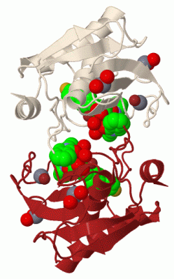
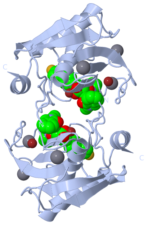
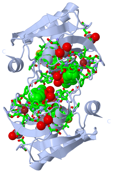
Asymmetric Unit (12, 12)
| No. | Name | Evidence | Residues | Description |
|---|
| 01 | AC1 | SOFTWARE | HIS A:201 , HIS A:205 , HIS A:211 , L04 A:256 | BINDING SITE FOR RESIDUE ZN A 257 |
| 02 | AC2 | SOFTWARE | HIS A:151 , ASP A:153 , HIS A:166 , HIS A:179 | BINDING SITE FOR RESIDUE ZN A 258 |
| 03 | AC3 | SOFTWARE | ASP A:158 , GLY A:159 , GLY A:161 , VAL A:163 , ASP A:181 , GLU A:184 | BINDING SITE FOR RESIDUE CA A 259 |
| 04 | AC4 | SOFTWARE | ASP A:141 , GLY A:173 , ASN A:175 , ASP A:177 , HOH A:306 , HOH A:312 | BINDING SITE FOR RESIDUE CA A 260 |
| 05 | AC5 | SOFTWARE | ASP A:107 , ASP A:182 , GLU A:184 , HOH A:313 , HOH A:332 | BINDING SITE FOR RESIDUE CA A 261 |
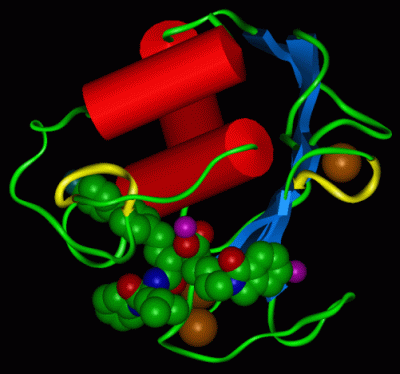
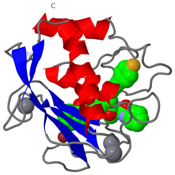
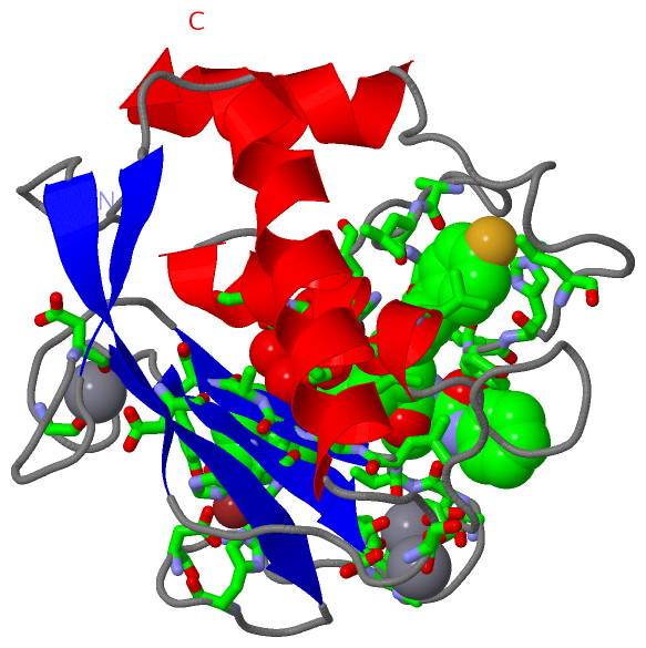
| 06 | AC6 | SOFTWARE | TYR A:155 , ASN A:162 , VAL A:163 , LEU A:164 , ALA A:165 , HIS A:166 , ALA A:167 , TYR A:168 , HIS A:201 , GLU A:202 , HIS A:205 , HIS A:211 , LEU A:218 , TYR A:220 , PRO A:221 , LEU A:222 , TYR A:223 , HIS A:224 , LEU A:226 , ZN A:257 , HOH A:301 , HOH A:307 , HOH A:323 , HOH A:329 | BINDING SITE FOR RESIDUE L04 A 256 |
| 07 | CA1 | UNKNOWN | ASP A:158 , GLY A:159 , GLY A:161 , VAL A:163 , ASP A:181 , GLU A:184 | LIGANDS OF THE CALCIUM ION CA 259. |
| 08 | CA2 | UNKNOWN | ASP A:141 , GLY A:173 , ASN A:175 , ASP A:177 , HOH A:306 , HOH A:312 | LIGANDS OF THE CALCIUM ION CA 260. |
| 09 | CA3 | UNKNOWN | ASP A:107 , ASP A:182 , GLU A:184 , HOH A:313 , HOH A:332 | LIGANDS OF THE CALCIUM ION CA 261. |
| 10 | L04 | UNKNOWN | TYR A:155 , ASN A:162 , VAL A:163 , LEU A:164 , ALA A:165 , HIS A:166 , TYR A:168 , LEU A:197 , VAL A:198 , HIS A:201 , GLU A:202 , HIS A:205 , HIS A:211 , ALA A:217 , LEU A:218 , TYR A:220 , PRO A:221 , LEU A:222 , TYR A:223 , HIS A:224 , LEU A:226 , ZN A:257 | BINDING SITE FOR THE INHIBITOR L-764,004, DENOTED L04 256. |
| 11 | ZN1 | UNKNOWN | HIS A:201 , HIS A:205 , HIS A:211 , L04 A:256 | LIGANDS OF THE CATALYTIC (ZN 257) ZINC ION. |
| 12 | ZN2 | UNKNOWN | HIS A:151 , ASP A:153 , HIS A:166 , HIS A:179 | LIGANDS OF THE STRUCTURAL (ZN 258) ZINC ION. |
|
 Description