Show PDB file:   
         Plain Text   HTML   (compressed file size)
QuickSearch:   
by PDB,NDB,UniProt,PROSITE Code or Search Term(s)  
(-)Asymmetric Unit
(-)Asym. Unit - sites
(-)Biological Unit 1
(-)Biol. Unit 1 - sites
collapse expand < >
Image Asymmetric Unit
Asymmetric Unit  (Jmol Viewer)
Image Asym. Unit - sites
Asym. Unit - sites  (Jmol Viewer)
Image Biological Unit 1
Biological Unit 1  (Jmol Viewer)
Image Biol. Unit 1 - sites
Biol. Unit 1 - sites  (Jmol Viewer)

(-) Description

Title :  CRYSTAL STRUCTURE OF THE LIGHT-ACTIVATED CONSTITUTIVELY ACTIVE N2C,M257Y,D282C RHODOPSIN MUTANT IN COMPLEX WITH A PEPTIDE RESEMBLING THE C-TERMINUS OF THE GALPHA-PROTEIN SUBUNIT (GACT)
 
Authors :  X. Deupi, P. Edwards, A. Singhal, B. Nickle, D. D. Oprian, G. F. X. Schert J. Standfuss
Date :  17 Oct 11  (Deposition) - 25 Jan 12  (Release) - 25 Jan 12  (Revision)
Method :  X-RAY DIFFRACTION
Resolution :  3.30
Chains :  Asym. Unit :  A,B
Biol. Unit 1:  A,B  (2x)
Keywords :  Signaling Protein, G-Protein, G-Protein-Coupled Receptors, Signal Tansduction, Visual System, Metarhodopsin-Ii (Keyword Search: [Gene Ontology, PubMed, Web (Google))
 
Reference :  X. Deupi, P. Edwards, A. Singhal, B. Nickle, D. Oprian, G. Schertler, J. Standfuss
Stabilized G Protein Binding Site In The Structure Of Constitutively Active Metarhodopsin-Ii.
Proc. Natl. Acad. Sci. Usa V. 109 119 2012
PubMed-ID: 22198838  |  Reference-DOI: 10.1073/PNAS.1114089108
(for further references see the PDB file header)

(-) Compounds

Molecule 1 - RHODOPSIN
    CellROD PHOTORECEPTOR
    ChainsA
    EngineeredYES
    Expression SystemHOMO SAPIENS
    Expression System Cell LineHEK293S GNTI-
    Expression System CommonHUMAN
    Expression System Taxid9606
    MutationYES
    OrganEYE
    Organism CommonBOVINE
    Organism ScientificBOS TAURUS
    Organism Taxid9913
    SynonymCONSTITUTIVELY ACTIVE RHODOPSIN MUTANT
    TissueRETINA
 
Molecule 2 - GUANINE NUCLEOTIDE-BINDING PROTEIN G(T) SUBUNIT ALPHA-3
    ChainsB
    FragmentRESIDUES 344-354
    MutationYES
    OrganEYE
    Organism CommonBOVINE
    Organism ScientificBOS TAURUS
    Organism Taxid9913
    SynonymGACT PEPTIDE, GUSTDUCIN ALPHA-3 CHAIN
    SyntheticYES
    TissueRETINA

 Structural Features

(-) Chains, Units

  12
Asymmetric Unit AB
Biological Unit 1 (2x)AB

Summary Information (see also Sequences/Alignments below)

(-) Ligands, Modified Residues, Ions  (6, 7)

Asymmetric Unit (6, 7)
No.NameCountTypeFull Name
1ACE1Mod. Amino AcidACETYL GROUP
2ACT1Ligand/IonACETATE ION
3BOG1Ligand/IonB-OCTYLGLUCOSIDE
4NAG2Ligand/IonN-ACETYL-D-GLUCOSAMINE
5PLM1Ligand/IonPALMITIC ACID
6RET1Ligand/IonRETINAL
Biological Unit 1 (6, 14)
No.NameCountTypeFull Name
1ACE2Mod. Amino AcidACETYL GROUP
2ACT2Ligand/IonACETATE ION
3BOG2Ligand/IonB-OCTYLGLUCOSIDE
4NAG4Ligand/IonN-ACETYL-D-GLUCOSAMINE
5PLM2Ligand/IonPALMITIC ACID
6RET2Ligand/IonRETINAL

(-) Sites  (5, 5)

Asymmetric Unit (5, 5)
No.NameEvidenceResiduesDescription
1AC1SOFTWAREALA A:117 , MET A:207 , HIS A:211 , TRP A:265 , TYR A:268 , ALA A:272 , LYS A:296BINDING SITE FOR RESIDUE RET A 401
2AC2SOFTWAREVAL A:204 , ILE A:205 , PHE A:208 , ALA A:272 , PHE A:273 , PHE A:276BINDING SITE FOR RESIDUE ACT A1402
3AC3SOFTWAREPHE A:220 , TYR A:223 , GLY A:224 , LYS A:231 , LYS A:248 , VAL A:254BINDING SITE FOR RESIDUE BOG A1403
4AC4SOFTWARELEU A:47 , ARG A:252 , ILE A:256 , THR A:297 , VAL A:300 , TYR A:301 , LEU A:321 , CYS A:322 , CYS A:323BINDING SITE FOR RESIDUE PLM A1404
5AC5SOFTWARETHR A:4 , ASN A:15 , GLY A:18 , VAL A:20 , ARG A:21BINDING SITE FOR CHAIN A OF SUGAR BOUND TO ASN A 15 RESIDUES 1405 TO 1406

(-) SS Bonds  (2, 2)

Asymmetric Unit
No.Residues
1A:2 -A:282
2A:110 -A:187

(-) Cis Peptide Bonds  (0, 0)

(no "Cis Peptide Bond" information available for 4A4M)

 Sequence-Structure Mapping

(-) SAPs(SNPs)/Variants  (0, 0)

(no "SAP(SNP)/Variant" information available for 4A4M)

(-) PROSITE Motifs  (2, 2)

Asymmetric Unit (2, 2)
 PROSITEUniProtKBPDB
No.IDACDescriptionIDLocationCountLocation
1G_PROTEIN_RECEP_F1_1PS00237 G-protein coupled receptors family 1 signature.OPSD_BOVIN123-139  1A:123-139
2OPSINPS00238 Visual pigments (opsins) retinal binding site.OPSD_BOVIN290-306  1A:290-306
Biological Unit 1 (2, 4)
 PROSITEUniProtKBPDB
No.IDACDescriptionIDLocationCountLocation
1G_PROTEIN_RECEP_F1_1PS00237 G-protein coupled receptors family 1 signature.OPSD_BOVIN123-139  2A:123-139
2OPSINPS00238 Visual pigments (opsins) retinal binding site.OPSD_BOVIN290-306  2A:290-306

(-) Exons   (5, 5)

Asymmetric Unit (5, 5)
 ENSEMBLUniProtKBPDB
No.Transcript IDExonExon IDGenome LocationLengthIDLocationLengthCountLocationLength
1.1ENSBTAT000000017301ENSBTAE00000013842chr22:57733705-57733336370OPSD_BOVIN1-1241241A:1-124124
1.2ENSBTAT000000017302ENSBTAE00000013843chr22:57731646-57731478169OPSD_BOVIN124-180571A:124-18057
1.3ENSBTAT000000017303ENSBTAE00000013844chr22:57730259-57730094166OPSD_BOVIN180-235561A:180-23556
1.4ENSBTAT000000017304ENSBTAE00000013845chr22:57729975-57729736240OPSD_BOVIN236-315801A:236-31580
1.5ENSBTAT000000017305ENSBTAE00000326840chr22:57728876-57728766111OPSD_BOVIN316-351361A:316-32611

(-) Sequences/Alignments

Asymmetric Unit
   Reformat: Number of residues per line =  ('0' or empty: single-line sequence representation)
  Number of residues per labelling interval =   
  UniProt sequence: complete  aligned part    
   Show mapping: SCOP domains CATH domains Pfam domains Secondary structure (by author)
SAPs(SNPs) PROSITE motifs Exons
(details for a mapped element are shown in a popup box when the mouse pointer rests over it)
Chain A from PDB  Type:PROTEIN  Length:327
 aligned with OPSD_BOVIN | P02699 from UniProtKB/Swiss-Prot  Length:348

    Alignment length:327
                             1                                                                                                                                                                                                                                                                                                                                     
                             |       9        19        29        39        49        59        69        79        89        99       109       119       129       139       149       159       169       179       189       199       209       219       229       239       249       259       269       279       289       299       309       319       
           OPSD_BOVIN     - -MNGTEGPNFYVPFSNKTGVVRSPFEAPQYYLAEPWQFSMLAAYMFLLIMLGFPINFLTLYVTVQHKKLRTPLNYILLNLAVADLFMVFGGFTTTLYTSLHGYFVFGPTGCNLEGFFATLGGEIALWSLVVLAIERYVVVCKPMSNFRFGENHAIMGVAFTWVMALACAAPPLVGWSRYIPEGMQCSCGIDYYTPHEETNNESFVIYMFVVHFIIPLIVIFFCYGQLVFTVKEAAAQQQESATTQKAEKEVTRMVIIMVIAFLICWLPYAGVAFYIFTHQGSDFGPIFMTIPAFFAKTSAVYNPVIYIMMNKQFRNCMVTTLCCGKN 326
               SCOP domains --------------------------------------------------------------------------------------------------------------------------------------------------------------------------------------------------------------------------------------------------------------------------------------------------------------------------------------- SCOP domains
               CATH domains --------------------------------------------------------------------------------------------------------------------------------------------------------------------------------------------------------------------------------------------------------------------------------------------------------------------------------------- CATH domains
               Pfam domains --------------------------------------------------------------------------------------------------------------------------------------------------------------------------------------------------------------------------------------------------------------------------------------------------------------------------------------- Pfam domains
         Sec.struct. author ....eee..eee..hhhhh..............hhhhhhhhhhhhhhhhhhhhhhhhhhhhhhhhh....hhhhhhhhhhhhhhhhhhhhhhhhhhhhhh.....hhhhhhhhhhhhhhhhhhhhhhhhhhhhhhhhhhhhh.......hhhhhhhhhhhhhhhhhhhhhhhhh....eeee....eeee.........hhhhhhhhhhhhhhhhhhhhhhhhhhhhhhhhhhhhhh....hhhhhhhhhhhhhhhhhhhhhhhhhhhhhhhhhhhhh...........hhhhhhhhhhhhhhhhhhh..hhhhhhhhhhhhh.... Sec.struct. author
                 SAPs(SNPs) --------------------------------------------------------------------------------------------------------------------------------------------------------------------------------------------------------------------------------------------------------------------------------------------------------------------------------------- SAPs(SNPs)
                    PROSITE ---------------------------------------------------------------------------------------------------------------------------G_PROTEIN_RECEP_F------------------------------------------------------------------------------------------------------------------------------------------------------OPSIN            -------------------- PROSITE
           Transcript 1 (1) -Exon 1.1  PDB: A:1-124 UniProt: 1-124                                                                                       -------------------------------------------------------Exon 1.3  PDB: A:180-235 UniProt: 180-235               Exon 1.4  PDB: A:236-315 UniProt: 236-315                                       Exon 1.5    Transcript 1 (1)
           Transcript 1 (2) ----------------------------------------------------------------------------------------------------------------------------Exon 1.2  PDB: A:124-180 UniProt: 124-180                -------------------------------------------------------------------------------------------------------------------------------------------------- Transcript 1 (2)
                 4a4m A   0 xMCGTEGPNFYVPFSNKTGVVRSPFEAPQYYLAEPWQFSMLAAYMFLLIMLGFPINFLTLYVTVQHKKLRTPLNYILLNLAVADLFMVFGGFTTTLYTSLHGYFVFGPTGCNLEGFFATLGGEIALWSLVVLAIERYVVVCKPMSNFRFGENHAIMGVAFTWVMALACAAPPLVGWSRYIPEGMQCSCGIDYYTPHEETNNESFVIYMFVVHFIIPLIVIFFCYGQLVFTVKEAAAQQQESATTQKAEKEVTRMVIIYVIAFLICWLPYAGVAFYIFTHQGSCFGPIFMTIPAFFAKTSAVYNPVIYIMMNKQFRNCMVTTLCCGKN 326
                            |        9        19        29        39        49        59        69        79        89        99       109       119       129       139       149       159       169       179       189       199       209       219       229       239       249       259       269       279       289       299       309       319       
                            0-ACE                                                                                                                                                                                                                                                                                                                                  

Chain B from PDB  Type:PROTEIN  Length:11
 aligned with GNAT3_BOVIN | P0C7Q4 from UniProtKB/Swiss-Prot  Length:354

    Alignment length:11
                                   353 
          GNAT3_BOVIN   344 IKENLKDCGLF 354
               SCOP domains ----------- SCOP domains
               CATH domains ----------- CATH domains
               Pfam domains ----------- Pfam domains
         Sec.struct. author hhhhhhhh... Sec.struct. author
                 SAPs(SNPs) ----------- SAPs(SNPs)
                    PROSITE ----------- PROSITE
                 Transcript ----------- Transcript
                 4a4m B 340 ILENLKDCGLF 350
                                   349 

   Legend:   → Mismatch (orange background)
  - → Gap (green background, '-', border residues have a numbering label)
    → Modified Residue (blue background, lower-case, 'x' indicates undefined single-letter code, labelled with number + name)
  x → Chemical Group (purple background, 'x', labelled with number + name, e.g. ACE or NH2)
  extra numbering lines below/above indicate numbering irregularities and modified residue names etc., number ends below/above '|'

 Classification and Annotation

(-) SCOP Domains  (0, 0)

(no "SCOP Domain" information available for 4A4M)

(-) CATH Domains  (0, 0)

(no "CATH Domain" information available for 4A4M)

(-) Pfam Domains  (0, 0)

(no "Pfam Domain" information available for 4A4M)

(-) Gene Ontology  (47, 51)

Asymmetric Unit(hide GO term definitions)
Chain A   (OPSD_BOVIN | P02699)
molecular function
    GO:0008020    G-protein coupled photoreceptor activity    Combining with incidental electromagnetic radiation, particularly visible light, and transmitting the signal across the membrane by activating an associated G-protein; promotes the exchange of GDP for GTP on the alpha subunit of a heterotrimeric G-protein complex.
    GO:0004930    G-protein coupled receptor activity    Combining with an extracellular signal and transmitting the signal across the membrane by activating an associated G-protein; promotes the exchange of GDP for GTP on the alpha subunit of a heterotrimeric G-protein complex.
    GO:0005085    guanyl-nucleotide exchange factor activity    Stimulates the exchange of guanyl nucleotides associated with a GTPase. Under normal cellular physiological conditions, the concentration of GTP is higher than that of GDP, favoring the replacement of GDP by GTP in association with the GTPase.
    GO:0046872    metal ion binding    Interacting selectively and non-covalently with any metal ion.
    GO:0009881    photoreceptor activity    The function of absorbing and responding to incidental electromagnetic radiation, particularly visible light. The response may involve a change in conformation.
    GO:0005515    protein binding    Interacting selectively and non-covalently with any protein or protein complex (a complex of two or more proteins that may include other nonprotein molecules).
    GO:0004871    signal transducer activity    Conveys a signal across a cell to trigger a change in cell function or state. A signal is a physical entity or change in state that is used to transfer information in order to trigger a response.
biological process
    GO:0007186    G-protein coupled receptor signaling pathway    A series of molecular signals that proceeds with an activated receptor promoting the exchange of GDP for GTP on the alpha-subunit of an associated heterotrimeric G-protein complex. The GTP-bound activated alpha-G-protein then dissociates from the beta- and gamma-subunits to further transmit the signal within the cell. The pathway begins with receptor-ligand interaction, or for basal GPCR signaling the pathway begins with the receptor activating its G protein in the absence of an agonist, and ends with regulation of a downstream cellular process, e.g. transcription. The pathway can start from the plasma membrane, Golgi or nuclear membrane (PMID:24568158 and PMID:16902576).
    GO:0016038    absorption of visible light    The reception of a (visible light) photon by a cell, visible light being defined as having a wavelength within the range 380-780 nm.
    GO:0071482    cellular response to light stimulus    Any process that results in a change in state or activity of a cell (in terms of movement, secretion, enzyme production, gene expression, etc.) as a result of a light stimulus, electromagnetic radiation of wavelengths classified as infrared, visible or ultraviolet light.
    GO:0009583    detection of light stimulus    The series of events in which a light stimulus (in the form of photons) is received and converted into a molecular signal.
    GO:0045494    photoreceptor cell maintenance    Any process preventing the degeneration of the photoreceptor, a specialized cell type that is sensitive to light.
    GO:0007602    phototransduction    The sequence of reactions within a cell required to convert absorbed photons into a molecular signal.
    GO:0007603    phototransduction, visible light    The sequence of reactions within a cell required to convert absorbed photons from visible light into a molecular signal. A visible light stimulus is electromagnetic radiation that can be perceived visually by an organism; for organisms lacking a visual system, this can be defined as light with a wavelength within the range 380 to 780 nm.
    GO:0043547    positive regulation of GTPase activity    Any process that activates or increases the activity of a GTPase.
    GO:0006468    protein phosphorylation    The process of introducing a phosphate group on to a protein.
    GO:0018298    protein-chromophore linkage    The covalent or noncovalent attachment of a chromophore to a protein.
    GO:0022400    regulation of rhodopsin mediated signaling pathway    Any process that modulates the frequency, rate or extent of rhodopsin-mediated signaling.
    GO:0009416    response to light stimulus    Any process that results in a change in state or activity of a cell or an organism (in terms of movement, secretion, enzyme production, gene expression, etc.) as a result of a light stimulus, electromagnetic radiation of wavelengths classified as infrared, visible or ultraviolet light.
    GO:0050896    response to stimulus    Any process that results in a change in state or activity of a cell or an organism (in terms of movement, secretion, enzyme production, gene expression, etc.) as a result of a stimulus. The process begins with detection of the stimulus and ends with a change in state or activity or the cell or organism.
    GO:0060041    retina development in camera-type eye    The process whose specific outcome is the progression of the retina over time, from its formation to the mature structure. The retina is the innermost layer or coating at the back of the eyeball, which is sensitive to light and in which the optic nerve terminates.
    GO:0016056    rhodopsin mediated signaling pathway    The series of molecular signals generated as a consequence of excitation of rhodopsin by a photon and the events that convert the absorbed photons into a cellular response.
    GO:0050953    sensory perception of light stimulus    The series of events required for an organism to receive a sensory light stimulus, convert it to a molecular signal, and recognize and characterize the signal. This is a neurological process.
    GO:0007165    signal transduction    The cellular process in which a signal is conveyed to trigger a change in the activity or state of a cell. Signal transduction begins with reception of a signal (e.g. a ligand binding to a receptor or receptor activation by a stimulus such as light), or for signal transduction in the absence of ligand, signal-withdrawal or the activity of a constitutively active receptor. Signal transduction ends with regulation of a downstream cellular process, e.g. regulation of transcription or regulation of a metabolic process. Signal transduction covers signaling from receptors located on the surface of the cell and signaling via molecules located within the cell. For signaling between cells, signal transduction is restricted to events at and within the receiving cell.
    GO:0007601    visual perception    The series of events required for an organism to receive a visual stimulus, convert it to a molecular signal, and recognize and characterize the signal. Visual stimuli are detected in the form of photons and are processed to form an image.
cellular component
    GO:0005794    Golgi apparatus    A compound membranous cytoplasmic organelle of eukaryotic cells, consisting of flattened, ribosome-free vesicles arranged in a more or less regular stack. The Golgi apparatus differs from the endoplasmic reticulum in often having slightly thicker membranes, appearing in sections as a characteristic shallow semicircle so that the convex side (cis or entry face) abuts the endoplasmic reticulum, secretory vesicles emerging from the concave side (trans or exit face). In vertebrate cells there is usually one such organelle, while in invertebrates and plants, where they are known usually as dictyosomes, there may be several scattered in the cytoplasm. The Golgi apparatus processes proteins produced on the ribosomes of the rough endoplasmic reticulum; such processing includes modification of the core oligosaccharides of glycoproteins, and the sorting and packaging of proteins for transport to a variety of cellular locations. Three different regions of the Golgi are now recognized both in terms of structure and function: cis, in the vicinity of the cis face, trans, in the vicinity of the trans face, and medial, lying between the cis and trans regions.
    GO:0000139    Golgi membrane    The lipid bilayer surrounding any of the compartments of the Golgi apparatus.
    GO:0005911    cell-cell junction    A cell junction that forms a connection between two or more cells in a multicellular organism; excludes direct cytoplasmic junctions such as ring canals.
    GO:0016021    integral component of membrane    The component of a membrane consisting of the gene products and protein complexes having at least some part of their peptide sequence embedded in the hydrophobic region of the membrane.
    GO:0005887    integral component of plasma membrane    The component of the plasma membrane consisting of the gene products and protein complexes having at least some part of their peptide sequence embedded in the hydrophobic region of the membrane.
    GO:0016020    membrane    A lipid bilayer along with all the proteins and protein complexes embedded in it an attached to it.
    GO:0097381    photoreceptor disc membrane    Ovally-shaped membranous stack located inside the photoreceptor outer segment, and containing densely packed molecules of the photoreceptor protein rhodopsin that traverse the lipid bilayer. Disc membranes are apparently derived from the plasma membrane in the region of the cilium that connects the photoreceptor outer segment to the inner segment.
    GO:0001917    photoreceptor inner segment    The inner segment of a vertebrate photoreceptor containing mitochondria, ribosomes and membranes where opsin molecules are assembled and passed to be part of the outer segment discs.
    GO:0060342    photoreceptor inner segment membrane    The membrane surrounding the inner segment of a vertebrate photoreceptor. The photoreceptor inner segment contains mitochondria, ribosomes and membranes where opsin molecules are assembled and passed to be part of the outer segment discs.
    GO:0001750    photoreceptor outer segment    The outer segment of a vertebrate photoreceptor that contains discs of photoreceptive membranes.
    GO:0042622    photoreceptor outer segment membrane    The membrane surrounding the outer segment of a vertebrate photoreceptor.
    GO:0005886    plasma membrane    The membrane surrounding a cell that separates the cell from its external environment. It consists of a phospholipid bilayer and associated proteins.
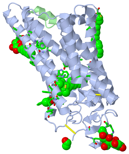
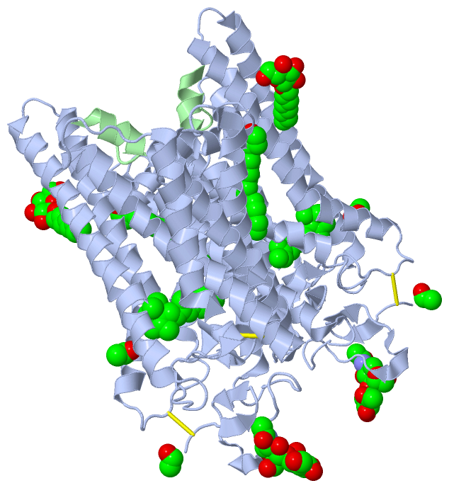
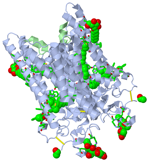
Chain B   (GNAT3_BOVIN | P0C7Q4)
molecular function
    GO:0031683    G-protein beta/gamma-subunit complex binding    Interacting selectively and non-covalently with a complex of G-protein beta/gamma subunits.
    GO:0001664    G-protein coupled receptor binding    Interacting selectively and non-covalently with a G-protein coupled receptor.
    GO:0005525    GTP binding    Interacting selectively and non-covalently with GTP, guanosine triphosphate.
    GO:0003924    GTPase activity    Catalysis of the reaction: GTP + H2O = GDP + phosphate.
    GO:0019001    guanyl nucleotide binding    Interacting selectively and non-covalently with guanyl nucleotides, any compound consisting of guanosine esterified with (ortho)phosphate.
    GO:0046872    metal ion binding    Interacting selectively and non-covalently with any metal ion.
    GO:0000166    nucleotide binding    Interacting selectively and non-covalently with a nucleotide, any compound consisting of a nucleoside that is esterified with (ortho)phosphate or an oligophosphate at any hydroxyl group on the ribose or deoxyribose.
    GO:0004871    signal transducer activity    Conveys a signal across a cell to trigger a change in cell function or state. A signal is a physical entity or change in state that is used to transfer information in order to trigger a response.
biological process
    GO:0007186    G-protein coupled receptor signaling pathway    A series of molecular signals that proceeds with an activated receptor promoting the exchange of GDP for GTP on the alpha-subunit of an associated heterotrimeric G-protein complex. The GTP-bound activated alpha-G-protein then dissociates from the beta- and gamma-subunits to further transmit the signal within the cell. The pathway begins with receptor-ligand interaction, or for basal GPCR signaling the pathway begins with the receptor activating its G protein in the absence of an agonist, and ends with regulation of a downstream cellular process, e.g. transcription. The pathway can start from the plasma membrane, Golgi or nuclear membrane (PMID:24568158 and PMID:16902576).
    GO:0007188    adenylate cyclase-modulating G-protein coupled receptor signaling pathway    The series of molecular signals generated as a consequence of a G-protein coupled receptor binding to its physiological ligand, where the pathway proceeds through activation or inhibition of adenylyl cyclase activity and a subsequent change in the concentration of cyclic AMP (cAMP).
    GO:0050916    sensory perception of sweet taste    The series of events required to receive a sweet taste stimulus, convert it to a molecular signal, and recognize and characterize the signal. This is a neurological process.
    GO:0007165    signal transduction    The cellular process in which a signal is conveyed to trigger a change in the activity or state of a cell. Signal transduction begins with reception of a signal (e.g. a ligand binding to a receptor or receptor activation by a stimulus such as light), or for signal transduction in the absence of ligand, signal-withdrawal or the activity of a constitutively active receptor. Signal transduction ends with regulation of a downstream cellular process, e.g. regulation of transcription or regulation of a metabolic process. Signal transduction covers signaling from receptors located on the surface of the cell and signaling via molecules located within the cell. For signaling between cells, signal transduction is restricted to events at and within the receiving cell.
cellular component
    GO:0005737    cytoplasm    All of the contents of a cell excluding the plasma membrane and nucleus, but including other subcellular structures.
    GO:0005834    heterotrimeric G-protein complex    Any of a family of heterotrimeric GTP-binding and hydrolyzing proteins; they belong to a superfamily of GTPases that includes monomeric proteins such as EF-Tu and RAS. Heterotrimeric G-proteins consist of three subunits; the alpha subunit contains the guanine nucleotide binding site and possesses GTPase activity; the beta and gamma subunits are tightly associated and function as a beta-gamma heterodimer; extrinsic plasma membrane proteins (cytoplasmic face) that function as a complex to transduce signals from G-protein coupled receptors to an effector protein.

 Visualization

(-) Interactive Views

Asymmetric Unit
  Complete Structure
    Jena3D(integrated viewing of ligand, site, SAP, PROSITE, SCOP information)
    WebMol | AstexViewer[tm]@PDBe
(Java Applets, require no local installation except for Java; loading may be slow)
    STRAP
(Java WebStart application, automatic local installation, requires Java; full application with system access!)
    RasMol
(require local installation)
    Molscript (VRML)
(requires installation of a VRML viewer; select preferred view via VRML and generate a mono or stereo PDF format file)
 
  Ligands, Modified Residues, Ions
    ACE  [ RasMol | Jena3D ]  +environment [ RasMol | Jena3D ]
    ACT  [ RasMol | Jena3D ]  +environment [ RasMol | Jena3D ]
    BOG  [ RasMol | Jena3D ]  +environment [ RasMol | Jena3D ]
    NAG  [ RasMol | Jena3D ]  +environment [ RasMol | Jena3D ]
    PLM  [ RasMol | Jena3D ]  +environment [ RasMol | Jena3D ]
    RET  [ RasMol | Jena3D ]  +environment [ RasMol | Jena3D ]
 
  Sites
    AC1  [ RasMol ]  +environment [ RasMol ]
    AC2  [ RasMol ]  +environment [ RasMol ]
    AC3  [ RasMol ]  +environment [ RasMol ]
    AC4  [ RasMol ]  +environment [ RasMol ]
    AC5  [ RasMol ]  +environment [ RasMol ]
 
  Cis Peptide Bonds
(no "Cis Peptide Bonds" information available for 4a4m)
 
Biological Unit
  Complete Structure
    Biological Unit 1  [ Jena3D ]

(-) Still Images

Jmol
  protein: cartoon or spacefill or dots and stick; nucleic acid: cartoon and stick; ligands: spacefill; active site: stick
Molscript
  protein, nucleic acid: cartoon; ligands: spacefill; active site: ball and stick

 Databases and Analysis Tools

(-) Databases

Access by PDB/NDB ID
  4a4m
    Family and Domain InformationProDom | SYSTERS
    General Structural InformationGlycoscienceDB | MMDB | NDB | OCA | PDB | PDBe | PDBj | PDBsum | PDBWiki | PQS | PROTEOPEDIA
    Orientation in MembranesOPM
    Protein SurfaceSURFACE
    Secondary StructureDSSP (structure derived) | HSSP (homology derived)
    Structural GenomicsGeneCensus
    Structural NeighboursCE | VAST
    Structure ClassificationCATH | Dali | SCOP
    Validation and Original DataBMRB Data View | BMRB Restraints Grid | EDS | PROCHECK | RECOORD | WHAT_CHECK
 
Access by UniProt ID/Accession number
  GNAT3_BOVIN | P0C7Q4
    Comparative Protein Structure ModelsModBase
    Genomic InformationEnsembl
    Protein-protein InteractionDIP
    Sequence, Family and Domain InformationInterPro | Pfam | SMART | UniProtKB/SwissProt
  OPSD_BOVIN | P02699
    Comparative Protein Structure ModelsModBase
    Genomic InformationEnsembl
    Protein-protein InteractionDIP
    Sequence, Family and Domain InformationInterPro | Pfam | SMART | UniProtKB/SwissProt
 
Access by Enzyme Classificator   (EC Number)
  (no 'Enzyme Classificator' available)
    General Enzyme InformationBRENDA | EC-PDB | Enzyme | IntEnz
    PathwayKEGG | MetaCyc
 
Access by Disease Identifier   (MIM ID)
  (no 'MIM ID' available)
    Disease InformationOMIM
 
Access by GenAge ID
  (no 'GenAge ID' available)
    Age Related InformationGenAge

(-) Analysis Tools

Access by PDB/NDB ID
    Domain InformationXDom
    Interatomic Contacts of Structural UnitsCSU
    Ligand-protein ContactsLPC
    Protein CavitiescastP
    Sequence and Secondary StructurePDBCartoon
    Structure AlignmentSTRAP(Java WebStart application, automatic local installation, requires Java; full application with system access!)
    Structure and Sequence BrowserSTING
 
Access by UniProt ID/Accession number
  GNAT3_BOVIN | P0C7Q4
    Protein Disorder PredictionDisEMBL | FoldIndex | GLOBPLOT (for more information see DisProt)
  OPSD_BOVIN | P02699
    Protein Disorder PredictionDisEMBL | FoldIndex | GLOBPLOT (for more information see DisProt)

 Related Entries

(-) Entries Sharing at Least One Protein Chain (UniProt ID)

UniProtKB/Swiss-Prot
        GNAT3_BOVIN | P0C7Q45en0
        OPSD_BOVIN | P026991boj 1bok 1eds 1edv 1edw 1edx 1f88 1fdf 1gzm 1hzx 1jfp 1l9h 1ln6 1n3m 1nzs 1ov0 1ov1 1u19 1vqx 1y6y 2g87 2hpy 2i35 2i36 2i37 2iqk 2j4y 2ped 2x72 3c9l 3c9m 3cap 3dqb 3oax 3pqr 3pxo 4bey 4bez 4j4q 4pxf 4x1h 5dys 5en0 5te3 5te5

(-) Related Entries Specified in the PDB File

1boj BOVINE RHODOPSIN (7-HELIX BUNDLE) WITH ALL-TRANS RETINAL, METARHODOPSIN II MODEL, THEORETICAL MODEL
1bok BOVINE RHODOPSIN (7-HELIX BUNDLE) WITH 11-CIS RETINAL , THEORETICAL MODEL
1eds SOLUTION STRUCTURE OF INTRADISKAL LOOP 1 OF BOVINE RHODOPSIN (RHODOPSIN RESIDUES 92-123)
1edv SOLUTION STRUCTURE OF 2ND INTRADISKAL LOOP OF BOVINE RHODOPSIN (RESIDUES 172-205)
1edw SOLUTION STRUCTURE OF THIRD INTRADISKAL LOOP OF BOVINE RHODOPSIN (RESIDUES 268-293)
1edx SOLUTION STRUCTURE OF AMINO TERMINUS OF BOVINE RHODOPSIN (RESIDUES 1- 40)
1f88 CRYSTAL STRUCTURE OF BOVINE RHODOPSIN
1fdf HELIX 7 BOVINE RHODOPSIN
1gzm STRUCTURE OF BOVINE RHODOPSIN IN A TRIGONAL CRYSTAL FORM
1hzx CRYSTAL STRUCTURE OF BOVINE RHODOPSIN
1jfp STRUCTURE OF BOVINE RHODOPSIN (DARK ADAPTED)
1l9h CRYSTAL STRUCTURE OF BOVINE RHODOPSIN AT 2.6 ANGSTROMSRESOLUTION
1ln6 STRUCTURE OF BOVINE RHODOPSIN (METARHODOPSIN II)
1n3m THEORETICAL MODEL OF RHODOPSIN OLIGOMER
1nzs NMR STRUCTURES OF PHOSPHORYLATED CARBOXY TERMINUS OF BOVINERHODOPSIN IN ARRESTIN-BOUND STATE
1ov0 CALCULATED 3D MODEL OF META-II RHODOPSIN BASED ON LIMITEDDATA OF SITE-DIRECTED SPIN-LABELING
1ov1 CALCULATED 3D MODEL OF AN ACTIVATED "STRAITJACKED" RHODOPSIN
1u19 CRYSTAL STRUCTURE OF BOVINE RHODOPSIN AT 2.2 ANGSTROMSRESOLUTION
1vqx ARRESTIN-BOUND NMR STRUCTURES OF THE PHOSPHORYLATED CARBOXY-TERMINAL DOMAIN OF RHODOPSIN, REFINED
2i35 CRYSTAL STRUCTURE OF RHOMBOHEDRAL CRYSTAL FORM OF GROUND -STATE RHODOPSIN
2i36 CRYSTAL STRUCTURE OF TRIGONAL CRYSTAL FORM OF GROUND- STATERHODOPSIN
2i37 CRYSTAL STRUCTURE OF A PHOTOACTIVATED RHODOPSIN
2j4y CRYSTAL STRUCTURE OF A RHODOPSIN STABILIZING MUTANT EXPRESSED IN MAMMALIAN CELLS
2x72 CRYSTAL STRUCTURE OF THE CONSTITUTIVELY ACTIVE E113Q,N2C ,D282C RHODOPSIN MUTANT WITH BOUND GALPHACT PEPTIDE.