Show PDB file:   
         Plain Text   HTML   (compressed file size)
QuickSearch:   
by PDB,NDB,UniProt,PROSITE Code or Search Term(s)  
(-)Asym./Biol. Unit
(-)Asym./Biol. Unit - sites
collapse expand < >
Image Asym./Biol. Unit
Asym./Biol. Unit  (Jmol Viewer)
Image Asym./Biol. Unit - sites
Asym./Biol. Unit - sites  (Jmol Viewer)

(-) Description

Title :  X-RAY STRUCTURE OF PCCEL45A N92D WITH CELLOPENTAOSE AT 95K.
 
Authors :  A. Nakamura, T. Ishida, M. Samejima, K. Igarashi
Date :  22 Dec 14  (Deposition) - 07 Oct 15  (Release) - 07 Oct 15  (Revision)
Method :  X-RAY DIFFRACTION
Resolution :  0.99
Chains :  Asym./Biol. Unit :  A
Keywords :  Hydrolase (Keyword Search: [Gene Ontology, PubMed, Web (Google))
 
Reference :  A. Nakamura, T. Ishida, K. Kusaka, T. Yamada, S. Fushinobu, I. Tanaka, S. Kaneko, K. Ohta, H. Tanaka, K. Inaka, Y. Higuchi, N. Niimura, M. Samejima, K. Igarashi
Newton S Cradle Proton Relay With Amide Imidic Acid Tautomerization In Inverting Cellulase Visualized By Neutro Crystallography
Sci Adv V. 1 00263 2015
PubMed: search  |  Reference-DOI: 10.1126/SCIADV.1500263

(-) Compounds

Molecule 1 - ENDOGLUCANASE V-LIKE PROTEIN
    ChainsA
    EngineeredYES
    Expression SystemPICHIA PASTORIS
    Expression System PlasmidPPICZA
    Expression System StrainKM71H
    Expression System Taxid4922
    Expression System Vector TypePLASMID
    FragmentUNP RESIDUES 27-206
    GeneEGV, PCCEL45A
    MutationYES
    Organism CommonWHITE-ROT FUNGUS
    Organism ScientificPHANEROCHAETE CHRYSOSPORIUM
    Organism Taxid5306
    StrainK-3

 Structural Features

(-) Chains, Units

  1
Asymmetric/Biological Unit A

Summary Information (see also Sequences/Alignments below)

(-) Ligands, Modified Residues, Ions  (4, 10)

Asymmetric/Biological Unit (4, 10)
No.NameCountTypeFull Name
140S3Ligand/Ion3-METHYLPENTANE-1,5-DIOL
2BGC4Ligand/IonBETA-D-GLUCOSE
3CE52Ligand/IonCELLOPENTAOSE
4GLC1Ligand/IonALPHA-D-GLUCOSE

(-) Sites  (10, 10)

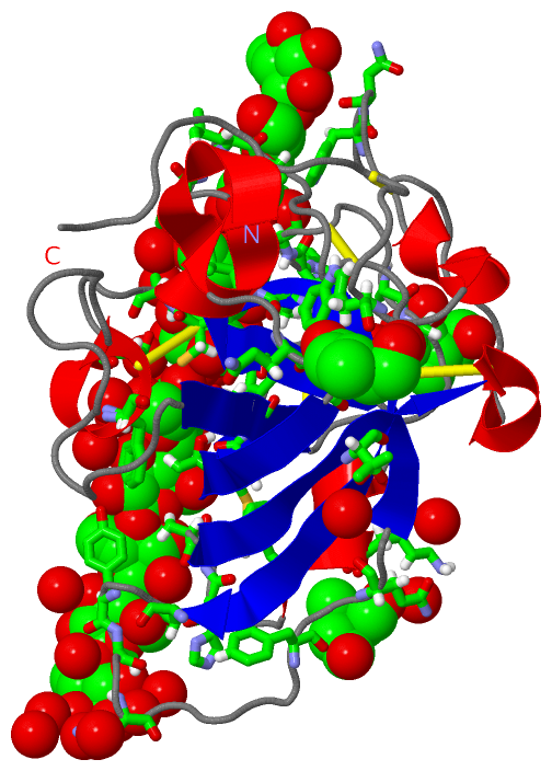
Asymmetric Unit (10, 10)
No.NameEvidenceResiduesDescription
01AC1SOFTWAREGLY A:22 , SER A:23 , SER A:68 , BGC A:202 , HOH A:373 , HOH A:422 , HOH A:427 , HOH A:440 , HOH A:451BINDING SITE FOR RESIDUE GLC A 201
02AC2SOFTWARESER A:23 , ASP A:65 , TYR A:67 , SER A:68 , GLY A:129 , GLC A:201 , BGC A:203 , HOH A:374 , HOH A:504BINDING SITE FOR RESIDUE BGC A 202
03AC3SOFTWARETYR A:67 , GLY A:129 , HIS A:130 , GLY A:131 , ALA A:132 , BGC A:202 , BGC A:204 , HOH A:358BINDING SITE FOR RESIDUE BGC A 203
04AC4SOFTWARETHR A:16 , MET A:17 , TYR A:67 , PHE A:95 , GLY A:131 , BGC A:203 , BGC A:205 , HOH A:453BINDING SITE FOR RESIDUE BGC A 204
05AC5SOFTWARETHR A:16 , MET A:17 , TYR A:18 , ASP A:92 , ASP A:114 , BGC A:204 , CE5 A:207 , HOH A:338 , HOH A:426 , HOH A:436 , HOH A:458 , HOH A:475BINDING SITE FOR RESIDUE BGC A 205
06AC6SOFTWARETHR A:16 , MET A:17 , TYR A:18 , GLY A:22 , SER A:23 , ASP A:65 , TYR A:67 , SER A:68 , ASP A:92 , PHE A:95 , ASP A:114 , GLY A:129 , HIS A:130 , GLY A:131 , ALA A:132 , CE5 A:207 , HOH A:338 , HOH A:373 , HOH A:374 , HOH A:422 , HOH A:426 , HOH A:436 , HOH A:440 , HOH A:453 , HOH A:458 , HOH A:475BINDING SITE FOR RESIDUE CE5 A 206
07AC7SOFTWARETYR A:18 , SER A:23 , PRO A:24 , ALA A:25 , ALA A:36 , PRO A:46 , GLY A:47 , LEU A:48 , GLY A:49 , ASP A:85 , LEU A:86 , CYS A:87 , PRO A:88 , ASP A:92 , ASP A:114 , TRP A:154 , ASN A:155 , BGC A:205 , CE5 A:206 , HOH A:350 , HOH A:370 , HOH A:423 , HOH A:442 , HOH A:462 , HOH A:482 , HOH A:497 , HOH A:502BINDING SITE FOR RESIDUE CE5 A 207
08AC8SOFTWAREGLN A:8 , ALA A:9 , ASN A:70 , TRP A:167 , HOH A:307 , HOH A:331 , HOH A:367 , HOH A:403BINDING SITE FOR RESIDUE 40S A 208
09AC9SOFTWAREPHE A:75 , GLN A:77 , ALA A:123 , LYS A:124 , PHE A:126 , HOH A:344BINDING SITE FOR RESIDUE 40S A 209
10BC1SOFTWAREALA A:29 , ALA A:30 , THR A:138 , HOH A:360 , HOH A:388 , HOH A:431BINDING SITE FOR RESIDUE 40S A 210

(-) SS Bonds  (5, 5)

Asymmetric/Biological Unit
No.Residues
1A:21 -A:116
2A:26 -A:54
3A:57 -A:142
4A:87 -A:96
5A:158 -A:172

(-) Cis Peptide Bonds  (2, 2)

Asymmetric/Biological Unit
No.Residues
1Gly A:73 -Pro A:74
2Trp A:167 -Pro A:168

 Sequence-Structure Mapping

(-) SAPs(SNPs)/Variants  (0, 0)

(no "SAP(SNP)/Variant" information available for 3X2H)

(-) PROSITE Motifs  (0, 0)

(no "PROSITE Motif" information available for 3X2H)

(-) Exons   (0, 0)

(no "Exon" information available for 3X2H)

(-) Sequences/Alignments

Asymmetric/Biological Unit
   Reformat: Number of residues per line =  ('0' or empty: single-line sequence representation)
  Number of residues per labelling interval =   
  UniProt sequence: complete  aligned part    
   Show mapping: SCOP domains CATH domains Pfam domains Secondary structure (by author)
SAPs(SNPs) PROSITE motifs Exons
(details for a mapped element are shown in a popup box when the mouse pointer rests over it)
Chain A from PDB  Type:PROTEIN  Length:180
                                                                                                                                                                                                                    
               SCOP domains ------------------------------------------------------------------------------------------------------------------------------------------------------------------------------------ SCOP domains
               CATH domains ------------------------------------------------------------------------------------------------------------------------------------------------------------------------------------ CATH domains
               Pfam domains ------------------------------------------------------------------------------------------------------------------------------------------------------------------------------------ Pfam domains
         Sec.struct. author hhhhh....eeeeeeeee...............eeeeehhhhh.............eeeeeeee..............eeeeeeeee....hhhhhh.............eeeeee...hhhhhhh......eeeeeeeehhhhh.............hhhhhh................ Sec.struct. author
                 SAPs(SNPs) ------------------------------------------------------------------------------------------------------------------------------------------------------------------------------------ SAPs(SNPs)
                    PROSITE ------------------------------------------------------------------------------------------------------------------------------------------------------------------------------------ PROSITE
                 Transcript ------------------------------------------------------------------------------------------------------------------------------------------------------------------------------------ Transcript
                 3x2h A   1 ATGGYVQQATGQASFTMYSGCGSPACGKAASGFTAAINQLAFGSAPGLGAGDACGRCFALTGNHDPYSPNYTGPFGQTIVVKVTDLCPVQGDQEFCGQTTSNPTNQHGMPFHFDICEDTGGSAKFFPSGHGALTGTFTEVSCSQWSGSDGGQLWNGACLSGETAPNWPSTACGNKGTAPS 180
                                    10        20        30        40        50        60        70        80        90       100       110       120       130       140       150       160       170       180

   Legend:   → Mismatch (orange background)
  - → Gap (green background, '-', border residues have a numbering label)
    → Modified Residue (blue background, lower-case, 'x' indicates undefined single-letter code, labelled with number + name)
  x → Chemical Group (purple background, 'x', labelled with number + name, e.g. ACE or NH2)
  extra numbering lines below/above indicate numbering irregularities and modified residue names etc., number ends below/above '|'

 Classification and Annotation

(-) SCOP Domains  (0, 0)

(no "SCOP Domain" information available for 3X2H)

(-) CATH Domains  (0, 0)

(no "CATH Domain" information available for 3X2H)

(-) Pfam Domains  (0, 0)

(no "Pfam Domain" information available for 3X2H)

(-) Gene Ontology  (0, 0)

Asymmetric/Biological Unit(hide GO term definitions)
    (no "Gene Ontology" information available for 3X2H)

 Visualization

(-) Interactive Views

Asymmetric/Biological Unit
  Complete Structure
    Jena3D(integrated viewing of ligand, site, SAP, PROSITE, SCOP information)
    WebMol | AstexViewer[tm]@PDBe
(Java Applets, require no local installation except for Java; loading may be slow)
    STRAP
(Java WebStart application, automatic local installation, requires Java; full application with system access!)
    RasMol
(require local installation)
    Molscript (VRML)
(requires installation of a VRML viewer; select preferred view via VRML and generate a mono or stereo PDF format file)
 
  Ligands, Modified Residues, Ions
    40S  [ RasMol | Jena3D ]  +environment [ RasMol | Jena3D ]
    BGC  [ RasMol | Jena3D ]  +environment [ RasMol | Jena3D ]
    CE5  [ RasMol | Jena3D ]  +environment [ RasMol | Jena3D ]
    GLC  [ RasMol | Jena3D ]  +environment [ RasMol | Jena3D ]
 
  Sites
    AC1  [ RasMol ]  +environment [ RasMol ]
    AC2  [ RasMol ]  +environment [ RasMol ]
    AC3  [ RasMol ]  +environment [ RasMol ]
    AC4  [ RasMol ]  +environment [ RasMol ]
    AC5  [ RasMol ]  +environment [ RasMol ]
    AC6  [ RasMol ]  +environment [ RasMol ]
    AC7  [ RasMol ]  +environment [ RasMol ]
    AC8  [ RasMol ]  +environment [ RasMol ]
    AC9  [ RasMol ]  +environment [ RasMol ]
    BC1  [ RasMol ]  +environment [ RasMol ]
 
  Cis Peptide Bonds
    Gly A:73 - Pro A:74   [ RasMol ]  
    Trp A:167 - Pro A:168   [ RasMol ]  
 

(-) Still Images

Jmol
  protein: cartoon or spacefill or dots and stick; nucleic acid: cartoon and stick; ligands: spacefill; active site: stick
Molscript
  protein, nucleic acid: cartoon; ligands: spacefill; active site: ball and stick

 Databases and Analysis Tools

(-) Databases

Access by PDB/NDB ID
  3x2h
    Family and Domain InformationProDom | SYSTERS
    General Structural InformationGlycoscienceDB | MMDB | NDB | OCA | PDB | PDBe | PDBj | PDBsum | PDBWiki | PQS | PROTEOPEDIA
    Orientation in MembranesOPM
    Protein SurfaceSURFACE
    Secondary StructureDSSP (structure derived) | HSSP (homology derived)
    Structural GenomicsGeneCensus
    Structural NeighboursCE | VAST
    Structure ClassificationCATH | Dali | SCOP
    Validation and Original DataBMRB Data View | BMRB Restraints Grid | EDS | PROCHECK | RECOORD | WHAT_CHECK
 
Access by UniProt ID/Accession number
  B3Y002_PHACH | B3Y002
    Comparative Protein Structure ModelsModBase
    Genomic InformationEnsembl
    Protein-protein InteractionDIP
    Sequence, Family and Domain InformationInterPro | Pfam | SMART | UniProtKB/TrEMBL
 
Access by Enzyme Classificator   (EC Number)
  (no 'Enzyme Classificator' available)
    General Enzyme InformationBRENDA | EC-PDB | Enzyme | IntEnz
    PathwayKEGG | MetaCyc
 
Access by Disease Identifier   (MIM ID)
  (no 'MIM ID' available)
    Disease InformationOMIM
 
Access by GenAge ID
  (no 'GenAge ID' available)
    Age Related InformationGenAge

(-) Analysis Tools

Access by PDB/NDB ID
    Domain InformationXDom
    Interatomic Contacts of Structural UnitsCSU
    Ligand-protein ContactsLPC
    Protein CavitiescastP
    Sequence and Secondary StructurePDBCartoon
    Structure AlignmentSTRAP(Java WebStart application, automatic local installation, requires Java; full application with system access!)
    Structure and Sequence BrowserSTING
 
Access by UniProt ID/Accession number
  B3Y002_PHACH | B3Y002
    Protein Disorder PredictionDisEMBL | FoldIndex | GLOBPLOT (for more information see DisProt)

 Related Entries

(-) Entries Sharing at Least One Protein Chain (UniProt ID)

UniProtKB/Swiss-Prot
        B3Y002_PHACH | B3Y0025kjo 5kjq
UniProtKB/TrEMBL
        B3Y002_PHACH | B3Y0023x2g 3x2i 3x2j 3x2k 3x2l 3x2m 3x2n 3x2o 3x2p 4zm7

(-) Related Entries Specified in the PDB File

3x2g PCCEL45A N92D APO FORM