|
|
|
|
 Description
|
|
Compounds
|
||||||||||||||||||||||||||||||||||||||||||||||||||||||||||||||||
Chains, Units
Summary Information (see also Sequences/Alignments below) |
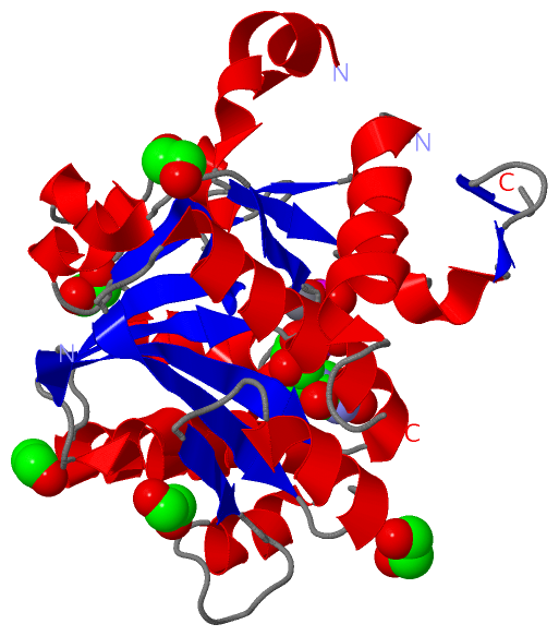
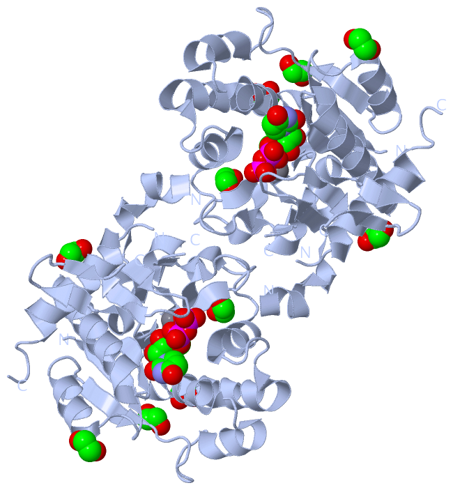
Ligands, Modified Residues, Ions (3, 7)| Asymmetric Unit (3, 7) Biological Unit 1 (2, 12) |
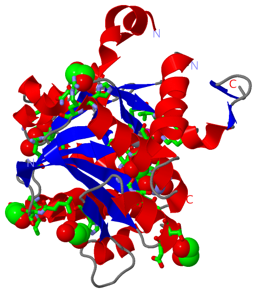
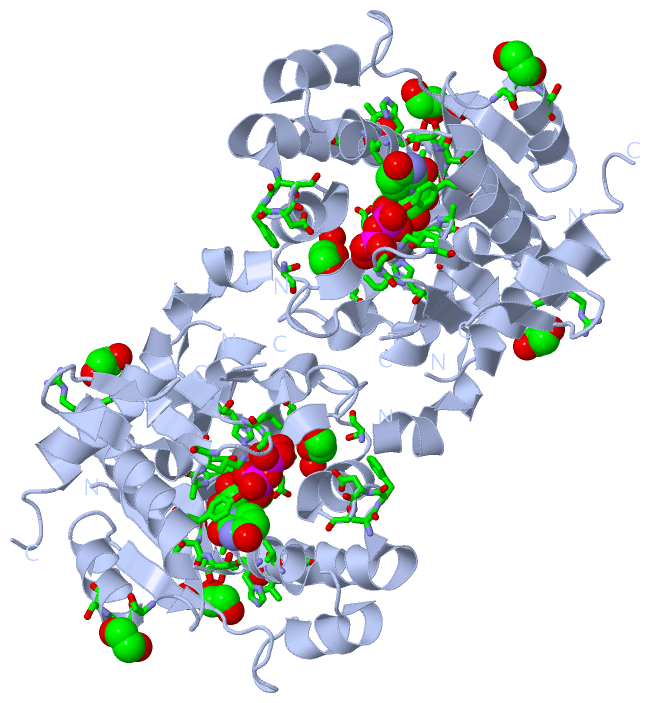
Sites (7, 7)
Asymmetric Unit (7, 7)
|
SS Bonds (0, 0)| (no "SS Bond" information available for 3QVB) |
Cis Peptide Bonds (1, 1)
Asymmetric Unit
|
||||||||
SAPs(SNPs)/Variants (3, 3)
Asymmetric Unit (3, 3)
|
||||||||||||||||||||||||||||||||||||||||||||||||||||||||||||||||||||||||||||||||||||||||||||||||||||||||||||||||||||||||||||||||||||||||||||||||||||||||||||||
PROSITE Motifs (0, 0)| (no "PROSITE Motif" information available for 3QVB) |
Exons (0, 0)| (no "Exon" information available for 3QVB) |
Sequences/Alignments
Asymmetric UnitChain A from PDB Type:PROTEIN Length:251 aligned with GLYG_HUMAN | P46976 from UniProtKB/Swiss-Prot Length:350 Alignment length:263 1 | 9 19 29 39 49 59 69 79 89 99 109 119 129 139 149 159 169 179 189 199 209 219 229 239 249 259 GLYG_HUMAN - -MTDQAFVTLTTNDAYAKGALVLGSSLKQHRTTRRLVVLATPQVSDSMRKVLETVFDEVIMVDVLDSGDSAHLTLMKRPELGVTLTKLHCWSLTQYSKCVFMDADTLVLANIDDLFDREELSAAPDPGWPDCFNSGVFVYQPSVETYNQLLHLASEQGSFDGGDQGILNTFFSSWATTDIRKHLPFIYNLSSISIYSYLPAFKVFGASAKVVHFLGRVKPWNYTYDPKTKSVKSEAHDPNMTHPEFLILWWNIFTTNVLPLLQ 262 SCOP domains d3qvba_ A: Glycogenin SCOP domains CATH domains ----------------------------------------------------------------------------------------------------------------------------------------------------------------------------------------------------------------------------------------------------------------------- CATH domains Pfam domains ------Glyco_transf_8-3qvbA01 A:6-224 --------- ---------------------- Pfam domains SAPs(SNPs) ----------------P------------------------------------------------------------------M------------------H---------------------------------------------------------------------------------------------------------------------------------------------------------------- SAPs(SNPs) PROSITE ----------------------------------------------------------------------------------------------------------------------------------------------------------------------------------------------------------------------------------------------------------------------- PROSITE Transcript ----------------------------------------------------------------------------------------------------------------------------------------------------------------------------------------------------------------------------------------------------------------------- Transcript 3qvb A 0 SMTDQAFVTLTTNDAYAKGALVLGSSLKQHRTTRRLVVLATPQVSDSMRKVLETVFDEVIMVDVLDSGDSAHLTLMKRPELGVTLTKLHCWSLTQYSKCVFMDADTLVLANIDDLFDREELSAAPDPGWPDCFNSGVFVYQPSVETYNQLLHLASEQGSFDGGDQGILNTFFSSWATTDIRKHLPFIYNLSSI-----LPAFKVFGASAKVVHFLGRVKPWNYTYDPKTKSVKS-------THPEFLILWWNIFTTNVLPLLQ 262 9 19 29 39 49 59 69 79 89 99 109 119 129 139 149 159 169 179 189 | 199 209 219 229 | - | 249 259 192 198 233 241
|
||||||||||||||||||||
SCOP Domains (1, 1)
Asymmetric Unit
 |
CATH Domains (0, 0)| (no "CATH Domain" information available for 3QVB) |
Pfam Domains (1, 1)
Asymmetric Unit
|
Gene Ontology (11, 11)|
Asymmetric Unit(hide GO term definitions) Chain A (GLYG_HUMAN | P46976)
|
||||||||||||||||||||||||||||||||||||||||||||||||||||||||||||||||||||||||||||||||||||
Interactive Views
|
|||||||||||||||||||||||||||||||||||||||||||||||||||||||||||||||||||||||||||||||||||||||||||||||||||||||||||||||||||||||||||||||||||||||||||||||||||||||||||||||||||||||||||||||||||||||||||||||||
Still Images
|
||||||||||||||||
Databases
|
|||||||||||||||||||||||||||||||||||||||||||||||||||||||||||||||||||||||||||||||||||||||||||||||||||||||||||||||||||||||||||||||||||||||||||||||||||||||||||||||||||||||||||
Analysis Tools
|
|||||||||||||||||||||||||||||||||||||||||||||||||||||||||||||
Entries Sharing at Least One Protein Chain (UniProt ID)
Related Entries Specified in the PDB File
|
|