Show PDB file:   
         Plain Text   HTML   (compressed file size)
QuickSearch:   
by PDB,NDB,UniProt,PROSITE Code or Search Term(s)  
(-)Asymmetric Unit
(-)Asym. Unit - sites
(-)Biological Unit 1
(-)Biol. Unit 1 - sites
collapse expand < >
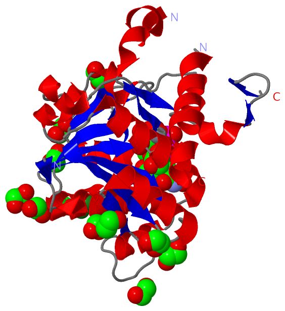
Image Asymmetric Unit
Asymmetric Unit  (Jmol Viewer)
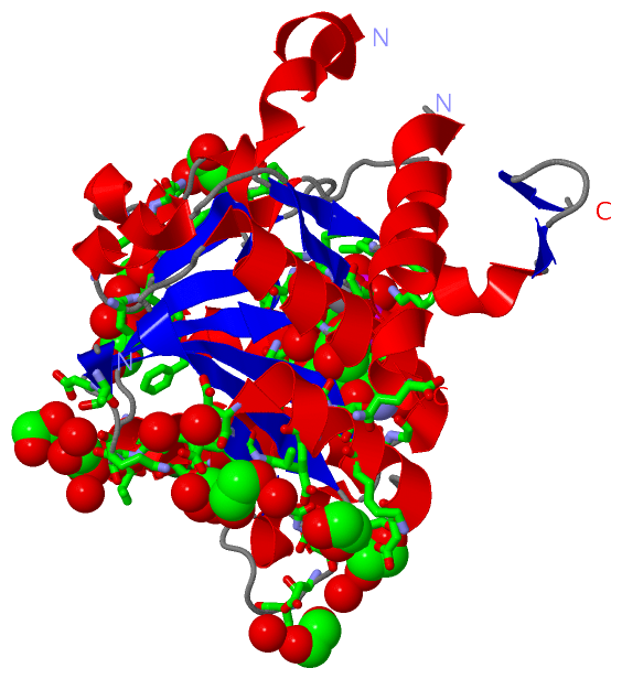
Image Asym. Unit - sites
Asym. Unit - sites  (Jmol Viewer)
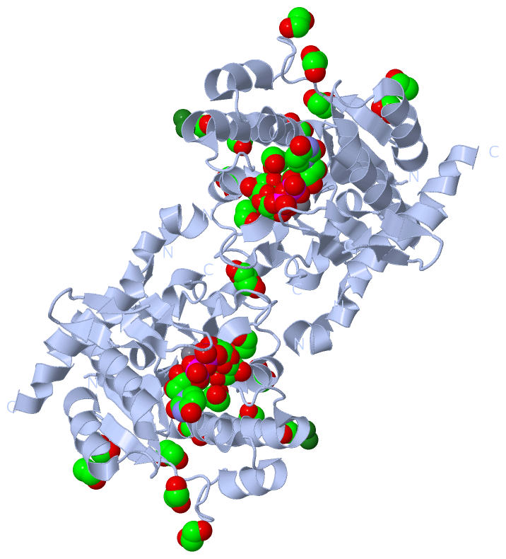
Image Biological Unit 1
Biological Unit 1  (Jmol Viewer)
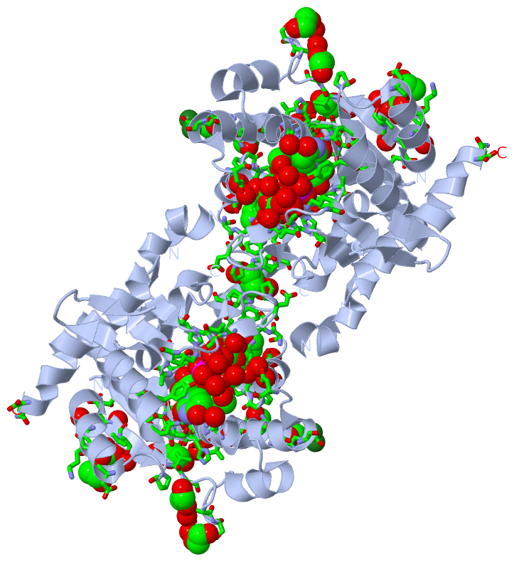
Image Biol. Unit 1 - sites
Biol. Unit 1 - sites  (Jmol Viewer)

(-) Description

Title :  CRYSTAL STRUCTURE OF HUMAN GLYCOGENIN-1 (GYG1) T83M MUTANT COMPLEXED WITH MANGANESE AND UDP-GLUCOSE
 
Authors :  A. Chaikuad, D. S. Froese, W. W. Yue, E. Krysztofinska, F. Von Delft, J. C. H. Arrowsmith, A. M. Edwards, C. Bountra, U. Oppermann, Structural Consortium (Sgc)
Date :  21 Apr 11  (Deposition) - 11 May 11  (Release) - 11 Jan 12  (Revision)
Method :  X-RAY DIFFRACTION
Resolution :  1.93
Chains :  Asym. Unit :  A
Biol. Unit 1:  A  (2x)
Keywords :  Structural Genomics, Structural Genomics Consortium, Sgc, Transferase, Glycosyltransferase, Glycogen Biosynthesis, Glycosylation (Keyword Search: [Gene Ontology, PubMed, Web (Google)] )
 
Reference :  A. Chaikuad, D. S. Froese, G. Berridge, F. Von Delft, U. Oppermann, W. W. Yue
Conformational Plasticity Of Glycogenin And Its Maltosaccharide Substrate During Glycogen Biogenesis.
Proc. Natl. Acad. Sci. Usa V. 108 21028 2011
PubMed-ID: 22160680  |  Reference-DOI: 10.1073/PNAS.1113921108

(-) Compounds

Molecule 1 - GLYCOGENIN-1
    Chains: A
    EC Number: 2.4.1.186
    Engineered: YES
    Expression System: ESCHERICHIA COLI
    Expression System Plasmid: PNIC28-BSA4
    Expression System Strain: BL21(DE3)-R3-PRARE2
    Expression System Taxid: 469008
    Expression System Vector Type: PLASMID
    Fragment: UNP RESIDUES 1-262
    Gene: GYG, GYG1
    Mutation: YES
    Organism Common: HUMAN
    Organism Scientific: HOMO SAPIENS
    Organism Taxid: 9606
    Synonym: GN-1, GN1

 Structural Features

(-) Chains, Units

  1
Asymmetric Unit : A
Biological Unit 1 (2x): A

Summary Information (see also Sequences/Alignments below)

(-) Ligands, Modified Residues, Ions  (4, 15)

Asymmetric Unit (4, 15)
No.NameCountTypeFull Name
1EDO12Ligand/Ion1,2-ETHANEDIOL
2MG1Ligand/IonMAGNESIUM ION
3MN1Ligand/IonMANGANESE (II) ION
4UPG1Ligand/IonURIDINE-5'-DIPHOSPHATE-GLUCOSE
Biological Unit 1 (2, 26)
No.NameCountTypeFull Name
1EDO24Ligand/Ion1,2-ETHANEDIOL
2MG-1Ligand/IonMAGNESIUM ION
3MN-1Ligand/IonMANGANESE (II) ION
4UPG2Ligand/IonURIDINE-5'-DIPHOSPHATE-GLUCOSE

(-) Sites  (15, 15)

Asymmetric Unit (15, 15)
No.NameEvidenceResiduesDescription
01AC1SOFTWARELEU A:9 , THR A:10 , THR A:11 , ASN A:12 , TYR A:15 , LYS A:86 , ASP A:102 , ALA A:103 , ASP A:104 , ASN A:133 , SER A:134 , GLY A:135 , ASP A:160 , GLY A:162 , ASP A:163 , GLN A:164 , HIS A:212 , LEU A:214 , GLY A:215 , LYS A:218 , MN A:264 , EDO A:270 , HOH A:309 , HOH A:343 , HOH A:352 , HOH A:399 , HOH A:400 , HOH A:404 , HOH A:405 , HOH A:461 , HOH A:466BINDING SITE FOR RESIDUE UPG A 263
02AC2SOFTWAREASP A:102 , ASP A:104 , HIS A:212 , UPG A:263BINDING SITE FOR RESIDUE MN A 264
03AC3SOFTWAREASP A:3 , GLN A:94 , HIS A:151 , GLU A:155 , HOH A:417 , HOH A:418BINDING SITE FOR RESIDUE MG A 265
04AC4SOFTWAREVAL A:61 , HIS A:88 , SER A:91 , LEU A:92 , HOH A:340BINDING SITE FOR RESIDUE EDO A 266
05AC5SOFTWARETHR A:93 , VAL A:143 , HOH A:293 , HOH A:446 , HOH A:447BINDING SITE FOR RESIDUE EDO A 267
06AC6SOFTWAREPRO A:124 , PRO A:129 , ASP A:130 , HIS A:182 , HOH A:348BINDING SITE FOR RESIDUE EDO A 268
07AC7SOFTWAREGLU A:119 , LEU A:120 , SER A:173 , TRP A:174 , LYS A:181 , HOH A:312BINDING SITE FOR RESIDUE EDO A 269
08AC8SOFTWAREVAL A:82 , LYS A:86 , GLY A:161 , ASP A:163 , UPG A:263BINDING SITE FOR RESIDUE EDO A 270
09AC9SOFTWARETHR A:2 , GLN A:94 , GLN A:148 , HIS A:151 , HOH A:334BINDING SITE FOR RESIDUE EDO A 271
10BC1SOFTWAREPHE A:170 , PHE A:171 , HOH A:283 , HOH A:356BINDING SITE FOR RESIDUE EDO A 272
11BC2SOFTWAREASP A:56 , GLU A:57 , VAL A:58 , HOH A:436 , HOH A:438BINDING SITE FOR RESIDUE EDO A 273
12BC3SOFTWAREPRO A:41 , ASP A:62 , GLN A:262 , HOH A:336BINDING SITE FOR RESIDUE EDO A 274
13BC4SOFTWAREASP A:45 , LYS A:49 , GLU A:52BINDING SITE FOR RESIDUE EDO A 275
14BC5SOFTWAREASP A:65 , SER A:66 , HOH A:371BINDING SITE FOR RESIDUE EDO A 276
15BC6SOFTWAREASP A:56 , GLU A:144 , HOH A:439BINDING SITE FOR RESIDUE EDO A 277

(-) SS Bonds  (0, 0)

(no "SS Bond" information available for 3RMW)

(-) Cis Peptide Bonds  (1, 1)

Asymmetric Unit
No.Residues
1Glu A:119 -Leu A:120

 Sequence-Structure Mapping

(-) SAPs(SNPs)/Variants  (3, 3)

Asymmetric Unit (3, 3)
  dbSNPPDB
No.SourceVariant IDVariantUniProt IDStatusIDChainVariant
1UniProtVAR_072706A16PGLYG_HUMANDisease (PGBM2)  ---AA16P
2UniProtVAR_063768T83MGLYG_HUMANDisease (GSD15)267606858AM83M
3UniProtVAR_072707D102HGLYG_HUMANDisease (PGBM2)143137713AD102H

  SNP/SAP Summary Statistics (UniProtKB/Swiss-Prot)
Biological Unit 1 (3, 6)
  dbSNPPDB
No.SourceVariant IDVariantUniProt IDStatusIDChainVariant
1UniProtVAR_072706A16PGLYG_HUMANDisease (PGBM2)  ---AA16P
2UniProtVAR_063768T83MGLYG_HUMANDisease (GSD15)267606858AM83M
3UniProtVAR_072707D102HGLYG_HUMANDisease (PGBM2)143137713AD102H

  SNP/SAP Summary Statistics (UniProtKB/Swiss-Prot)

(-) PROSITE Motifs  (0, 0)

(no "PROSITE Motif" information available for 3RMW)

(-) Exons   (0, 0)

(no "Exon" information available for 3RMW)

(-) Sequences/Alignments

Asymmetric Unit
   Reformat: Number of residues per line =  ('0' or empty: single-line sequence representation)
  Number of residues per labelling interval =   
  UniProt sequence: complete  aligned part    
   Show mapping: SCOP domains CATH domains Pfam domains Secondary structure (by author)
SAPs(SNPs) PROSITE motifs Exons
(details for a mapped element are shown in a popup box when the mouse pointer rests over it)
Chain A from PDB  Type:PROTEIN  Length:250
 aligned with GLYG_HUMAN | P46976 from UniProtKB/Swiss-Prot  Length:350

    Alignment length:263
                             1                                                                                                                                                                                                                                                                     
                             |       9        19        29        39        49        59        69        79        89        99       109       119       129       139       149       159       169       179       189       199       209       219       229       239       249       259   
           GLYG_HUMAN     - -MTDQAFVTLTTNDAYAKGALVLGSSLKQHRTTRRLVVLATPQVSDSMRKVLETVFDEVIMVDVLDSGDSAHLTLMKRPELGVTLTKLHCWSLTQYSKCVFMDADTLVLANIDDLFDREELSAAPDPGWPDCFNSGVFVYQPSVETYNQLLHLASEQGSFDGGDQGILNTFFSSWATTDIRKHLPFIYNLSSISIYSYLPAFKVFGASAKVVHFLGRVKPWNYTYDPKTKSVKSEAHDPNMTHPEFLILWWNIFTTNVLPLLQ 262
               SCOP domains d3rmwa_ A: Glycogenin                                                                                                                                                                                                                                                   SCOP domains
               CATH domains ----------------------------------------------------------------------------------------------------------------------------------------------------------------------------------------------------------------------------------------------------------------------- CATH domains
               Pfam domains ------Glyco_transf_8-3rmwA01 A:6-224                                                                                                                                                                                             ---------        --------------------- Pfam domains
         Sec.struct. author .eeeeeeeeee.hhhhhhhhhhhhhhhhhh...eeeeeee....hhhhhhhhhhhh.eeee.........hhhhhhhh..hhhhhhhhhhhhh....eeeee...eee...hhhhhhh...eeee.......eeeeeeee..hhhhhhhhhhhhhhhh.....hhhhhhhhh......hhhhh.hhhhh.....-----hhhhhhhhhh.eee.....hhhhh.ee....ee..--------.hhhhhhhhhhhhhhhhhhhh Sec.struct. author
                 SAPs(SNPs) ----------------P------------------------------------------------------------------M------------------H---------------------------------------------------------------------------------------------------------------------------------------------------------------- SAPs(SNPs)
                    PROSITE ----------------------------------------------------------------------------------------------------------------------------------------------------------------------------------------------------------------------------------------------------------------------- PROSITE
                 Transcript ----------------------------------------------------------------------------------------------------------------------------------------------------------------------------------------------------------------------------------------------------------------------- Transcript
                 3rmw A   0 SMTDQAFVTLTTNDAYAKGALVLGSSLKQHRTTRRLVVLATPQVSDSMRKVLETVFDEVIMVDVLDSGDSAHLTLMKRPELGVMLTKLHCWSLTQYSKCVFMDADTLVLANIDDLFDREELSAAPDPGWPDCFNSGVFVYQPSVETYNQLLHLASEQGSFDGGDQGILNTFFSSWATTDIRKHLPFIYNLSSIS-----PAFKVFGASAKVVHFLGRVKPWNYTYDPKTKSVKS--------HPEFLILWWNIFTTNVLPLLQ 262
                                     9        19        29        39        49        59        69        79        89        99       109       119       129       139       149       159       169       179       189   |   199       209       219       229   |     -  |    249       259   
                                                                                                                                                                                                                           193   199                               233      242                    

   Legend:   → Mismatch (orange background)
  - → Gap (green background, '-', border residues have a numbering label)
    → Modified Residue (blue background, lower-case, 'x' indicates undefined single-letter code, labelled with number + name)
  x → Chemical Group (purple background, 'x', labelled with number + name, e.g. ACE or NH2)
  extra numbering lines below/above indicate numbering irregularities and modified residue names etc., number ends below/above '|'

 Classification and Annotation

(-) SCOP Domains  (1, 1)

Asymmetric Unit

(-) CATH Domains  (0, 0)

(no "CATH Domain" information available for 3RMW)

(-) Pfam Domains  (1, 1)

Asymmetric Unit
(-)
Clan: GT-A (172)

(-) Gene Ontology  (11, 11)

Asymmetric Unit(hide GO term definitions)
Chain A   (GLYG_HUMAN | P46976)
molecular function
    GO:0008466    glycogenin glucosyltransferase activity    Catalysis of the reaction: UDP-glucose + glycogenin = UDP + glucosylglycogenin.
    GO:0046872    metal ion binding    Interacting selectively and non-covalently with any metal ion.
    GO:0005515    protein binding    Interacting selectively and non-covalently with any protein or protein complex (a complex of two or more proteins that may include other nonprotein molecules).
    GO:0016740    transferase activity    Catalysis of the transfer of a group, e.g. a methyl group, glycosyl group, acyl group, phosphorus-containing, or other groups, from one compound (generally regarded as the donor) to another compound (generally regarded as the acceptor). Transferase is the systematic name for any enzyme of EC class 2.
    GO:0016757    transferase activity, transferring glycosyl groups    Catalysis of the transfer of a glycosyl group from one compound (donor) to another (acceptor).
biological process
    GO:0005978    glycogen biosynthetic process    The chemical reactions and pathways resulting in the formation of glycogen, a polydisperse, highly branched glucan composed of chains of D-glucose residues.
    GO:0005980    glycogen catabolic process    The chemical reactions and pathways resulting in the breakdown of glycogen, a polydisperse, highly branched glucan composed of chains of D-glucose residues.
cellular component
    GO:0005829    cytosol    The part of the cytoplasm that does not contain organelles but which does contain other particulate matter, such as protein complexes.
    GO:0070062    extracellular exosome    A vesicle that is released into the extracellular region by fusion of the limiting endosomal membrane of a multivesicular body with the plasma membrane. Extracellular exosomes, also simply called exosomes, have a diameter of about 40-100 nm.
    GO:0043202    lysosomal lumen    The volume enclosed within the lysosomal membrane.
    GO:0016020    membrane    A lipid bilayer along with all the proteins and protein complexes embedded in it an attached to it.

 Visualization

(-) Interactive Views

Asymmetric Unit
  Complete Structure
    Jena3D(integrated viewing of ligand, site, SAP, PROSITE, SCOP information)
    WebMol | AstexViewer[tm]@PDBe
(Java Applets, require no local installation except for Java; loading may be slow)
    STRAP
(Java WebStart application, automatic local installation, requires Java; full application with system access!)
    RasMol
(require local installation)
    Molscript (VRML)
(requires installation of a VRML viewer; select preferred view via VRML and generate a mono or stereo PDF format file)
 
  Ligands, Modified Residues, Ions
    EDO  [ RasMol | Jena3D ]  +environment [ RasMol | Jena3D ]
    MG  [ RasMol | Jena3D ]  +environment [ RasMol | Jena3D ]
    MN  [ RasMol | Jena3D ]  +environment [ RasMol | Jena3D ]
    UPG  [ RasMol | Jena3D ]  +environment [ RasMol | Jena3D ]
 
  Sites
    AC1  [ RasMol ]  +environment [ RasMol ]
    AC2  [ RasMol ]  +environment [ RasMol ]
    AC3  [ RasMol ]  +environment [ RasMol ]
    AC4  [ RasMol ]  +environment [ RasMol ]
    AC5  [ RasMol ]  +environment [ RasMol ]
    AC6  [ RasMol ]  +environment [ RasMol ]
    AC7  [ RasMol ]  +environment [ RasMol ]
    AC8  [ RasMol ]  +environment [ RasMol ]
    AC9  [ RasMol ]  +environment [ RasMol ]
    BC1  [ RasMol ]  +environment [ RasMol ]
    BC2  [ RasMol ]  +environment [ RasMol ]
    BC3  [ RasMol ]  +environment [ RasMol ]
    BC4  [ RasMol ]  +environment [ RasMol ]
    BC5  [ RasMol ]  +environment [ RasMol ]
    BC6  [ RasMol ]  +environment [ RasMol ]
 
  Cis Peptide Bonds
    Glu A:119 - Leu A:120   [ RasMol ]  
 
Biological Unit
  Complete Structure
    Biological Unit 1  [ Jena3D ]

(-) Still Images

Jmol
  protein: cartoon or spacefill or dots and stick; nucleic acid: cartoon and stick; ligands: spacefill; active site: stick
Molscript
  protein, nucleic acid: cartoon; ligands: spacefill; active site: ball and stick

 Databases and Analysis Tools

(-) Databases

Access by PDB/NDB ID
  3rmw
    Family and Domain Information: ProDom | SYSTERS
    General Structural Information: GlycoscienceDB | MMDB | NDB | OCA | PDB | PDBe | PDBj | PDBsum | PDBWiki | PQS | PROTEOPEDIA
    Orientation in Membranes: OPM
    Protein Surface: SURFACE
    Secondary Structure: DSSP (structure derived) | HSSP (homology derived)
    Structural Genomics: GeneCensus
    Structural Neighbours: CE | VAST
    Structure Classification: CATH | Dali | SCOP
    Validation and Original Data: BMRB Data View | BMRB Restraints Grid | EDS | PROCHECK | RECOORD | WHAT_CHECK
 
Access by UniProt ID/Accession number
  GLYG_HUMAN | P46976
    Comparative Protein Structure Models: ModBase
    Genomic Information: Ensembl
    Protein-protein Interaction: DIP
    Sequence, Family and Domain Information: InterPro | Pfam | SMART | UniProtKB/SwissProt
 
Access by Enzyme Classificator   (EC Number)
  2.4.1.186
    General Enzyme Information: BRENDA | EC-PDB | Enzyme | IntEnz
    Pathway: KEGG | MetaCyc
 
Access by Disease Identifier   (MIM ID)
  613507
    Disease Information: OMIM
  616199
    Disease Information: OMIM
 
Access by GenAge ID
  (no 'GenAge ID' available)
    Age Related Information: GenAge

(-) Analysis Tools

Access by PDB/NDB ID
    Domain Information: XDom
    Interatomic Contacts of Structural Units: CSU
    Ligand-protein Contacts: LPC
    Protein Cavities: castP
    Sequence and Secondary Structure: PDBCartoon
    Structure Alignment: STRAP(Java WebStart application, automatic local installation, requires Java; full application with system access!)
    Structure and Sequence Browser: STING
 
Access by UniProt ID/Accession number
  GLYG_HUMAN | P46976
    Protein Disorder Prediction: DisEMBL | FoldIndex | GLOBPLOT (for more information see DisProt)

 Related Entries

(-) Entries Sharing at Least One Protein Chain (UniProt ID)

UniProtKB/Swiss-Prot
        GLYG_HUMAN | P46976: 3q4s 3qvb 3rmv 3t7m 3t7n 3t7o 3u2t 3u2u 3u2v 3u2w 3u2x

(-) Related Entries Specified in the PDB File

3q4s HGYG1 (Y195F) APO FORM
3qvb HGYG1 (Y195F) WITH UDP,MN
3rmv HGYG1 (T83M) WITH UDP,MN