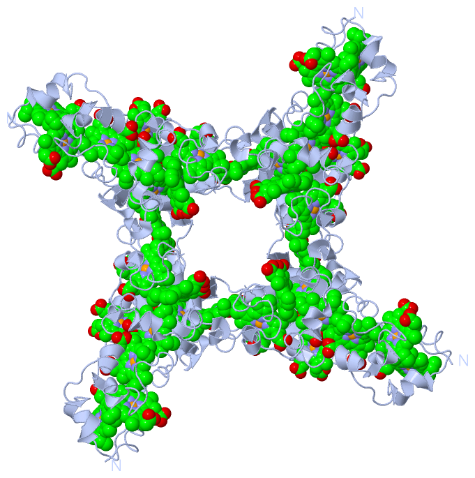
Asymmetric Unit (12, 12)
| No. | Name | Evidence | Residues | Description |
|---|
| 01 | AC1 | SOFTWARE | LYS A:4 , VAL A:6 , PHE A:8 , PHE A:18 , HIS A:20 , HIS A:23 , LEU A:24 , CYS A:31 , CYS A:34 , HIS A:35 , LEU A:41 , PRO A:44 , HEM A:602 | BINDING SITE FOR RESIDUE HEM A 601 |
| 02 | AC2 | SOFTWARE | PHE A:18 , ILE A:22 , HIS A:23 , TYR A:27 , PHE A:39 , CYS A:58 , CYS A:61 , HIS A:62 , ALA A:67 , ARG A:78 , CYS A:79 , HEM A:601 , HEM A:603 | BINDING SITE FOR RESIDUE HEM A 602 |
| 03 | AC3 | SOFTWARE | PHE A:8 , ARG A:46 , TYR A:47 , THR A:48 , MET A:49 , VAL A:70 , CYS A:76 , CYS A:79 , HIS A:80 , HEM A:602 , HEM A:604 | BINDING SITE FOR RESIDUE HEM A 603 |
| 04 | AC4 | SOFTWARE | LEU A:10 , ASN A:12 , HIS A:80 , PHE A:99 , HIS A:101 , VAL A:105 , CYS A:112 , CYS A:115 , HIS A:116 , HEM A:603 | BINDING SITE FOR RESIDUE HEM A 604 |
| 05 | AC5 | SOFTWARE | VAL A:98 , PHE A:99 , HIS A:104 , ILE A:121 , CYS A:138 , CYS A:141 , HIS A:142 , MET A:146 , ALA A:147 , HEM A:606 | BINDING SITE FOR RESIDUE HEM A 605 |
| 06 | AC6 | SOFTWARE | TYR A:89 , ASN A:126 , VAL A:127 , MET A:129 , GLY A:152 , CYS A:154 , CYS A:157 , HIS A:158 , CYS A:190 , HEM A:605 | BINDING SITE FOR RESIDUE HEM A 606 |
| 07 | AC7 | SOFTWARE | MET A:161 , PRO A:163 , VAL A:167 , PHE A:179 , HIS A:181 , HIS A:184 , LEU A:185 , TYR A:188 , CYS A:190 , CYS A:193 , HIS A:194 , PHE A:198 , TYR A:200 , LYS A:201 , ALA A:202 , HEM A:608 | BINDING SITE FOR RESIDUE HEM A 607 |
| 08 | AC8 | SOFTWARE | GLU A:96 , PHE A:179 , HIS A:184 , MET A:187 , TYR A:188 , PHE A:198 , CYS A:218 , CYS A:221 , HIS A:222 , ALA A:227 , HEM A:607 , HEM A:609 | BINDING SITE FOR RESIDUE HEM A 608 |
| 09 | AC9 | SOFTWARE | PHE A:169 , VAL A:174 , ARG A:206 , PHE A:207 , THR A:208 , MET A:209 , CYS A:218 , HIS A:222 , PHE A:228 , ALA A:231 , CYS A:234 , CYS A:237 , HIS A:238 , LEU A:241 , TYR A:278 , HEM A:608 | BINDING SITE FOR RESIDUE HEM A 609 |
| 10 | BC1 | SOFTWARE | LEU A:241 , LYS A:242 , PRO A:243 , ALA A:244 , LEU A:246 , TYR A:248 , PHE A:257 , HIS A:259 , HIS A:262 , LEU A:263 , CYS A:268 , CYS A:271 , HIS A:272 , PHE A:276 , TYR A:278 , SER A:282 | BINDING SITE FOR RESIDUE HEM A 610 |
| 11 | BC2 | SOFTWARE | TYR A:248 , PHE A:257 , HIS A:262 , PHE A:266 , CYS A:296 , CYS A:299 , HIS A:300 , ALA A:305 | BINDING SITE FOR RESIDUE HEM A 611 |
| 12 | BC3 | SOFTWARE | GLU A:53 , ARG A:113 , TYR A:248 , THR A:250 , VAL A:252 , PRO A:284 , ALA A:285 , MET A:287 , CYS A:296 , VAL A:308 , ALA A:309 , CYS A:312 , CYS A:315 , HIS A:316 | BINDING SITE FOR RESIDUE HEM A 612 |
|
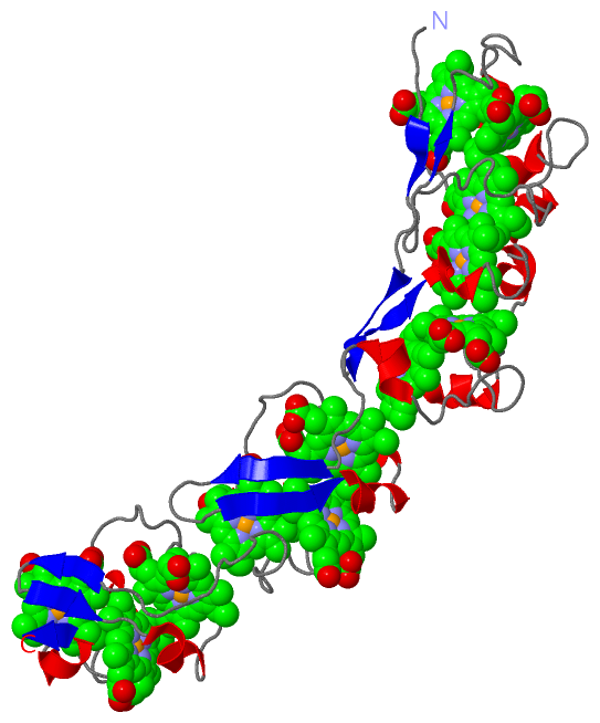
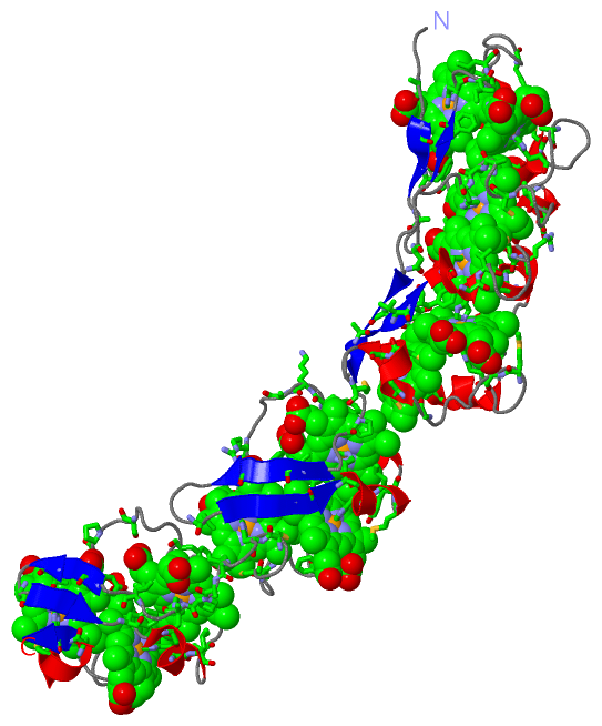
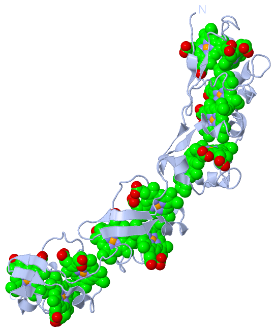
 Description