|
|
|
|
 Description
|
|
Compounds
|
||||||||||||||||||||||||||||||||||||||||||||
Chains, Units
Summary Information (see also Sequences/Alignments below) |
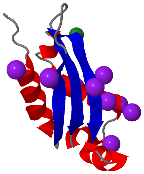
Ligands, Modified Residues, Ions (2, 10)| Asymmetric Unit (2, 10) Biological Unit 1 (1, 9) Biological Unit 2 (1, 18) |
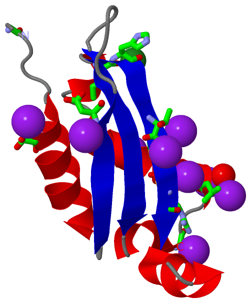
Sites (10, 10)
Asymmetric Unit (10, 10)
|
SS Bonds (1, 1)
Asymmetric Unit
|
||||||||
Cis Peptide Bonds (0, 0)| (no "Cis Peptide Bond" information available for 3O2E) |
SAPs(SNPs)/Variants (0, 0)| (no "SAP(SNP)/Variant" information available for 3O2E) |
PROSITE Motifs (0, 0)| (no "PROSITE Motif" information available for 3O2E) |
Exons (0, 0)| (no "Exon" information available for 3O2E) |
Sequences/Alignments
Asymmetric UnitChain A from PDB Type:PROTEIN Length:86 aligned with A7ATL3_BABBO | A7ATL3 from UniProtKB/TrEMBL Length:86 Alignment length:86 1 | 7 17 27 37 47 57 67 77 A7ATL3_BABBO - ---MVSKSIVEERLRSMLSPQFLKVTDNSGGCGAAFNAYIVSQQFEGKGLLDRQRLVNSAIAAEMPQIHAFTMKCLTPGEWEAKNR 83 SCOP domains -------------------------------------------------------------------------------------- SCOP domains CATH domains -------------------------------------------------------------------------------------- CATH domains Pfam domains -----------BolA-3o2eA01 A:9-78 ----- Pfam domains SAPs(SNPs) -------------------------------------------------------------------------------------- SAPs(SNPs) PROSITE -------------------------------------------------------------------------------------- PROSITE Transcript -------------------------------------------------------------------------------------- Transcript 3o2e A -2 PGSMVSKSIVEERLRSMLSPQFLKVTDNSGGCGAAFNAYIVSQQFEGKGLLDRQRLVNSAIAAEMPQIHAFTMKCLTPGEWEAKNR 83 7 17 27 37 47 57 67 77
|
||||||||||||||||||||
SCOP Domains (0, 0)| (no "SCOP Domain" information available for 3O2E) |
CATH Domains (0, 0)| (no "CATH Domain" information available for 3O2E) |
Pfam Domains (1, 1)
Asymmetric Unit

|
Gene Ontology (0, 0)|
Asymmetric Unit(hide GO term definitions)
(no "Gene Ontology" information available for 3O2E)
|
Interactive Views
|
|||||||||||||||||||||||||||||||||||||||||||||||||||||||||||||||||||||||||||||||||||||||||||||||||||||||||||||||||||||||||||||||||||||||||||||||||||||||||||||||||||||||||||||||||||||||||||||||||||||||||||||||||||
Still Images
|
||||||||||||||||
Databases
|
||||||||||||||||||||||||||||||||||||||||||||||||||||||||||||||||||||||||||||||||||||||||||||||||||||||||||||||||||||||||||||||||||||||||||||||||||||||||||||||||
Analysis Tools
|
|||||||||||||||||||||||||||||||||||||||||||||||||||||||||||||
Entries Sharing at Least One Protein Chain (UniProt ID)
Related Entries Specified in the PDB File
|
|