|
|
|
|
 Description
|
|
Compounds
|
||||||||||||||||||||||||||||||||||||||||||||||||||||
Chains, Units
Summary Information (see also Sequences/Alignments below) |
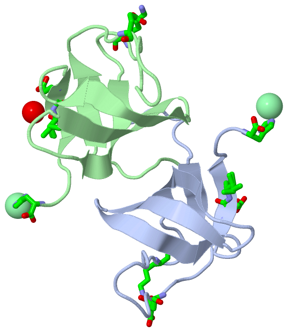
Ligands, Modified Residues, Ions (1, 2)
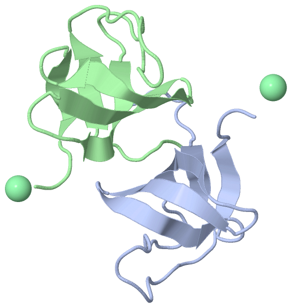
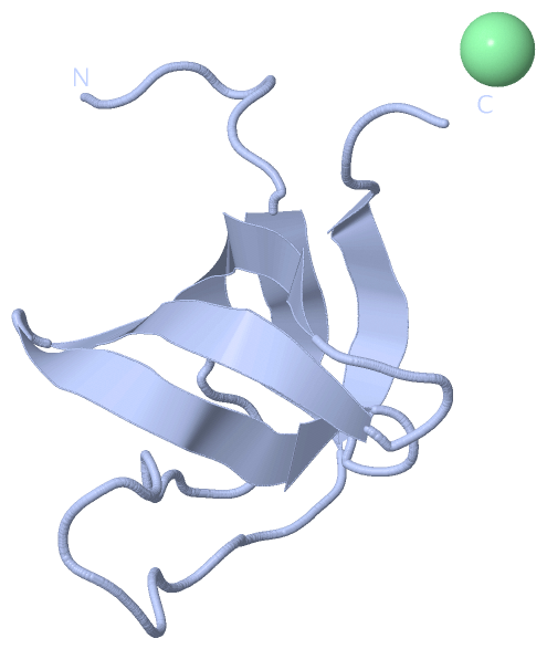
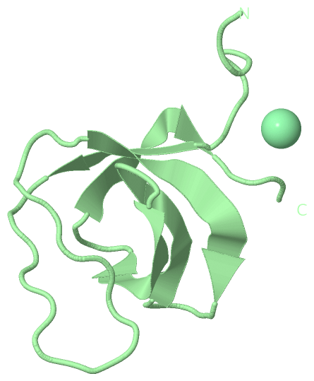
Asymmetric Unit (1, 2)
|
Sites (2, 2)
Asymmetric Unit (2, 2)
|
SS Bonds (0, 0)| (no "SS Bond" information available for 3IQL) |
Cis Peptide Bonds (0, 0)| (no "Cis Peptide Bond" information available for 3IQL) |
SAPs(SNPs)/Variants (0, 0)| (no "SAP(SNP)/Variant" information available for 3IQL) |
PROSITE Motifs (1, 2)
Asymmetric Unit (1, 2)
|
||||||||||||||||||||||||
Exons (1, 2)
Asymmetric Unit (1, 2)
|
||||||||||||||||||||||||||||||||||||||||||||||||||||||||||||||||||||||||||||||||||||||||||||||||||||||||||||||||||||||||
Sequences/Alignments
Asymmetric UnitChain A from PDB Type:PROTEIN Length:66 aligned with SH3G2_RAT | O35179 from UniProtKB/Swiss-Prot Length:352 Alignment length:66 296 306 316 326 336 346 SH3G2_RAT 287 GVQMDQPCCRALYDFEPENEGELGFKEGDIITLTNQIDENWYEGMLHGQSGFFPINYVEILVALPH 352 SCOP domains d3iqla_ A: automated matches SCOP domains CATH domains ------------------------------------------------------------------ CATH domains Pfam domains ------------------------------------------------------------------ Pfam domains SAPs(SNPs) ------------------------------------------------------------------ SAPs(SNPs) PROSITE ---SH3 PDB: A:291-349 UniProt: 290-349 --- PROSITE Transcript 1 Exon 1.9 PDB: A:287-338 UniProt: 273-338 -------------- Transcript 1 3iql A 287 SVGSDQPCCRALYDFEPENEGELGFKEGDIITLTNQIDENWYEGMLHGQSGFFPINYVEILVALPH 352 296 306 316 326 336 346 Chain B from PDB Type:PROTEIN Length:67 aligned with SH3G2_RAT | O35179 from UniProtKB/Swiss-Prot Length:352 Alignment length:67 295 305 315 325 335 345 SH3G2_RAT 286 AGVQMDQPCCRALYDFEPENEGELGFKEGDIITLTNQIDENWYEGMLHGQSGFFPINYVEILVALPH 352 SCOP domains d3iqlb_ B: automated matches SCOP domains CATH domains ------------------------------------------------------------------- CATH domains Pfam domains ------------------------------------------------------------------- Pfam domains SAPs(SNPs) ------------------------------------------------------------------- SAPs(SNPs) PROSITE ----SH3 PDB: B:291-349 UniProt: 290-349 --- PROSITE Transcript 1 Exon 1.9 PDB: B:286-338 UniProt: 273-338 -------------- Transcript 1 3iql B 286 ASVGSDQPCCRALYDFEPENEGELGFKEGDIITLTNQIDENWYEGMLHGQSGFFPINYVEILVALPH 352 295 305 315 325 335 345
|
||||||||||||||||||||
SCOP Domains (1, 2)
Asymmetric Unit
 |
CATH Domains (0, 0)| (no "CATH Domain" information available for 3IQL) |
Pfam Domains (0, 0)| (no "Pfam Domain" information available for 3IQL) |
Gene Ontology (11, 11)|
Asymmetric Unit(hide GO term definitions) Chain A,B (SH3G2_RAT | O35179)
|
||||||||||||||||||||||||||||||||||||||||||||||||||||||||||||||||||||||||||||||||||||
Interactive Views
|
||||||||||||||||||||||||||||||||||||||||||||||||||||||||||||||||||||||||||||||||||||||||||||||||||||||||||||||||||||||||||||||||||||||||||||||||||||
Still Images
|
||||||||||||||||
Databases
|
||||||||||||||||||||||||||||||||||||||||||||||||||||||||||||||||||||||||||||||||||||||||||||||||||||||||||||||||||||||||||||||||||||||||||||||||||||||||||||||||
Analysis Tools
|
|||||||||||||||||||||||||||||||||||||||||||||||||||||||||||||
Entries Sharing at Least One Protein Chain (UniProt ID)
Related Entries Specified in the PDB File
|
|