Show PDB file:   
         Plain Text   HTML   (compressed file size)
QuickSearch:   
by PDB,NDB,UniProt,PROSITE Code or Search Term(s)  
(-)Asymmetric Unit
(-)Asym. Unit - sites
(-)Biological Unit 1
(-)Biol. Unit 1 - sites
(-)Biological Unit 2
(-)Biological Unit 3
collapse expand < >
Image Asymmetric Unit
Asymmetric Unit  (Jmol Viewer)
Image Asym. Unit - sites
Asym. Unit - sites  (Jmol Viewer)
Image Biological Unit 1
Biological Unit 1  (Jmol Viewer)
Image Biol. Unit 1 - sites
Biol. Unit 1 - sites  (Jmol Viewer)
Image Biological Unit 2
Biological Unit 2  (Jmol Viewer)
Image Biological Unit 3
Biological Unit 3  (Jmol Viewer)

(-) Description

Title :  THE STRUCTURE OF THE M53A MUTANT OF CAULOBACTER CRESCENTUS CLPS PROTEASE ADAPTOR PROTEIN IN COMPLEX WITH WLFVQRDSKE PEPTIDE
 
Authors :  T. A. Baker, G. Roman-Hernandez, R. T. Sauer, R. A. Grant
Date :  29 Jan 09  (Deposition) - 28 Apr 09  (Release) - 30 Jun 09  (Revision)
Method :  X-RAY DIFFRACTION
Resolution :  1.45
Chains :  Asym. Unit :  A,B,C,D
Biol. Unit 1:  A,B,C,D  (1x)
Biol. Unit 2:  A,C  (1x)
Biol. Unit 3:  B,D  (1x)
Keywords :  Adaptor, Protein-Peptide Complex, Peptide-Binding Protein, Peptide Binding Protein (Keyword Search: [Gene Ontology, PubMed, Web (Google))
 
Reference :  G. Roman-Hernandez, R. A. Grant, R. T. Sauer, T. A. Baker
Molecular Basis Of Substrate Selection By The N-End Rule Adaptor Protein Clps.
Proc. Natl. Acad. Sci. Usa V. 106 8888 2009
PubMed-ID: 19451643  |  Reference-DOI: 10.1073/PNAS.0903614106
(for further references see the PDB file header)

(-) Compounds

Molecule 1 - ATP-DEPENDENT CLP PROTEASE ADAPTER PROTEIN CLPS
    ChainsA, B
    EngineeredYES
    Expression SystemESCHERICHIA COLI
    Expression System PlasmidPET23B
    Expression System Taxid562
    Expression System Vector TypePLASMID
    GeneCC_2467, CLPS
    MutationYES
    Organism CommonCAULOBACTER VIBRIOIDES
    Organism ScientificCAULOBACTER VIBRIOIDES
    Organism Taxid155892
    StrainCB15
 
Molecule 2 - 10-RESIDUE PEPTIDE
    ChainsC, D
    EngineeredYES
    Other DetailsSYNTHETIC PEPTIDE
    SyntheticYES

 Structural Features

(-) Chains, Units

  1234
Asymmetric Unit ABCD
Biological Unit 1 (1x)ABCD
Biological Unit 2 (1x)A C 
Biological Unit 3 (1x) B D

Summary Information (see also Sequences/Alignments below)

(-) Ligands, Modified Residues, Ions  (1, 2)

Asymmetric Unit (1, 2)
No.NameCountTypeFull Name
1MG2Ligand/IonMAGNESIUM ION
Biological Unit 1 (0, 0)
No.NameCountTypeFull Name
1MG-1Ligand/IonMAGNESIUM ION
Biological Unit 2 (0, 0)
No.NameCountTypeFull Name
1MG-1Ligand/IonMAGNESIUM ION
Biological Unit 3 (0, 0)
No.NameCountTypeFull Name
1MG-1Ligand/IonMAGNESIUM ION

(-) Sites  (2, 2)

Asymmetric Unit (2, 2)
No.NameEvidenceResiduesDescription
1AC1SOFTWAREASP A:119 , HOH A:192 , HOH A:202 , ASP B:119 , HOH B:140 , HOH B:203BINDING SITE FOR RESIDUE MG A 1
2AC2SOFTWAREARG A:42 , GLU A:117 , ARG B:42 , LEU B:44 , GLU B:117BINDING SITE FOR RESIDUE MG B 1

(-) SS Bonds  (0, 0)

(no "SS Bond" information available for 3G1B)

(-) Cis Peptide Bonds  (0, 0)

(no "Cis Peptide Bond" information available for 3G1B)

 Sequence-Structure Mapping

(-) SAPs(SNPs)/Variants  (0, 0)

(no "SAP(SNP)/Variant" information available for 3G1B)

(-) PROSITE Motifs  (0, 0)

(no "PROSITE Motif" information available for 3G1B)

(-) Exons   (0, 0)

(no "Exon" information available for 3G1B)

(-) Sequences/Alignments

Asymmetric Unit
   Reformat: Number of residues per line =  ('0' or empty: single-line sequence representation)
  Number of residues per labelling interval =   
  UniProt sequence: complete  aligned part    
   Show mapping: SCOP domains CATH domains Pfam domains Secondary structure (by author)
SAPs(SNPs) PROSITE motifs Exons
(details for a mapped element are shown in a popup box when the mouse pointer rests over it)
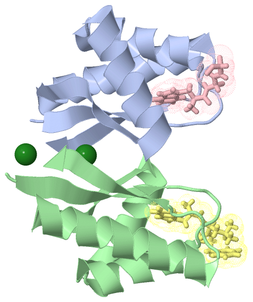
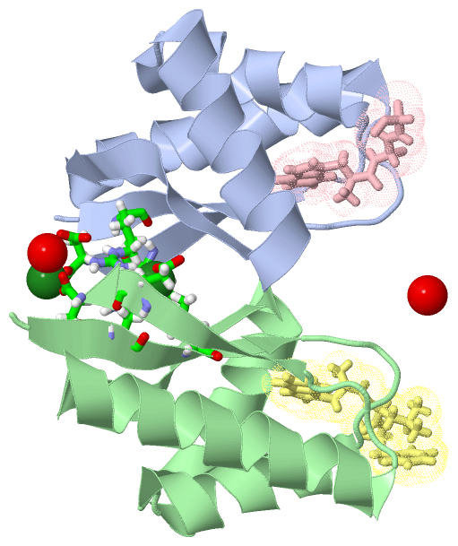
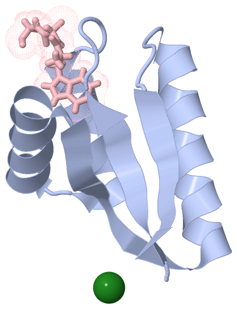
Chain A from PDB  Type:PROTEIN  Length:80
 aligned with CLPS_CAUCR | Q9A5I0 from UniProtKB/Swiss-Prot  Length:119

    Alignment length:80
                                    49        59        69        79        89        99       109       119
           CLPS_CAUCR    40 LYRVLILNDDYTPMEFVVYVLERFFNKSREDATRIMLHVHQNGVGVCGVYTYEVAETKVAQVIDSARRHQHPLQCTMEKD 119
               SCOP domains d3g1ba_ A: automated matches                                                     SCOP domains
               CATH domains 3g1bA00 A:40-119  [code=3.30.1390.10, no name defined]                           CATH domains
               Pfam domains -------------------------------------------------------------------------------- Pfam domains
         Sec.struct. author .eeeeee.....hhhhhhhhhhhhhh.hhhhhhhhhhhhhhhheeeeeeehhhhhhhhhhhhhhhhhhh....eeeeee. Sec.struct. author
                 SAPs(SNPs) -------------------------------------------------------------------------------- SAPs(SNPs)
                    PROSITE -------------------------------------------------------------------------------- PROSITE
                 Transcript -------------------------------------------------------------------------------- Transcript
                 3g1b A  40 LYRVLILNDDYTPAEFVVYVLERFFNKSREDATRIMLHVHQNGVGVCGVYTYEVAETKVAQVIDSARRHQHPLQCTMEKD 119
                                    49        59        69        79        89        99       109       119

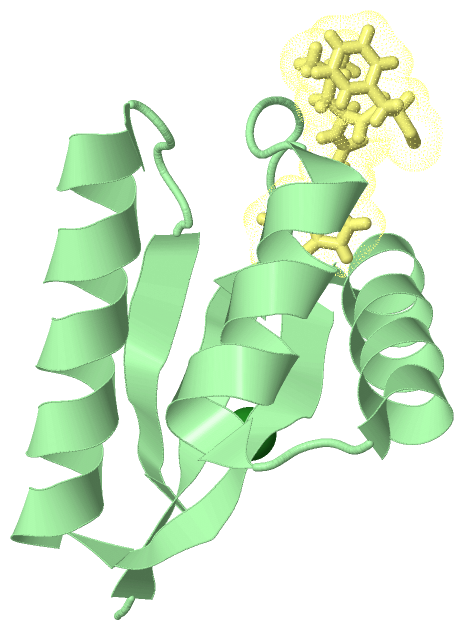
Chain B from PDB  Type:PROTEIN  Length:81
 aligned with CLPS_CAUCR | Q9A5I0 from UniProtKB/Swiss-Prot  Length:119

    Alignment length:81
                                    48        58        68        78        88        98       108       118 
           CLPS_CAUCR    39 SLYRVLILNDDYTPMEFVVYVLERFFNKSREDATRIMLHVHQNGVGVCGVYTYEVAETKVAQVIDSARRHQHPLQCTMEKD 119
               SCOP domains d3g1bb_ B: automated matches                                                      SCOP domains
               CATH domains 3g1bB00 B:39-119  [code=3.30.1390.10, no name defined]                            CATH domains
               Pfam domains --------------------------------------------------------------------------------- Pfam domains
         Sec.struct. author .eeeeeee.....hhhhhhhhhhhhh..hhhhhhhhhhhhhhhheeeeeeeehhhhhhhhhhhhhhhhhh....eeeeee. Sec.struct. author
                 SAPs(SNPs) --------------------------------------------------------------------------------- SAPs(SNPs)
                    PROSITE --------------------------------------------------------------------------------- PROSITE
                 Transcript --------------------------------------------------------------------------------- Transcript
                 3g1b B  39 SLYRVLILNDDYTPAEFVVYVLERFFNKSREDATRIMLHVHQNGVGVCGVYTYEVAETKVAQVIDSARRHQHPLQCTMEKD 119
                                    48        58        68        78        88        98       108       118 

Chain C from PDB  Type:PROTEIN  Length:3
                                   
               SCOP domains --- SCOP domains
               CATH domains --- CATH domains
               Pfam domains --- Pfam domains
         Sec.struct. author ... Sec.struct. author
                 SAPs(SNPs) --- SAPs(SNPs)
                    PROSITE --- PROSITE
                 Transcript --- Transcript
                 3g1b C   1 WLF   3

Chain D from PDB  Type:PROTEIN  Length:3
                                   
               SCOP domains --- SCOP domains
               CATH domains --- CATH domains
               Pfam domains --- Pfam domains
         Sec.struct. author ... Sec.struct. author
                 SAPs(SNPs) --- SAPs(SNPs)
                    PROSITE --- PROSITE
                 Transcript --- Transcript
                 3g1b D   1 WLF   3

   Legend:   → Mismatch (orange background)
  - → Gap (green background, '-', border residues have a numbering label)
    → Modified Residue (blue background, lower-case, 'x' indicates undefined single-letter code, labelled with number + name)
  x → Chemical Group (purple background, 'x', labelled with number + name, e.g. ACE or NH2)
  extra numbering lines below/above indicate numbering irregularities and modified residue names etc., number ends below/above '|'

 Classification and Annotation

(-) SCOP Domains  (1, 2)

Asymmetric Unit

(-) CATH Domains  (1, 2)

Asymmetric Unit
(-)
Class: Alpha Beta (26913)

(-) Pfam Domains  (0, 0)

(no "Pfam Domain" information available for 3G1B)

(-) Gene Ontology  (2, 2)

Asymmetric Unit(hide GO term definitions)
Chain A,B   (CLPS_CAUCR | Q9A5I0)
biological process
    GO:0030163    protein catabolic process    The chemical reactions and pathways resulting in the breakdown of a protein by the destruction of the native, active configuration, with or without the hydrolysis of peptide bonds.
    GO:0006508    proteolysis    The hydrolysis of proteins into smaller polypeptides and/or amino acids by cleavage of their peptide bonds.

 Visualization

(-) Interactive Views

Asymmetric Unit
  Complete Structure
    Jena3D(integrated viewing of ligand, site, SAP, PROSITE, SCOP information)
    WebMol | AstexViewer[tm]@PDBe
(Java Applets, require no local installation except for Java; loading may be slow)
    STRAP
(Java WebStart application, automatic local installation, requires Java; full application with system access!)
    RasMol
(require local installation)
    Molscript (VRML)
(requires installation of a VRML viewer; select preferred view via VRML and generate a mono or stereo PDF format file)
 
  Ligands, Modified Residues, Ions
    MG  [ RasMol | Jena3D ]  +environment [ RasMol | Jena3D ]
 
  Sites
    AC1  [ RasMol ]  +environment [ RasMol ]
    AC2  [ RasMol ]  +environment [ RasMol ]
 
  Cis Peptide Bonds
(no "Cis Peptide Bonds" information available for 3g1b)
 
Biological Units
  Complete Structure
    Biological Unit 1  [ Jena3D ]
    Biological Unit 2  [ Jena3D ]
    Biological Unit 3  [ Jena3D ]

(-) Still Images

Jmol
  protein: cartoon or spacefill or dots and stick; nucleic acid: cartoon and stick; ligands: spacefill; active site: stick
Molscript
  protein, nucleic acid: cartoon; ligands: spacefill; active site: ball and stick

 Databases and Analysis Tools

(-) Databases

Access by PDB/NDB ID
  3g1b
    Family and Domain InformationProDom | SYSTERS
    General Structural InformationGlycoscienceDB | MMDB | NDB | OCA | PDB | PDBe | PDBj | PDBsum | PDBWiki | PQS | PROTEOPEDIA
    Orientation in MembranesOPM
    Protein SurfaceSURFACE
    Secondary StructureDSSP (structure derived) | HSSP (homology derived)
    Structural GenomicsGeneCensus
    Structural NeighboursCE | VAST
    Structure ClassificationCATH | Dali | SCOP
    Validation and Original DataBMRB Data View | BMRB Restraints Grid | EDS | PROCHECK | RECOORD | WHAT_CHECK
 
Access by UniProt ID/Accession number
  CLPS_CAUCR | Q9A5I0
    Comparative Protein Structure ModelsModBase
    Genomic InformationEnsembl
    Protein-protein InteractionDIP
    Sequence, Family and Domain InformationInterPro | Pfam | SMART | UniProtKB/SwissProt
 
Access by Enzyme Classificator   (EC Number)
  (no 'Enzyme Classificator' available)
    General Enzyme InformationBRENDA | EC-PDB | Enzyme | IntEnz
    PathwayKEGG | MetaCyc
 
Access by Disease Identifier   (MIM ID)
  (no 'MIM ID' available)
    Disease InformationOMIM
 
Access by GenAge ID
  (no 'GenAge ID' available)
    Age Related InformationGenAge

(-) Analysis Tools

Access by PDB/NDB ID
    Domain InformationXDom
    Interatomic Contacts of Structural UnitsCSU
    Ligand-protein ContactsLPC
    Protein CavitiescastP
    Sequence and Secondary StructurePDBCartoon
    Structure AlignmentSTRAP(Java WebStart application, automatic local installation, requires Java; full application with system access!)
    Structure and Sequence BrowserSTING
 
Access by UniProt ID/Accession number
  CLPS_CAUCR | Q9A5I0
    Protein Disorder PredictionDisEMBL | FoldIndex | GLOBPLOT (for more information see DisProt)

 Related Entries

(-) Entries Sharing at Least One Protein Chain (UniProt ID)

UniProtKB/Swiss-Prot
        CLPS_CAUCR | Q9A5I03dnj 3g19 3g3p 3gq0 3gq1 3gw1

(-) Related Entries Specified in the PDB File

3dnj WILD TYPE PROTEIN WITH DIFFERENT PEPTIDE
3g19