|
|
|
|
 Description
|
|
Compounds
|
||||||||||||||||||||||||||||||||||||||||||||||||
Chains, Units
Summary Information (see also Sequences/Alignments below) |
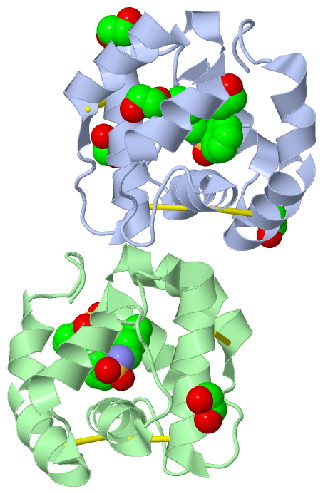
Ligands, Modified Residues, Ions (2, 9)| Asymmetric Unit (2, 9) Biological Unit 1 (2, 9) Biological Unit 2 (2, 6) Biological Unit 3 (2, 3) |
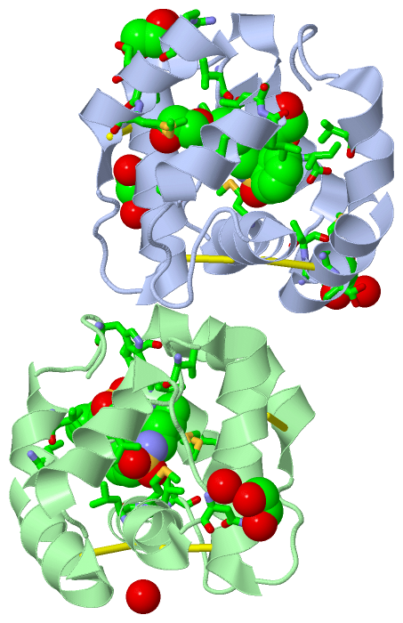
Sites (9, 9)
Asymmetric Unit (9, 9)
|
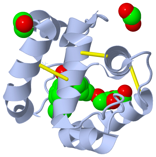
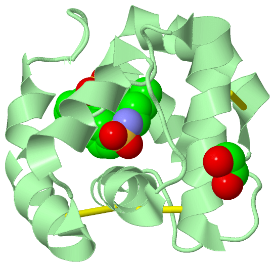
SS Bonds (6, 6)
Asymmetric Unit
|
||||||||||||||||||||||||||||
Cis Peptide Bonds (2, 2)
Asymmetric Unit
|
||||||||||||
SAPs(SNPs)/Variants (0, 0)| (no "SAP(SNP)/Variant" information available for 3D78) |
PROSITE Motifs (0, 0)| (no "PROSITE Motif" information available for 3D78) |
Exons (0, 0)| (no "Exon" information available for 3D78) |
Sequences/Alignments
Asymmetric UnitChain A from PDB Type:PROTEIN Length:119 aligned with Q9U9J6_APIME | Q9U9J6 from UniProtKB/TrEMBL Length:144 Alignment length:119 35 45 55 65 75 85 95 105 115 125 135 Q9U9J6_APIME 26 APDWVPPEVFDLVAEDKARCMSEHGTTQAQIDDVDKGNLVNEPSITCYMYCLLEAFSLVDDEANVDEDIMLGLLPDQLQERAQSVMGKCLPTSGSDNCNKIYNLAKCVQESAPDVWFVI 144 SCOP domains d3d78a_ A: automated matches SCOP domains CATH domains 3d78A00 A:1-119 [code=1.10.238.20, no name defined] CATH domains Pfam domains ----------------------------------------------------------------------------------------------------------------------- Pfam domains SAPs(SNPs) ----------------------------------------------------------------------------------------------------------------------- SAPs(SNPs) PROSITE ----------------------------------------------------------------------------------------------------------------------- PROSITE Transcript ----------------------------------------------------------------------------------------------------------------------- Transcript 3d78 A 1 APDWVPPEVFDLVAEDKARCMSEHGTTQAQIDDVNKGNLVNEPSITCYMYCLLEAFSLVDDEANVDEDIMLGLLPDQLQERAQSVMGKCLPTSGSDNCNKIYNLAKCVQESAPDVWFVI 119 10 20 30 40 50 60 70 80 90 100 110 Chain B from PDB Type:PROTEIN Length:119 aligned with Q9U9J6_APIME | Q9U9J6 from UniProtKB/TrEMBL Length:144 Alignment length:119 35 45 55 65 75 85 95 105 115 125 135 Q9U9J6_APIME 26 APDWVPPEVFDLVAEDKARCMSEHGTTQAQIDDVDKGNLVNEPSITCYMYCLLEAFSLVDDEANVDEDIMLGLLPDQLQERAQSVMGKCLPTSGSDNCNKIYNLAKCVQESAPDVWFVI 144 SCOP domains d3d78b_ B: automated matches SCOP domains CATH domains 3d78B00 B:1-119 [code=1.10.238.20, no name defined] CATH domains Pfam domains ----------------------------------------------------------------------------------------------------------------------- Pfam domains SAPs(SNPs) ----------------------------------------------------------------------------------------------------------------------- SAPs(SNPs) PROSITE ----------------------------------------------------------------------------------------------------------------------- PROSITE Transcript ----------------------------------------------------------------------------------------------------------------------- Transcript 3d78 B 1 APDWVPPEVFDLVAEDKARCMSEHGTTQAQIDDVNKGNLVNEPSITCYMYCLLEAFSLVDDEANVDEDIMLGLLPDQLQERAQSVMGKCLPTSGSDNCNKIYNLAKCVQESAPDVWFVI 119 10 20 30 40 50 60 70 80 90 100 110
|
||||||||||||||||||||
SCOP Domains (1, 2)
Asymmetric Unit
 |
CATH Domains (1, 2)
Asymmetric Unit
|
Pfam Domains (0, 0)| (no "Pfam Domain" information available for 3D78) |
Gene Ontology (1, 1)|
Asymmetric Unit(hide GO term definitions) Chain A,B (Q9U9J6_APIME | Q9U9J6)
|
||||||||||||
Interactive Views
|
|||||||||||||||||||||||||||||||||||||||||||||||||||||||||||||||||||||||||||||||||||||||||||||||||||||||||||||||||||||||||||||||||||||||||||||||||||||||||||||||||||||||||||||||||||||||||||||||||||||||||||||||||||||||||
Still Images
|
||||||||||||||||
Databases
|
||||||||||||||||||||||||||||||||||||||||||||||||||||||||||||||||||||||||||||||||||||||||||||||||||||||||||||||||||||||||||||||||||||||||||||||||||||||||||||||||
Analysis Tools
|
|||||||||||||||||||||||||||||||||||||||||||||||||||||||||||||
Entries Sharing at Least One Protein Chain (UniProt ID)
Related Entries Specified in the PDB File
|
|