Show PDB file:   
         Plain Text   HTML   (compressed file size)
QuickSearch:   
by PDB,NDB,UniProt,PROSITE Code or Search Term(s)  
(-)Asym./Biol. Unit
(-)Asym./Biol. Unit - sites
collapse expand < >
Image Asym./Biol. Unit
Asym./Biol. Unit  (Jmol Viewer)
Image Asym./Biol. Unit - sites
Asym./Biol. Unit - sites  (Jmol Viewer)

(-) Description

Title :  DIMERIC CRYSTAL STRUCTURE OF A PHEROMONE BINDING PROTEIN FROM APIS MELLIFERA AT PH 7.0
 
Authors :  M. E. Pesenti, S. Spinelli, V. Bezirard, L. Briand, J. C. Pernollet, M. Tegoni, C. Cambillau
Date :  27 Apr 08  (Deposition) - 28 Apr 09  (Release) - 28 Jul 09  (Revision)
Method :  X-RAY DIFFRACTION
Resolution :  2.50
Chains :  Asym./Biol. Unit :  A,B
Keywords :  Honey Bee, Apis Mellifera, Signal Transduction, Queen Mandibular Protein, Pheromone Binding Protein (Keyword Search: [Gene Ontology, PubMed, Web (Google))
 
Reference :  M. E. Pesenti, S. Spinelli, V. Bezirard, L. Briand, J. C. Pernollet, V. Campanacci, M. Tegoni, C. Cambillau
Queen Bee Pheromone Binding Protein Ph-Induced Domain Swapping Favors Pheromone Release
J. Mol. Biol. V. 390 981 2009
PubMed-ID: 19481550  |  Reference-DOI: 10.1016/J.JMB.2009.05.067
(for further references see the PDB file header)

(-) Compounds

Molecule 1 - PHEROMONE-BINDING PROTEIN ASP1
    ChainsA, B
    EngineeredYES
    Expression SystemPICHIA PASTORIS
    Expression System PlasmidPHIL-D2
    Expression System Taxid4922
    Expression System Vector TypePLASMID
    FragmentUNP RESIDUES 26-144
    Organism CommonHONEYBEE
    Organism ScientificAPIS MELLIFERA
    Organism Taxid7460

 Structural Features

(-) Chains, Units

  12
Asymmetric/Biological Unit AB

Summary Information (see also Sequences/Alignments below)

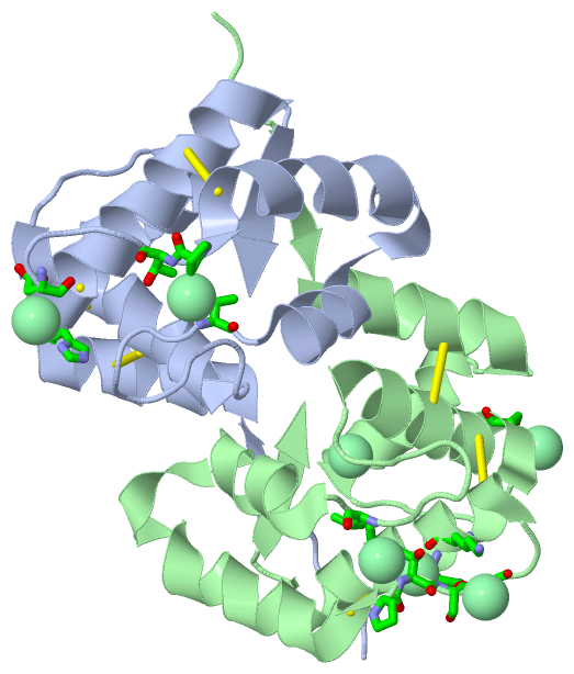
(-) Ligands, Modified Residues, Ions  (1, 7)

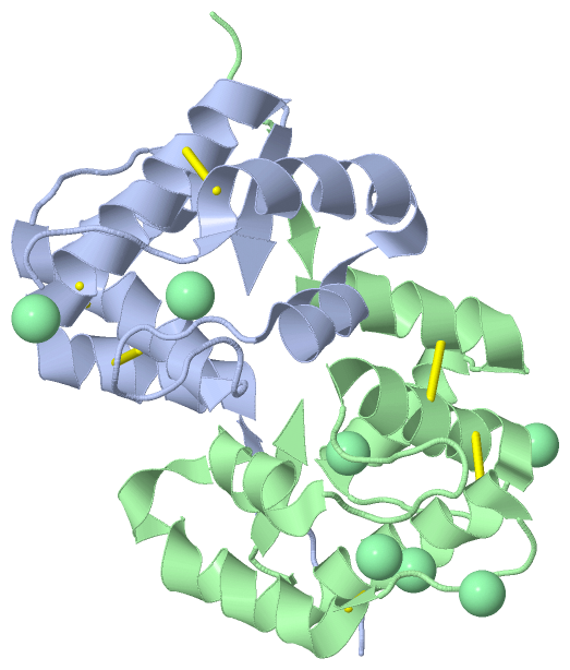
Asymmetric/Biological Unit (1, 7)
No.NameCountTypeFull Name
1CL7Ligand/IonCHLORIDE ION

(-) Sites  (6, 6)

Asymmetric Unit (6, 6)
No.NameEvidenceResiduesDescription
1AC1SOFTWAREVAL A:65 , PRO A:91 , THR A:92BINDING SITE FOR RESIDUE CL A 120
2AC2SOFTWAREASP A:96BINDING SITE FOR RESIDUE CL A 121
3AC3SOFTWAREHIS A:24 , SER B:93 , GLY B:94BINDING SITE FOR RESIDUE CL B 120
4AC4SOFTWAREVAL B:65 , PRO B:91 , THR B:92BINDING SITE FOR RESIDUE CL B 121
5AC5SOFTWARESER B:44BINDING SITE FOR RESIDUE CL B 122
6AC6SOFTWARETHR B:92 , SER B:93 , LYS B:100 , ASN B:103 , LEU B:104BINDING SITE FOR RESIDUE CL B 123

(-) SS Bonds  (6, 6)

Asymmetric/Biological Unit
No.Residues
1A:20 -A:51
2A:47 -A:98
3A:89 -A:107
4B:20 -B:51
5B:47 -B:98
6B:89 -B:107

(-) Cis Peptide Bonds  (2, 2)

Asymmetric/Biological Unit
No.Residues
1Leu A:90 -Pro A:91
2Leu B:90 -Pro B:91

 Sequence-Structure Mapping

(-) SAPs(SNPs)/Variants  (0, 0)

(no "SAP(SNP)/Variant" information available for 3CZ2)

(-) PROSITE Motifs  (0, 0)

(no "PROSITE Motif" information available for 3CZ2)

(-) Exons   (0, 0)

(no "Exon" information available for 3CZ2)

(-) Sequences/Alignments

Asymmetric/Biological Unit
   Reformat: Number of residues per line =  ('0' or empty: single-line sequence representation)
  Number of residues per labelling interval =   
  UniProt sequence: complete  aligned part    
   Show mapping: SCOP domains CATH domains Pfam domains Secondary structure (by author)
SAPs(SNPs) PROSITE motifs Exons
(details for a mapped element are shown in a popup box when the mouse pointer rests over it)
Chain A from PDB  Type:PROTEIN  Length:118
 aligned with Q9U9J6_APIME | Q9U9J6 from UniProtKB/TrEMBL  Length:144

    Alignment length:118
                                    36        46        56        66        76        86        96       106       116       126       136        
         Q9U9J6_APIME    27 PDWVPPEVFDLVAEDKARCMSEHGTTQAQIDDVDKGNLVNEPSITCYMYCLLEAFSLVDDEANVDEDIMLGLLPDQLQERAQSVMGKCLPTSGSDNCNKIYNLAKCVQESAPDVWFVI 144
               SCOP domains d3cz2a_ A: Pheromone-binding protein asp1                                                                              SCOP domains
               CATH domains 3cz2A00 A:2-119  [code=1.10.238.20, no name defined]                                                                   CATH domains
               Pfam domains ---------------------------------------------------------------------------------------------------------------------- Pfam domains
         Sec.struct. author ......eeeehhhhhhhhhhhhhh.hhhhhhhhhhh....hhhhhhhhhhhhhh..........hhhhhhhhhhhhhhhhhhhhhhhhh.....hhhhhhhhhhhhhhhhh..eeee. Sec.struct. author
                 SAPs(SNPs) ---------------------------------------------------------------------------------------------------------------------- SAPs(SNPs)
                    PROSITE ---------------------------------------------------------------------------------------------------------------------- PROSITE
                 Transcript ---------------------------------------------------------------------------------------------------------------------- Transcript
                 3cz2 A   2 PDWVPPEVFDLVAEDKARCMSEHGTTQAQIDDVDKGNLVNEPSITCYMYCLLEAFSLVDDEANVDEDIMLGLLPDQLQERAQSVMGKCLPTSGSDNCNKIYNLAKCVQESAPDVWFVI 119
                                    11        21        31        41        51        61        71        81        91       101       111        

Chain B from PDB  Type:PROTEIN  Length:116
 aligned with Q9U9J6_APIME | Q9U9J6 from UniProtKB/TrEMBL  Length:144

    Alignment length:116
                                    38        48        58        68        78        88        98       108       118       128       138      
         Q9U9J6_APIME    29 WVPPEVFDLVAEDKARCMSEHGTTQAQIDDVDKGNLVNEPSITCYMYCLLEAFSLVDDEANVDEDIMLGLLPDQLQERAQSVMGKCLPTSGSDNCNKIYNLAKCVQESAPDVWFVI 144
               SCOP domains d3cz2b_ B: Pheromone-binding protein asp1                                                                            SCOP domains
               CATH domains 3cz2B00 B:4-119  [code=1.10.238.20, no name defined]                                                                 CATH domains
               Pfam domains -------------------------------------------------------------------------------------------------------------------- Pfam domains
         Sec.struct. author ....eeeehhhhhhhhhhhhhh.hhhhhhhhhh.....hhhhhhhhhhhhhhh.........hhhhhhhhhhhhhhhhhhhhhhhhh.....hhhhhhhhhhhhhhhhh..eeee. Sec.struct. author
                 SAPs(SNPs) -------------------------------------------------------------------------------------------------------------------- SAPs(SNPs)
                    PROSITE -------------------------------------------------------------------------------------------------------------------- PROSITE
                 Transcript -------------------------------------------------------------------------------------------------------------------- Transcript
                 3cz2 B   4 WVPPEVFDLVAEDKARCMSEHGTTQAQIDDVDKGNLVNEPSITCYMYCLLEAFSLVDDEANVDEDIMLGLLPDQLQERAQSVMGKCLPTSGSDNCNKIYNLAKCVQESAPDVWFVI 119
                                    13        23        33        43        53        63        73        83        93       103       113      

   Legend:   → Mismatch (orange background)
  - → Gap (green background, '-', border residues have a numbering label)
    → Modified Residue (blue background, lower-case, 'x' indicates undefined single-letter code, labelled with number + name)
  x → Chemical Group (purple background, 'x', labelled with number + name, e.g. ACE or NH2)
  extra numbering lines below/above indicate numbering irregularities and modified residue names etc., number ends below/above '|'

 Classification and Annotation

(-) SCOP Domains  (1, 2)

Asymmetric/Biological Unit

(-) CATH Domains  (1, 2)

Asymmetric/Biological Unit

(-) Pfam Domains  (0, 0)

(no "Pfam Domain" information available for 3CZ2)

(-) Gene Ontology  (1, 1)

Asymmetric/Biological Unit(hide GO term definitions)
Chain A,B   (Q9U9J6_APIME | Q9U9J6)
molecular function
    GO:0005549    odorant binding    Interacting selectively and non-covalently with an odorant, any substance capable of stimulating the sense of smell.

 Visualization

(-) Interactive Views

Asymmetric/Biological Unit
  Complete Structure
    Jena3D(integrated viewing of ligand, site, SAP, PROSITE, SCOP information)
    WebMol | AstexViewer[tm]@PDBe
(Java Applets, require no local installation except for Java; loading may be slow)
    STRAP
(Java WebStart application, automatic local installation, requires Java; full application with system access!)
    RasMol
(require local installation)
    Molscript (VRML)
(requires installation of a VRML viewer; select preferred view via VRML and generate a mono or stereo PDF format file)
 
  Ligands, Modified Residues, Ions
    CL  [ RasMol | Jena3D ]  +environment [ RasMol | Jena3D ]
 
  Sites
    AC1  [ RasMol ]  +environment [ RasMol ]
    AC2  [ RasMol ]  +environment [ RasMol ]
    AC3  [ RasMol ]  +environment [ RasMol ]
    AC4  [ RasMol ]  +environment [ RasMol ]
    AC5  [ RasMol ]  +environment [ RasMol ]
    AC6  [ RasMol ]  +environment [ RasMol ]
 
  Cis Peptide Bonds
    Leu A:90 - Pro A:91   [ RasMol ]  
    Leu B:90 - Pro B:91   [ RasMol ]  
 

(-) Still Images

Jmol
  protein: cartoon or spacefill or dots and stick; nucleic acid: cartoon and stick; ligands: spacefill; active site: stick
Molscript
  protein, nucleic acid: cartoon; ligands: spacefill; active site: ball and stick

 Databases and Analysis Tools

(-) Databases

Access by PDB/NDB ID
  3cz2
    Family and Domain InformationProDom | SYSTERS
    General Structural InformationGlycoscienceDB | MMDB | NDB | OCA | PDB | PDBe | PDBj | PDBsum | PDBWiki | PQS | PROTEOPEDIA
    Orientation in MembranesOPM
    Protein SurfaceSURFACE
    Secondary StructureDSSP (structure derived) | HSSP (homology derived)
    Structural GenomicsGeneCensus
    Structural NeighboursCE | VAST
    Structure ClassificationCATH | Dali | SCOP
    Validation and Original DataBMRB Data View | BMRB Restraints Grid | EDS | PROCHECK | RECOORD | WHAT_CHECK
 
Access by UniProt ID/Accession number
  Q9U9J6_APIME | Q9U9J6
    Comparative Protein Structure ModelsModBase
    Genomic InformationEnsembl
    Protein-protein InteractionDIP
    Sequence, Family and Domain InformationInterPro | Pfam | SMART | UniProtKB/TrEMBL
 
Access by Enzyme Classificator   (EC Number)
  (no 'Enzyme Classificator' available)
    General Enzyme InformationBRENDA | EC-PDB | Enzyme | IntEnz
    PathwayKEGG | MetaCyc
 
Access by Disease Identifier   (MIM ID)
  (no 'MIM ID' available)
    Disease InformationOMIM
 
Access by GenAge ID
  (no 'GenAge ID' available)
    Age Related InformationGenAge

(-) Analysis Tools

Access by PDB/NDB ID
    Domain InformationXDom
    Interatomic Contacts of Structural UnitsCSU
    Ligand-protein ContactsLPC
    Protein CavitiescastP
    Sequence and Secondary StructurePDBCartoon
    Structure AlignmentSTRAP(Java WebStart application, automatic local installation, requires Java; full application with system access!)
    Structure and Sequence BrowserSTING
 
Access by UniProt ID/Accession number
  Q9U9J6_APIME | Q9U9J6
    Protein Disorder PredictionDisEMBL | FoldIndex | GLOBPLOT (for more information see DisProt)

 Related Entries

(-) Entries Sharing at Least One Protein Chain (UniProt ID)

UniProtKB/Swiss-Prot
        Q9U9J6_APIME | Q9U9J62h8v 3bfa 3bjh 3cab 3cdn
UniProtKB/TrEMBL
        Q9U9J6_APIME | Q9U9J63bfb 3bfh 3cyz 3cz0 3cz1 3d73 3d74 3d75 3d76 3d77 3d78 3fe6 3fe8 3fe9

(-) Related Entries Specified in the PDB File

2h8v THE APO FORM OF THE SAME PROTEIN AT PH 5.5
3bfa THE SAME PROTEIN IN COMPLEX WITH THE QUEEN MANDIBULAR PHEROMONE AT PH 5.5
3bfb THE SAME PROTEIN IN COMPLEX WITH THE 9-KETO-2(E)-DECENOIC ACID AT PH 5.5
3bfh THE SAME PROTEIN IN COMPLEX WITH THE HEXADECANOIC ACID AT PH 5.5
3bjh THE SAME PROTEIN IN COMPLEX WITH THE N-BUTYL BENZENE SULFONAMIDE AT PH 5.5
3cab THE SAME PROTEIN IN COMPLEX WITH THE N-BUTYL BENZENE SULFONAMIDE SOAKED AT PH 7.0
3cdn THE APO FORM OF THE SAME PROTEIN SOAKED AT PH 4.0
3cyz THE SAME PROTEIN IN COMPLEX WITH THE 9-KETO-2(E)-DECENOIC ACID AT PH 7.0
3cz0 THE SAME PROTEIN IN COMPLEX WITH THE QUEEN MANDIBULAR PHEROMONE AT PH 7.0
3cz1 THE SAME PROTEIN IN COMPLEX WITH THE N BUTYL BENZENE SULFONAMIDE AT PH 7.0