Show PDB file:   
         Plain Text   HTML   (compressed file size)
QuickSearch:   
by PDB,NDB,UniProt,PROSITE Code or Search Term(s)  
(-)Asymmetric Unit
(-)Asym. Unit - sites
(-)Biological Unit 1
(-)Biol. Unit 1 - sites
(-)Biological Unit 2
collapse expand < >
Image Asymmetric Unit
Asymmetric Unit  (Jmol Viewer)
Image Asym. Unit - sites
Asym. Unit - sites  (Jmol Viewer)
Image Biological Unit 1
Biological Unit 1  (Jmol Viewer)
Image Biol. Unit 1 - sites
Biol. Unit 1 - sites  (Jmol Viewer)
Image Biological Unit 2
Biological Unit 2  (Jmol Viewer)

(-) Description

Title :  CRYSTAL STRUCTURE OF A PHEROMONE BINDING PROTEIN FROM APIS MELLIFERA IN COMPLEX WITH THE 9-KETO-2(E)-DECENOIC ACID
 
Authors :  M. E. Pesenti, S. Spinelli, V. Bezirard, L. Briand, J. C. Pernollet, M. T C. Cambillau
Date :  21 Nov 07  (Deposition) - 10 Jun 08  (Release) - 13 Jul 11  (Revision)
Method :  X-RAY DIFFRACTION
Resolution :  2.15
Chains :  Asym. Unit :  A
Biol. Unit 1:  A  (1x)
Biol. Unit 2:  A  (2x)
Keywords :  Honeybee, Apis Mellifera, Pheromone Binding Protein, Signal Transduction, Queen Mandibular Pheromone, Pheromone-Binding Protein (Keyword Search: [Gene Ontology, PubMed, Web (Google))
 
Reference :  M. E. Pesenti, S. Spinelli, V. Bezirard, L. Briand, J. C. Pernollet, M. Tegoni, C. Cambillau
Structural Basis Of The Honey Bee Pbp Pheromone And Ph-Induced Conformational Change
J. Mol. Biol. V. 380 158 2008
PubMed-ID: 18508083  |  Reference-DOI: 10.1016/J.JMB.2008.04.048

(-) Compounds

Molecule 1 - PHEROMONE-BINDING PROTEIN ASP1
    ChainsA
    EngineeredYES
    Expression SystemPICHIA PASTORIS
    Expression System PlasmidPHIL-D2
    Expression System Vector TypePLASMID
    FragmentUNP RESIDUES 26-144
    Organism CommonHONEYBEE
    Organism ScientificAPIS MELLIFERA

 Structural Features

(-) Chains, Units

  1
Asymmetric Unit A
Biological Unit 1 (1x)A
Biological Unit 2 (2x)A

Summary Information (see also Sequences/Alignments below)

(-) Ligands, Modified Residues, Ions  (3, 4)

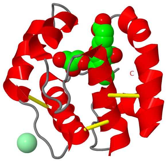
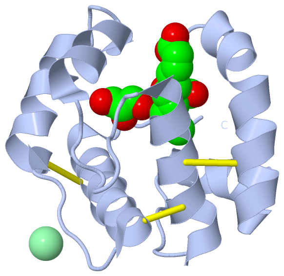
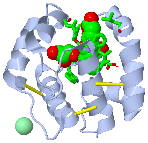
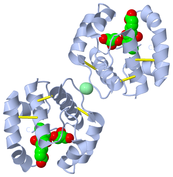
Asymmetric Unit (3, 4)
No.NameCountTypeFull Name
19OD1Ligand/Ion(2Z)-9-OXODEC-2-ENOIC ACID
2CL1Ligand/IonCHLORIDE ION
3GOL2Ligand/IonGLYCEROL
Biological Unit 1 (2, 3)
No.NameCountTypeFull Name
19OD1Ligand/Ion(2Z)-9-OXODEC-2-ENOIC ACID
2CL-1Ligand/IonCHLORIDE ION
3GOL2Ligand/IonGLYCEROL
Biological Unit 2 (2, 6)
No.NameCountTypeFull Name
19OD2Ligand/Ion(2Z)-9-OXODEC-2-ENOIC ACID
2CL-1Ligand/IonCHLORIDE ION
3GOL4Ligand/IonGLYCEROL

(-) Sites  (3, 3)

Asymmetric Unit (3, 3)
No.NameEvidenceResiduesDescription
1AC1SOFTWARELEU A:12 , LEU A:52 , PHE A:56 , LEU A:73 , ILE A:119BINDING SITE FOR RESIDUE 9OD A 121
2AC2SOFTWARETRP A:4 , TRP A:116 , PHE A:117BINDING SITE FOR RESIDUE GOL A 122
3AC3SOFTWAREASP A:3 , TRP A:4 , PHE A:117BINDING SITE FOR RESIDUE GOL A 123

(-) SS Bonds  (3, 3)

Asymmetric Unit
No.Residues
1A:20 -A:51
2A:47 -A:98
3A:89 -A:107

(-) Cis Peptide Bonds  (2, 2)

Asymmetric Unit
No.Residues
1Asp A:3 -Trp A:4
2Leu A:90 -Pro A:91

 Sequence-Structure Mapping

(-) SAPs(SNPs)/Variants  (0, 0)

(no "SAP(SNP)/Variant" information available for 3BFB)

(-) PROSITE Motifs  (0, 0)

(no "PROSITE Motif" information available for 3BFB)

(-) Exons   (0, 0)

(no "Exon" information available for 3BFB)

(-) Sequences/Alignments

Asymmetric Unit
   Reformat: Number of residues per line =  ('0' or empty: single-line sequence representation)
  Number of residues per labelling interval =   
  UniProt sequence: complete  aligned part    
   Show mapping: SCOP domains CATH domains Pfam domains Secondary structure (by author)
SAPs(SNPs) PROSITE motifs Exons
(details for a mapped element are shown in a popup box when the mouse pointer rests over it)
Chain A from PDB  Type:PROTEIN  Length:117
 aligned with Q9U9J6_APIME | Q9U9J6 from UniProtKB/TrEMBL  Length:144

    Alignment length:117
                                    37        47        57        67        77        87        97       107       117       127       137       
         Q9U9J6_APIME    28 DWVPPEVFDLVAEDKARCMSEHGTTQAQIDDVDKGNLVNEPSITCYMYCLLEAFSLVDDEANVDEDIMLGLLPDQLQERAQSVMGKCLPTSGSDNCNKIYNLAKCVQESAPDVWFVI 144
               SCOP domains d3bfba_ A: Pheromone-binding protein asp1                                                                             SCOP domains
               CATH domains 3bfbA00 A:3-119  [code=1.10.238.20, no name defined]                                                                  CATH domains
               Pfam domains --------------------------------------------------------------------------------------------------------------------- Pfam domains
         Sec.struct. author ....hhhhhhhhhhhhhhhhhhh.hhhhhhhhhh.....hhhhhhhhhhhhhhh.........hhhhhhhhh...hhhhhhhhhhhhh.....hhhhhhhhhhhhhhhhh....... Sec.struct. author
                 SAPs(SNPs) --------------------------------------------------------------------------------------------------------------------- SAPs(SNPs)
                    PROSITE --------------------------------------------------------------------------------------------------------------------- PROSITE
                 Transcript --------------------------------------------------------------------------------------------------------------------- Transcript
                 3bfb A   3 DWVPPEVFDLVAEDKARCMSEHGTTQAQIDDVDKGNLVNEPSITCYMYCLLEAFSLVDDEANVDEDIMLGLLPDQLQERAQSVMGKCLPTSGSDNCNKIYNLAKCVQESAPDVWFVI 119
                                    12        22        32        42        52        62        72        82        92       102       112       

   Legend:   → Mismatch (orange background)
  - → Gap (green background, '-', border residues have a numbering label)
    → Modified Residue (blue background, lower-case, 'x' indicates undefined single-letter code, labelled with number + name)
  x → Chemical Group (purple background, 'x', labelled with number + name, e.g. ACE or NH2)
  extra numbering lines below/above indicate numbering irregularities and modified residue names etc., number ends below/above '|'

 Classification and Annotation

(-) SCOP Domains  (1, 1)

Asymmetric Unit

(-) CATH Domains  (1, 1)

Asymmetric Unit

(-) Pfam Domains  (0, 0)

(no "Pfam Domain" information available for 3BFB)

(-) Gene Ontology  (1, 1)

Asymmetric Unit(hide GO term definitions)
Chain A   (Q9U9J6_APIME | Q9U9J6)
molecular function
    GO:0005549    odorant binding    Interacting selectively and non-covalently with an odorant, any substance capable of stimulating the sense of smell.

 Visualization

(-) Interactive Views

Asymmetric Unit
  Complete Structure
    Jena3D(integrated viewing of ligand, site, SAP, PROSITE, SCOP information)
    WebMol | AstexViewer[tm]@PDBe
(Java Applets, require no local installation except for Java; loading may be slow)
    STRAP
(Java WebStart application, automatic local installation, requires Java; full application with system access!)
    RasMol
(require local installation)
    Molscript (VRML)
(requires installation of a VRML viewer; select preferred view via VRML and generate a mono or stereo PDF format file)
 
  Ligands, Modified Residues, Ions
    9OD  [ RasMol | Jena3D ]  +environment [ RasMol | Jena3D ]
    CL  [ RasMol | Jena3D ]  +environment [ RasMol | Jena3D ]
    GOL  [ RasMol | Jena3D ]  +environment [ RasMol | Jena3D ]
 
  Sites
    AC1  [ RasMol ]  +environment [ RasMol ]
    AC2  [ RasMol ]  +environment [ RasMol ]
    AC3  [ RasMol ]  +environment [ RasMol ]
 
  Cis Peptide Bonds
    Asp A:3 - Trp A:4   [ RasMol ]  
    Leu A:90 - Pro A:91   [ RasMol ]  
 
Biological Units
  Complete Structure
    Biological Unit 1  [ Jena3D ]
    Biological Unit 2  [ Jena3D ]

(-) Still Images

Jmol
  protein: cartoon or spacefill or dots and stick; nucleic acid: cartoon and stick; ligands: spacefill; active site: stick
Molscript
  protein, nucleic acid: cartoon; ligands: spacefill; active site: ball and stick

 Databases and Analysis Tools

(-) Databases

Access by PDB/NDB ID
  3bfb
    Family and Domain InformationProDom | SYSTERS
    General Structural InformationGlycoscienceDB | MMDB | NDB | OCA | PDB | PDBe | PDBj | PDBsum | PDBWiki | PQS | PROTEOPEDIA
    Orientation in MembranesOPM
    Protein SurfaceSURFACE
    Secondary StructureDSSP (structure derived) | HSSP (homology derived)
    Structural GenomicsGeneCensus
    Structural NeighboursCE | VAST
    Structure ClassificationCATH | Dali | SCOP
    Validation and Original DataBMRB Data View | BMRB Restraints Grid | EDS | PROCHECK | RECOORD | WHAT_CHECK
 
Access by UniProt ID/Accession number
  Q9U9J6_APIME | Q9U9J6
    Comparative Protein Structure ModelsModBase
    Genomic InformationEnsembl
    Protein-protein InteractionDIP
    Sequence, Family and Domain InformationInterPro | Pfam | SMART | UniProtKB/TrEMBL
 
Access by Enzyme Classificator   (EC Number)
  (no 'Enzyme Classificator' available)
    General Enzyme InformationBRENDA | EC-PDB | Enzyme | IntEnz
    PathwayKEGG | MetaCyc
 
Access by Disease Identifier   (MIM ID)
  (no 'MIM ID' available)
    Disease InformationOMIM
 
Access by GenAge ID
  (no 'GenAge ID' available)
    Age Related InformationGenAge

(-) Analysis Tools

Access by PDB/NDB ID
    Domain InformationXDom
    Interatomic Contacts of Structural UnitsCSU
    Ligand-protein ContactsLPC
    Protein CavitiescastP
    Sequence and Secondary StructurePDBCartoon
    Structure AlignmentSTRAP(Java WebStart application, automatic local installation, requires Java; full application with system access!)
    Structure and Sequence BrowserSTING
 
Access by UniProt ID/Accession number
  Q9U9J6_APIME | Q9U9J6
    Protein Disorder PredictionDisEMBL | FoldIndex | GLOBPLOT (for more information see DisProt)

 Related Entries

(-) Entries Sharing at Least One Protein Chain (UniProt ID)

UniProtKB/Swiss-Prot
        Q9U9J6_APIME | Q9U9J62h8v 3bfa 3bjh 3cab 3cdn
UniProtKB/TrEMBL
        Q9U9J6_APIME | Q9U9J63bfh 3cyz 3cz0 3cz1 3cz2 3d73 3d74 3d75 3d76 3d77 3d78 3fe6 3fe8 3fe9

(-) Related Entries Specified in the PDB File

1r5r THE SAME PROTEIN COMPLEXED WITH N-BUTYL BENZENESULFANAMIDE
2h8v UNCOMPLEXED FORM OF THE SAME PROTEIN
3bfa THE SAME PROTEIN COMPLEXED WITH THE QUEEN MANDIBULAR PHEROMONE
3bfh THE SAME PROEIN COMPLEXED WITH HEXADECANOIC ACID