|
|
|
|
 Description
|
|
Compounds
|
||||||||||||||||||||||||||||||||||||||||||||||||||||||||||||||||||||||||||||||||||||||||||||||
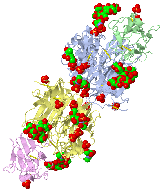
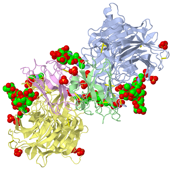
Chains, Units
Summary Information (see also Sequences/Alignments below) |
Ligands, Modified Residues, Ions (12, 52)
Asymmetric Unit (12, 52)
|
Sites (32, 32)
Asymmetric Unit (32, 32)
|
SS Bonds (18, 18)
Asymmetric Unit
|
||||||||||||||||||||||||||||||||||||||||||||||||||||||||||||||||||||||||||||
Cis Peptide Bonds (0, 0)| (no "Cis Peptide Bond" information available for 3D12) |
SAPs(SNPs)/Variants (8, 16)
Asymmetric Unit (8, 16)
|
||||||||||||||||||||||||||||||||||||||||||||||||||||||||||||||||||||||||||||||||||||||||||||||||||||||||||||||||||||||||||||||||||||||||||||||||||||||||||||||||||||||||||||||||||||||||||||||||||||||||||||||||||||||||||||||||||||||||||||||||||||||||||||||||||||||||||||||||||||||||||||||||||||||||||||||||||||||||||||||||||||||||||||||||||||||||||||||||||||||||||||||||||||||||||||||||||||||||||||||||||||||||||||||||||||||||||||||||||||||||||||||||||||||||||||||||||||||||||||||||||||||||||||||||||||||||||||||||||||||||||||||||||||||||||||||||||||||||||||||||||||||||||||||||||||||||||||||||||||
PROSITE Motifs (1, 2)
Asymmetric Unit (1, 2)
|
||||||||||||||||||||||||||||||||||||||||||||||||||||||||||||||||||||||||||||||||||||||||||||||||
Exons (0, 0)| (no "Exon" information available for 3D12) |
Sequences/Alignments
Asymmetric UnitChain A from PDB Type:PROTEIN Length:428 aligned with GLYCP_NIPAV | Q9IH62 from UniProtKB/Swiss-Prot Length:602 Alignment length:428 602 185 195 205 215 225 235 245 255 265 275 285 295 305 315 325 335 345 355 365 375 385 395 405 415 425 435 445 455 465 475 485 495 505 515 525 535 545 555 565 575 585 595 | GLYCP_NIPAV 176 EGVSNLVGLPNNICLQKTSNQILKPKLISYTLPVVGQSGTCITDPLLAMDEGYFAYSHLERIGSCSRGVSKQRIIGVGEVLDRGDEVPSLFMTNVWTPPNPNTVYHCSAVYNNEFYYVLCAVSTVGDPILNSTYWSGSLMMTRLAVKPKSNGGGYNQHQLALRSIEKGRYDKVMPYGPSGIKQGDTLYFPAVGFLVRTEFKYNDSNCPITKCQYSKPENCRLSMGIRPNSHYILRSGLLKYNLSDGENPKVVFIEISDQRLSIGSPSKIYDSLGQPVFYQASFSWDTMIKFGDVLTVNPLVVNWRNNTVISRPGQSQCPRFNTCPEICWEGVYNDAFLIDRINWISAGVFLDSNQTAENPVFTVFKDNEILYRAQLASEDTNAQKTITNCFLLKNKIWCISLVEIYDTGDNVIRPKLFAVKIPEQCT- - SCOP domains -------------------------------------------------------------------------------------------------------------------------------------------------------------------------------------------------------------------------------------------------------------------------------------------------------------------------------------------------------------------------------------------------------------------------------------------- SCOP domains CATH domains -------------------------------------------------------------------------------------------------------------------------------------------------------------------------------------------------------------------------------------------------------------------------------------------------------------------------------------------------------------------------------------------------------------------------------------------- CATH domains Pfam domains -------------------------------------------------------------------------------------------------------------------------------------------------------------------------------------------------------------------------------------------------------------------------------------------------------------------------------------------------------------------------------------------------------------------------------------------- Pfam domains SAPs(SNPs) ------------------------------------------------------------------------K-----------------------A------------------------------------------------------D--------------------------------------------------------------------------------V-----------------I-------------------------------------------Q-------S--D-------------------------------------------------------------------------------------------------------------------------- SAPs(SNPs) PROSITE -------------------------------------------------------------------------------------------------------------------------------------------------------------------------------------------------------------------------------------------------------------------------------------------------------------------------------------------------------------------------------------------------------------------------------------------- PROSITE Transcript -------------------------------------------------------------------------------------------------------------------------------------------------------------------------------------------------------------------------------------------------------------------------------------------------------------------------------------------------------------------------------------------------------------------------------------------- Transcript 3d12 A 176 EGVSNLVGLPNNICLQKTSNQILKPKLISYTLPVVGQSGTCITDPLLAMDEGYFAYSHLERIGSCSRGVSKQRIIGVGEVLDRGDEVPSLFMTNVWTPPNPNTVYHCSAVYNNEFYYVLCAVSTVGDPILNSTYWSGSLMMTRLAVKPKSNGGGYNQHQLALRSIEKGRYDKVMPYGPSGIKQGDTLYFPAVGFLVRTEFKYNDSNCPITKCQYSKPENCRLSMGIRPNSHYILRSGLLKYNLSDGENPKVVFIEISDQRLSIGSPSKIYDSLGQPVFYQASFSWDTMIKFGDVLTVNPLVVNWRNNTVISRPGQSQCPRFNTCPEICWEGVYNDAFLIDRINWISAGVFLDSNQTAENPVFTVFKDNEILYRAQLASEDTNAQKTITNCFLLKNKIWCISLVEIYDTGDNVIRPKLFAVKIPEQCTA 603 185 195 205 215 225 235 245 255 265 275 285 295 305 315 325 335 345 355 365 375 385 395 405 415 425 435 445 455 465 475 485 495 505 515 525 535 545 555 565 575 585 595 Chain B from PDB Type:PROTEIN Length:141 aligned with EFNB3_MOUSE | O35393 from UniProtKB/Swiss-Prot Length:340 Alignment length:141 38 48 58 68 78 88 98 108 118 128 138 148 158 168 EFNB3_MOUSE 29 SLEPVYWNSANKRFQAEGGYVLYPQIGDRLDLLCPRARPPGPHSSPSYEFYKLYLVEGAQGRRCEAPPAPNLLLTCDRPDLDLRFTIKFQEYSPNLWGHEFRSHHDYYIIATSDGTREGLESLQGGVCLTRGMKVLLRVGQ 169 SCOP domains d3d12b_ B: automated matches SCOP domains CATH domains --------------------------------------------------------------------------------------------------------------------------------------------- CATH domains Pfam domains --------------------------------------------------------------------------------------------------------------------------------------------- Pfam domains SAPs(SNPs) --------------------------------------------------------------------------------------------------------------------------------------------- SAPs(SNPs) PROSITE -----------------------------------------------------------------------------------EPHRIN_RBD_1 PDB: B:112-139------------------------------ PROSITE Transcript --------------------------------------------------------------------------------------------------------------------------------------------- Transcript 3d12 B 29 SLEPVYWNSANKRFQAEGGYVLYPQIGDRLDLLCPRARPPGPHSSPSYEFYKLYLVEGAQGRRCEAPPAPNLLLTCDRPDLDLRFTIKFQEYSPNLWGHEFRSHHDYYIIATSDGTREGLESLQGGVCLTRGMKVLLRVGQ 169 38 48 58 68 78 88 98 108 118 128 138 148 158 168 Chain D from PDB Type:PROTEIN Length:428 aligned with GLYCP_NIPAV | Q9IH62 from UniProtKB/Swiss-Prot Length:602 Alignment length:428 602 185 195 205 215 225 235 245 255 265 275 285 295 305 315 325 335 345 355 365 375 385 395 405 415 425 435 445 455 465 475 485 495 505 515 525 535 545 555 565 575 585 595 | GLYCP_NIPAV 176 EGVSNLVGLPNNICLQKTSNQILKPKLISYTLPVVGQSGTCITDPLLAMDEGYFAYSHLERIGSCSRGVSKQRIIGVGEVLDRGDEVPSLFMTNVWTPPNPNTVYHCSAVYNNEFYYVLCAVSTVGDPILNSTYWSGSLMMTRLAVKPKSNGGGYNQHQLALRSIEKGRYDKVMPYGPSGIKQGDTLYFPAVGFLVRTEFKYNDSNCPITKCQYSKPENCRLSMGIRPNSHYILRSGLLKYNLSDGENPKVVFIEISDQRLSIGSPSKIYDSLGQPVFYQASFSWDTMIKFGDVLTVNPLVVNWRNNTVISRPGQSQCPRFNTCPEICWEGVYNDAFLIDRINWISAGVFLDSNQTAENPVFTVFKDNEILYRAQLASEDTNAQKTITNCFLLKNKIWCISLVEIYDTGDNVIRPKLFAVKIPEQCT- - SCOP domains -------------------------------------------------------------------------------------------------------------------------------------------------------------------------------------------------------------------------------------------------------------------------------------------------------------------------------------------------------------------------------------------------------------------------------------------- SCOP domains CATH domains -------------------------------------------------------------------------------------------------------------------------------------------------------------------------------------------------------------------------------------------------------------------------------------------------------------------------------------------------------------------------------------------------------------------------------------------- CATH domains Pfam domains -------------------------------------------------------------------------------------------------------------------------------------------------------------------------------------------------------------------------------------------------------------------------------------------------------------------------------------------------------------------------------------------------------------------------------------------- Pfam domains SAPs(SNPs) ------------------------------------------------------------------------K-----------------------A------------------------------------------------------D--------------------------------------------------------------------------------V-----------------I-------------------------------------------Q-------S--D-------------------------------------------------------------------------------------------------------------------------- SAPs(SNPs) PROSITE -------------------------------------------------------------------------------------------------------------------------------------------------------------------------------------------------------------------------------------------------------------------------------------------------------------------------------------------------------------------------------------------------------------------------------------------- PROSITE Transcript -------------------------------------------------------------------------------------------------------------------------------------------------------------------------------------------------------------------------------------------------------------------------------------------------------------------------------------------------------------------------------------------------------------------------------------------- Transcript 3d12 D 176 EGVSNLVGLPNNICLQKTSNQILKPKLISYTLPVVGQSGTCITDPLLAMDEGYFAYSHLERIGSCSRGVSKQRIIGVGEVLDRGDEVPSLFMTNVWTPPNPNTVYHCSAVYNNEFYYVLCAVSTVGDPILNSTYWSGSLMMTRLAVKPKSNGGGYNQHQLALRSIEKGRYDKVMPYGPSGIKQGDTLYFPAVGFLVRTEFKYNDSNCPITKCQYSKPENCRLSMGIRPNSHYILRSGLLKYNLSDGENPKVVFIEISDQRLSIGSPSKIYDSLGQPVFYQASFSWDTMIKFGDVLTVNPLVVNWRNNTVISRPGQSQCPRFNTCPEICWEGVYNDAFLIDRINWISAGVFLDSNQTAENPVFTVFKDNEILYRAQLASEDTNAQKTITNCFLLKNKIWCISLVEIYDTGDNVIRPKLFAVKIPEQCTA 603 185 195 205 215 225 235 245 255 265 275 285 295 305 315 325 335 345 355 365 375 385 395 405 415 425 435 445 455 465 475 485 495 505 515 525 535 545 555 565 575 585 595 Chain E from PDB Type:PROTEIN Length:141 aligned with EFNB3_MOUSE | O35393 from UniProtKB/Swiss-Prot Length:340 Alignment length:141 38 48 58 68 78 88 98 108 118 128 138 148 158 168 EFNB3_MOUSE 29 SLEPVYWNSANKRFQAEGGYVLYPQIGDRLDLLCPRARPPGPHSSPSYEFYKLYLVEGAQGRRCEAPPAPNLLLTCDRPDLDLRFTIKFQEYSPNLWGHEFRSHHDYYIIATSDGTREGLESLQGGVCLTRGMKVLLRVGQ 169 SCOP domains d3d12e_ E: automated matches SCOP domains CATH domains --------------------------------------------------------------------------------------------------------------------------------------------- CATH domains Pfam domains --------------------------------------------------------------------------------------------------------------------------------------------- Pfam domains SAPs(SNPs) --------------------------------------------------------------------------------------------------------------------------------------------- SAPs(SNPs) PROSITE -----------------------------------------------------------------------------------EPHRIN_RBD_1 PDB: E:112-139------------------------------ PROSITE Transcript --------------------------------------------------------------------------------------------------------------------------------------------- Transcript 3d12 E 29 SLEPVYWNSANKRFQAEGGYVLYPQIGDRLDLLCPRARPPGPHSSPSYEFYKLYLVEGAQGRRCEAPPAPNLLLTCDRPDLDLRFTIKFQEYSPNLWGHEFRSHHDYYIIATSDGTREGLESLQGGVCLTRGMKVLLRVGQ 169 38 48 58 68 78 88 98 108 118 128 138 148 158 168
|
||||||||||||||||||||
SCOP Domains (1, 2)
Asymmetric Unit
 |
CATH Domains (0, 0)| (no "CATH Domain" information available for 3D12) |
Pfam Domains (0, 0)| (no "Pfam Domain" information available for 3D12) |
Gene Ontology (25, 27)|
Asymmetric Unit(hide GO term definitions) Chain A,D (GLYCP_NIPAV | Q9IH62)
Chain B,E (EFNB3_MOUSE | O35393)
|
||||||||||||||||||||||||||||||||||||||||||||||||||||||||||||||||||||||||||||||||||||||||||||||||||||||||||||||||||||||||||||||||||||||||||||||||||||||||||||||||||||||||||||||||||||||||||||||||||||||
Interactive Views
|
||||||||||||||||||||||||||||||||||||||||||||||||||||||||||||||||||||||||||||||||||||||||||||||||||||||||||||||||||||||||||||||||||||||||||||||||||||||||||||||||||||||||||||||||||||||||||||||||||||||||||||||||||||||||||||||||||||||||||||||||||||||||||||||||||||||||||||||||||||||||||||||||||||||||||||||||||||||||||||||||||||||||||||||||||||||||||||||||||||||||||||||||||||||||||||||||||||||||||||||||||||||||||||||||||||||||||||||||||||||||
Still Images
|
||||||||||||||||
Databases
|
||||||||||||||||||||||||||||||||||||||||||||||||||||||||||||||||||||||||||||||||||||||||||||||||||||||||||||||||||||||||||||||||||||||||||||||||||||||||||||||||||||||||||||||||||||||||||
Analysis Tools
|
||||||||||||||||||||||||||||||||||||||||||||||||||||||||||||||||||||||||
Entries Sharing at Least One Protein Chain (UniProt ID)
Related Entries Specified in the PDB File
|
|