Show PDB file:   
         Plain Text   HTML   (compressed file size)
QuickSearch:   
by PDB,NDB,UniProt,PROSITE Code or Search Term(s)  
(-)Asymmetric Unit
(-)Asym. Unit - sites
(-)Biological Unit 1
(-)Biol. Unit 1 - sites
(-)Biological Unit 2
collapse expand < >
Image Asymmetric Unit
Asymmetric Unit  (Jmol Viewer)
Image Asym. Unit - sites
Asym. Unit - sites  (Jmol Viewer)
Image Biological Unit 1
Biological Unit 1  (Jmol Viewer)
Image Biol. Unit 1 - sites
Biol. Unit 1 - sites  (Jmol Viewer)
Image Biological Unit 2
Biological Unit 2  (Jmol Viewer)

(-) Description

Title :  STRUCTURE OF AN ANTI-METHOTREXATE CDR1-3 GRAFT VHH ANTIBODY IN COMPLEX WITH METHOTREXATE
 
Authors :  S. W. Fanning, J. R. Horn
Date :  02 Mar 11  (Deposition) - 06 Jul 11  (Release) - 11 Sep 13  (Revision)
Method :  X-RAY DIFFRACTION
Resolution :  1.70
Chains :  Asym. Unit :  A,B
Biol. Unit 1:  A  (1x)
Biol. Unit 2:  B  (1x)
Keywords :  Camelid Single Domain Antibody, Heavy Chain Only, Vhh, Antibody, Anti-Hapten Antibody, Cdr, Hapten Binding, Small Molecule Sensing, Ligand Binding, Low Molecular Weight Compound, Methotrexate, Immune System (Keyword Search: [Gene Ontology, PubMed, Web (Google))
 
Reference :  S. W. Fanning, J. R. Horn
An Anti-Hapten Camelid Antibody Reveals A Cryptic Binding Site With Significant Energetic Contributions From A Nonhypervariable Loop.
Protein Sci. V. 20 1196 2011
PubMed-ID: 21557375  |  Reference-DOI: 10.1002/PRO.648

(-) Compounds

Molecule 1 - ANTI-METHOTREXATE CDR1-3 GRAFT VHH
    ChainsA, B
    EngineeredYES
    Expression SystemESCHERICHIA COLI
    Expression System PlasmidPET21A(+)
    Expression System StrainBL21(DE3)
    Expression System Taxid469008
    Expression System Vector TypePLASMID
    Organism CommonLLAMA
    Organism ScientificLAMA GLAMA
    Organism Taxid9844

 Structural Features

(-) Chains, Units

  12
Asymmetric Unit AB
Biological Unit 1 (1x)A 
Biological Unit 2 (1x) B

Summary Information (see also Sequences/Alignments below)
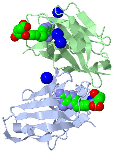
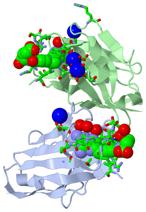
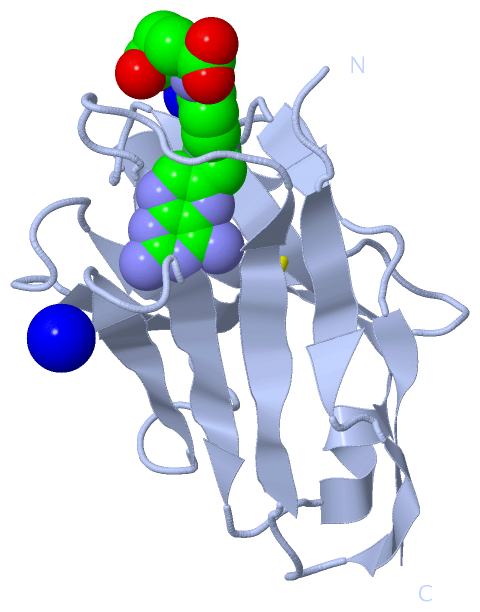
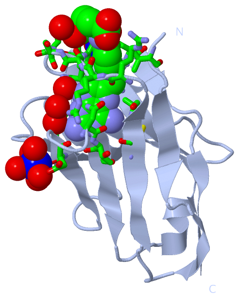
(-) Ligands, Modified Residues, Ions  (2, 7)

Asymmetric Unit (2, 7)
No.NameCountTypeFull Name
1MTX2Ligand/IonMETHOTREXATE
2NA5Ligand/IonSODIUM ION
Biological Unit 1 (1, 1)
No.NameCountTypeFull Name
1MTX1Ligand/IonMETHOTREXATE
2NA-1Ligand/IonSODIUM ION
Biological Unit 2 (1, 1)
No.NameCountTypeFull Name
1MTX1Ligand/IonMETHOTREXATE
2NA-1Ligand/IonSODIUM ION

(-) Sites  (7, 7)

Asymmetric Unit (7, 7)
No.NameEvidenceResiduesDescription
1AC1SOFTWAREVAL A:4 , CYS A:24 , ALA A:26 , ARG A:28 , ARG A:29 , SER A:30 , SER A:31 , ARG A:32 , TRP A:34 , ARG A:74 , ASP A:75 , ASN A:79 , THR A:80 , VAL A:81 , ALA A:100 , HOH A:1005 , HOH A:1064 , HOH A:1118 , ARG B:58BINDING SITE FOR RESIDUE MTX A 2000
2AC2SOFTWAREARG A:74 , HOH A:1023 , HOH A:1026 , HOH A:1098 , HOH A:1106 , HOH A:1120BINDING SITE FOR RESIDUE NA A 132
3AC3SOFTWAREASP A:101 , ASP A:102 , GLY A:112 , ASP A:119 , HOH A:1018 , HOH A:1066BINDING SITE FOR RESIDUE NA A 133
4AC4SOFTWAREVAL B:4 , CYS B:24 , ALA B:26 , ARG B:28 , ARG B:29 , SER B:30 , SER B:31 , ARG B:32 , TRP B:34 , MET B:36 , ARG B:74 , ASP B:75 , ASN B:79 , THR B:80 , VAL B:81 , ALA B:100 , HOH B:1022BINDING SITE FOR RESIDUE MTX B 2000
5AC5SOFTWAREASP B:101 , ASP B:102 , GLY B:112 , ASP B:119 , NA B:133 , HOH B:1044 , HOH B:1087BINDING SITE FOR RESIDUE NA B 132
6AC6SOFTWARESER B:111 , GLY B:112 , NA B:132 , HOH B:1017 , HOH B:1044 , HOH B:1055 , HOH B:1083BINDING SITE FOR RESIDUE NA B 133
7AC7SOFTWAREARG B:74 , HOH B:1018 , HOH B:1034 , HOH B:1048 , HOH B:1075BINDING SITE FOR RESIDUE NA B 134

(-) SS Bonds  (2, 2)

Asymmetric Unit
No.Residues
1A:24 -A:98
2B:24 -B:98

(-) Cis Peptide Bonds  (0, 0)

(no "Cis Peptide Bond" information available for 3QXT)

 Sequence-Structure Mapping

(-) SAPs(SNPs)/Variants  (0, 0)

(no "SAP(SNP)/Variant" information available for 3QXT)

(-) PROSITE Motifs  (0, 0)

(no "PROSITE Motif" information available for 3QXT)

(-) Exons   (0, 0)

(no "Exon" information available for 3QXT)

(-) Sequences/Alignments

Asymmetric Unit
   Reformat: Number of residues per line =  ('0' or empty: single-line sequence representation)
  Number of residues per labelling interval =   
  UniProt sequence: complete  aligned part    
   Show mapping: SCOP domains CATH domains Pfam domains Secondary structure (by author)
SAPs(SNPs) PROSITE motifs Exons
(details for a mapped element are shown in a popup box when the mouse pointer rests over it)
Chain A from PDB  Type:PROTEIN  Length:124
                                                                                                                                                            
               SCOP domains d3qxta_ A: automated matches                                                                                                 SCOP domains
               CATH domains ---------------------------------------------------------------------------------------------------------------------------- CATH domains
               Pfam domains ---------------------------------------------------------------------------------------------------------------------------- Pfam domains
         Sec.struct. author ...eeee..eeee....eeeeeee........eeeeeee......eeeeee......eee.......eeeeee....eeeeee...hhhhheeeeeeee...hhhhh.....ee...eeeeee. Sec.struct. author
                 SAPs(SNPs) ---------------------------------------------------------------------------------------------------------------------------- SAPs(SNPs)
                    PROSITE ---------------------------------------------------------------------------------------------------------------------------- PROSITE
                 Transcript ---------------------------------------------------------------------------------------------------------------------------- Transcript
                 3qxt A   3 QVQLVESGGGLVQAGGSLRLSCAASRRSSRSWAMAWFRQAPGKEREFVAKISGDGRLTTYGDSVKGRFTISRDKGKNTVYLQMDSLKPEDTAVYYCAADDNYVTASWRSGPDYWGQGTQVTVSS 131
                                    12        22        32        42        52        62        72        82        92       102       112||     127    
                                                                                                                                        113|            
                                                                                                                                         119            

Chain B from PDB  Type:PROTEIN  Length:124
                                                                                                                                                            
               SCOP domains d3qxtb_ B: automated matches                                                                                                 SCOP domains
               CATH domains ---------------------------------------------------------------------------------------------------------------------------- CATH domains
               Pfam domains ---------------------------------------------------------------------------------------------------------------------------- Pfam domains
         Sec.struct. author ...eeee..eeee....eeeeeee........eeeeeee......eeeeee......eee.......eeeeee....eeeeee...hhhhheeeeeeee..hhhhhh.....ee...eeeeee. Sec.struct. author
                 SAPs(SNPs) ---------------------------------------------------------------------------------------------------------------------------- SAPs(SNPs)
                    PROSITE ---------------------------------------------------------------------------------------------------------------------------- PROSITE
                 Transcript ---------------------------------------------------------------------------------------------------------------------------- Transcript
                 3qxt B   3 QVQLVESGGGLVQAGGSLRLSCAASRRSSRSWAMAWFRQAPGKEREFVAKISGDGRLTTYGDSVKGRFTISRDKGKNTVYLQMDSLKPEDTAVYYCAADDNYVTASWRSGPDYWGQGTQVTVSS 131
                                    12        22        32        42        52        62        72        82        92       102       112||     127    
                                                                                                                                        113|            
                                                                                                                                         119            

   Legend:   → Mismatch (orange background)
  - → Gap (green background, '-', border residues have a numbering label)
    → Modified Residue (blue background, lower-case, 'x' indicates undefined single-letter code, labelled with number + name)
  x → Chemical Group (purple background, 'x', labelled with number + name, e.g. ACE or NH2)
  extra numbering lines below/above indicate numbering irregularities and modified residue names etc., number ends below/above '|'

 Classification and Annotation

(-) SCOP Domains  (1, 2)

Asymmetric Unit

(-) CATH Domains  (0, 0)

(no "CATH Domain" information available for 3QXT)

(-) Pfam Domains  (0, 0)

(no "Pfam Domain" information available for 3QXT)

(-) Gene Ontology  (0, 0)

Asymmetric Unit(hide GO term definitions)
    (no "Gene Ontology" information available for 3QXT)

 Visualization

(-) Interactive Views

Asymmetric Unit
  Complete Structure
    Jena3D(integrated viewing of ligand, site, SAP, PROSITE, SCOP information)
    WebMol | AstexViewer[tm]@PDBe
(Java Applets, require no local installation except for Java; loading may be slow)
    STRAP
(Java WebStart application, automatic local installation, requires Java; full application with system access!)
    RasMol
(require local installation)
    Molscript (VRML)
(requires installation of a VRML viewer; select preferred view via VRML and generate a mono or stereo PDF format file)
 
  Ligands, Modified Residues, Ions
    MTX  [ RasMol | Jena3D ]  +environment [ RasMol | Jena3D ]
    NA  [ RasMol | Jena3D ]  +environment [ RasMol | Jena3D ]
 
  Sites
    AC1  [ RasMol ]  +environment [ RasMol ]
    AC2  [ RasMol ]  +environment [ RasMol ]
    AC3  [ RasMol ]  +environment [ RasMol ]
    AC4  [ RasMol ]  +environment [ RasMol ]
    AC5  [ RasMol ]  +environment [ RasMol ]
    AC6  [ RasMol ]  +environment [ RasMol ]
    AC7  [ RasMol ]  +environment [ RasMol ]
 
  Cis Peptide Bonds
(no "Cis Peptide Bonds" information available for 3qxt)
 
Biological Units
  Complete Structure
    Biological Unit 1  [ Jena3D ]
    Biological Unit 2  [ Jena3D ]

(-) Still Images

Jmol
  protein: cartoon or spacefill or dots and stick; nucleic acid: cartoon and stick; ligands: spacefill; active site: stick
Molscript
  protein, nucleic acid: cartoon; ligands: spacefill; active site: ball and stick

 Databases and Analysis Tools

(-) Databases

Access by PDB/NDB ID
  3qxt
    Family and Domain InformationProDom | SYSTERS
    General Structural InformationGlycoscienceDB | MMDB | NDB | OCA | PDB | PDBe | PDBj | PDBsum | PDBWiki | PQS | PROTEOPEDIA
    Orientation in MembranesOPM
    Protein SurfaceSURFACE
    Secondary StructureDSSP (structure derived) | HSSP (homology derived)
    Structural GenomicsGeneCensus
    Structural NeighboursCE | VAST
    Structure ClassificationCATH | Dali | SCOP
    Validation and Original DataBMRB Data View | BMRB Restraints Grid | EDS | PROCHECK | RECOORD | WHAT_CHECK
 
Access by UniProt ID/Accession number
  (no 'UniProt ID/Accession number' available) |
    Comparative Protein Structure ModelsModBase
    Genomic InformationEnsembl
    Protein-protein InteractionDIP
    Sequence, Family and Domain InformationInterPro | Pfam | SMART | UniProt
 
Access by Enzyme Classificator   (EC Number)
  (no 'Enzyme Classificator' available)
    General Enzyme InformationBRENDA | EC-PDB | Enzyme | IntEnz
    PathwayKEGG | MetaCyc
 
Access by Disease Identifier   (MIM ID)
  (no 'MIM ID' available)
    Disease InformationOMIM
 
Access by GenAge ID
  (no 'GenAge ID' available)
    Age Related InformationGenAge

(-) Analysis Tools

Access by PDB/NDB ID
    Domain InformationXDom
    Interatomic Contacts of Structural UnitsCSU
    Ligand-protein ContactsLPC
    Protein CavitiescastP
    Sequence and Secondary StructurePDBCartoon
    Structure AlignmentSTRAP(Java WebStart application, automatic local installation, requires Java; full application with system access!)
    Structure and Sequence BrowserSTING
 
Access by UniProt ID/Accession number
  (no 'UniProt ID/Accession number' available) |
    Protein Disorder PredictionDisEMBL | FoldIndex | GLOBPLOT

 Related Entries

(-) Entries Sharing at Least One Protein Chain (UniProt ID)

(no "Entries Sharing at Least One Protein Chain" available for 3QXT)

(-) Related Entries Specified in the PDB File

1i3u THREE-DIMENSIONAL STRUCTURE OF A LLAMA VHH DOMAIN COMPLEXED WITH THE DYE RR1
2x6m STRUCTURE OF A SINGLE DOMAIN CAMELID ANTIBODY FRAGMENT IN COMPLEX WITH A C-TERMINAL PEPTIDE OF ALPHA-SYNUCLEIN
3qxu
3qxv
3qxw