|
|
|
|
 Description
|
|
Compounds
|
||||||||||||||||||||||||||||||||||||||||||||
Chains, Units
Summary Information (see also Sequences/Alignments below) |
Ligands, Modified Residues, Ions (1, 8)
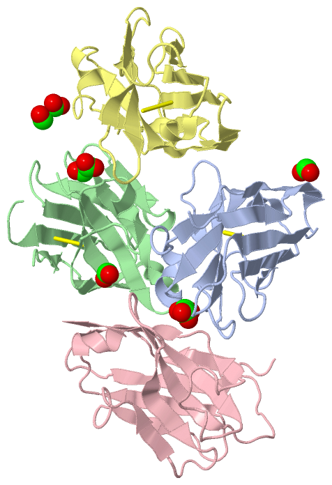
Asymmetric Unit (1, 8)
|
Sites (8, 8)
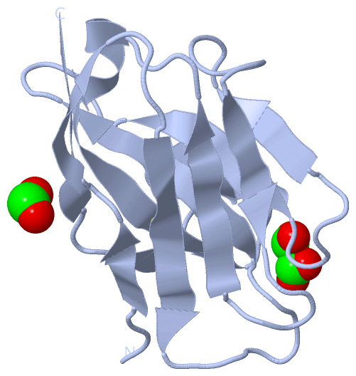
Asymmetric Unit (8, 8)
|
SS Bonds (4, 4)
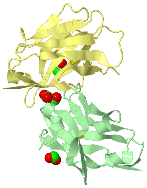
Asymmetric Unit
|
||||||||||||||||||||
Cis Peptide Bonds (0, 0)| (no "Cis Peptide Bond" information available for 3QXU) |
SAPs(SNPs)/Variants (0, 0)| (no "SAP(SNP)/Variant" information available for 3QXU) |
PROSITE Motifs (0, 0)| (no "PROSITE Motif" information available for 3QXU) |
Exons (0, 0)| (no "Exon" information available for 3QXU) |
Sequences/Alignments
Asymmetric Unit
Chain A from PDB Type:PROTEIN Length:124
SCOP domains d3qxua_ A: automated matches SCOP domains
CATH domains ---------------------------------------------------------------------------------------------------------------------------- CATH domains
Pfam domains ---------------------------------------------------------------------------------------------------------------------------- Pfam domains
SAPs(SNPs) ---------------------------------------------------------------------------------------------------------------------------- SAPs(SNPs)
PROSITE ---------------------------------------------------------------------------------------------------------------------------- PROSITE
Transcript ---------------------------------------------------------------------------------------------------------------------------- Transcript
3qxu A 3 QVQLVESGGGLVQAGGSLRLSCAASRRSSRSWAMAWFRQAPGKEREFVAKISGDGRLTTYGDSVKGRFTISRDKGKNTVYLQMDSLKPEDTAVYYCAADDNYVTASWRSGPDYWGQGTQVTVSS 131
12 22 32 42 52 62 72 82 92 102 112|| 127
113|
119
Chain B from PDB Type:PROTEIN Length:122
SCOP domains d3qxub_ B: automated matches SCOP domains
CATH domains -------------------------------------------------------------------------------------------------------------------------- CATH domains
Pfam domains -------------------------------------------------------------------------------------------------------------------------- Pfam domains
SAPs(SNPs) -------------------------------------------------------------------------------------------------------------------------- SAPs(SNPs)
PROSITE -------------------------------------------------------------------------------------------------------------------------- PROSITE
Transcript -------------------------------------------------------------------------------------------------------------------------- Transcript
3qxu B 3 QVQLVESGGGLVQAGGSLRLSCAASRRSSWAMAWFRQAPGKEREFVAKISGDGRLTTYGDSVKGRFTISRDKGKNTVYLQMDSLKPEDTAVYYCAADDNYVTASWRSGPDYWGQGTQVTVSS 131
12 22 |34 44 54 64 74 84 94 104 119 129
30| 113|
33 119
Chain C from PDB Type:PROTEIN Length:124
SCOP domains d3qxuc_ C: automated matches SCOP domains
CATH domains ---------------------------------------------------------------------------------------------------------------------------- CATH domains
Pfam domains ---------------------------------------------------------------------------------------------------------------------------- Pfam domains
SAPs(SNPs) ---------------------------------------------------------------------------------------------------------------------------- SAPs(SNPs)
PROSITE ---------------------------------------------------------------------------------------------------------------------------- PROSITE
Transcript ---------------------------------------------------------------------------------------------------------------------------- Transcript
3qxu C 3 QVQLVESGGGLVQAGGSLRLSCAASRRSSRSWAMAWFRQAPGKEREFVAKISGDGRLTTYGDSVKGRFTISRDKGKNTVYLQMDSLKPEDTAVYYCAADDNYVTASWRSGPDYWGQGTQVTVSS 131
12 22 32 42 52 62 72 82 92 102 112|| 127
113|
119
Chain D from PDB Type:PROTEIN Length:124
SCOP domains d3qxud_ D: automated matches SCOP domains
CATH domains ---------------------------------------------------------------------------------------------------------------------------- CATH domains
Pfam domains ---------------------------------------------------------------------------------------------------------------------------- Pfam domains
SAPs(SNPs) ---------------------------------------------------------------------------------------------------------------------------- SAPs(SNPs)
PROSITE ---------------------------------------------------------------------------------------------------------------------------- PROSITE
Transcript ---------------------------------------------------------------------------------------------------------------------------- Transcript
3qxu D 3 QVQLVESGGGLVQAGGSLRLSCAASRRSSRSWAMAWFRQAPGKEREFVAKISGDGRLTTYGDSVKGRFTISRDKGKNTVYLQMDSLKPEDTAVYYCAADDNYVTASWRSGPDYWGQGTQVTVSS 131
12 22 32 42 52 62 72 82 92 102 112|| 127
113|
119
|
||||||||||||||||||||
SCOP Domains (1, 4)
Asymmetric Unit
 |
CATH Domains (0, 0)| (no "CATH Domain" information available for 3QXU) |
Pfam Domains (0, 0)| (no "Pfam Domain" information available for 3QXU) |
Gene Ontology (0, 0)|
Asymmetric Unit(hide GO term definitions)
(no "Gene Ontology" information available for 3QXU)
|
Interactive Views
|
||||||||||||||||||||||||||||||||||||||||||||||||||||||||||||||||||||||||||||||||||||||||||||||||||||||||||||||||||||||||||||||||||||||||||||||||||||||||||||||||||||||||||||||||||||||||||||||||||||||||
Still Images
|
||||||||||||||||
Databases
|
||||||||||||||||||||||||||||||||||||||||||||||||||||||||||||||||||||||||||||||||||||||||||||||||||||||||||||||||||||||||||||||||||||||||||||||||||||||||||||||||
Analysis Tools
|
|||||||||||||||||||||||||||||||||||||||||||||||||||||||||||||
Entries Sharing at Least One Protein Chain (UniProt ID)
Related Entries Specified in the PDB File
|
|