|
|
|
|
 Description
|
|
Compounds
|
||||||||||||||||||||||||||||||||||||
Chains, Units
Summary Information (see also Sequences/Alignments below) |
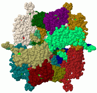
Ligands, Modified Residues, Ions (2, 5)| Asymmetric Unit (2, 5) Biological Unit 1 (2, 60) |
Sites (1, 1)
Asymmetric Unit (1, 1)
|
SS Bonds (1, 1)
Asymmetric Unit
|
||||||||
Cis Peptide Bonds (2, 2)
Asymmetric Unit
|
||||||||||||
SAPs(SNPs)/Variants (0, 0)| (no "SAP(SNP)/Variant" information available for 3JW6) |
PROSITE Motifs (0, 0)| (no "PROSITE Motif" information available for 3JW6) |
Exons (0, 0)| (no "Exon" information available for 3JW6) |
Sequences/Alignments
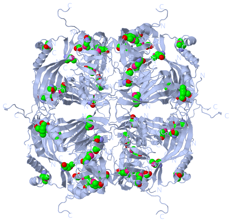
Asymmetric UnitChain A from PDB Type:PROTEIN Length:191 aligned with PYHD_NPVAC | P04871 from UniProtKB/Swiss-Prot Length:245 Alignment length:235 20 30 40 50 60 70 80 90 100 110 120 130 140 150 160 170 180 190 200 210 220 230 240 PYHD_NPVAC 11 GRTYVYDNKYYKNLGAVIKNAKRKKHFAEHEIEEATLDPLDNYLVAEDPFLGPGKNQKLTLFKEIRNVKPDTMKLVVGWKGKEFYRETWTRFMEDSFPIVNDQEVMDVFLVVNMRPTRPNRCYKFLAQHALRCDPDYVPHDVIRIVEPSWVGSNNEYRISLAKKGGGCPIMNLHSEYTNSFEQFIDRVIWENFYKPIVYIGTDSAEEEEILLEVSLVFKVKEFAPDAPLFTGPAY 245 SCOP domains ------------------------------------------------------------------------------------------------------------------------------------------------------------------------------------------------------------------------------------------- SCOP domains CATH domains ------------------------------------------------------------------------------------------------------------------------------------------------------------------------------------------------------------------------------------------- CATH domains Pfam domains Polyhedrin-3jw6A01 A:11-244 - Pfam domains SAPs(SNPs) ------------------------------------------------------------------------------------------------------------------------------------------------------------------------------------------------------------------------------------------- SAPs(SNPs) PROSITE ------------------------------------------------------------------------------------------------------------------------------------------------------------------------------------------------------------------------------------------- PROSITE Transcript ------------------------------------------------------------------------------------------------------------------------------------------------------------------------------------------------------------------------------------------- Transcript 3jw6 A 11 GRTYVYDNKYYKNLDAVI--------------------PLDNYLVAEDPFLGPGKNQKLTLFKEIRNVKPDTmKLVVGWKGKEFYRETWTRFmEDSFPIVNDQEVmDVFLVVNmRPTRPNRCYKFLAQHAL-----YVPHDVIRIVEPSWVGSNNEYRISLA---------------TNSFEQFIDRV----FYKPIVYIGTDSAEEEEILLEVSLVFKVKEFAPDAPLFTGPAY 245 20 | - - 50 60 70 80 | 90 100 | 110 | 120 | 130 140| |150 160 170 | - 190 | - | 210 220 230 240 28 49 83-MSE 103-MSE 116-MSE 124-MSE 141 147 172 188 198 203
|
||||||||||||||||||||
SCOP Domains (0, 0)| (no "SCOP Domain" information available for 3JW6) |
CATH Domains (0, 0)| (no "CATH Domain" information available for 3JW6) |
Pfam Domains (1, 1)
Asymmetric Unit
|
Gene Ontology (3, 3)|
Asymmetric Unit(hide GO term definitions) Chain A (PYHD_NPVAC | P04871)
|
||||||||||||||||||||||||||||||
Interactive Views
|
|||||||||||||||||||||||||||||||||||||||||||||||||||||||||||||||||||||||||||||||||||||||||||||||||||||||||||||||||||||||||||||||||||||||||||||||||||||||
Still Images
|
||||||||||||||||
Databases
|
||||||||||||||||||||||||||||||||||||||||||||||||||||||||||||||||||||||||||||||||||||||||||||||||||||||||||||||||||||||||||||||||||||||||||||||||||||||||||||||||
Analysis Tools
|
|||||||||||||||||||||||||||||||||||||||||||||||||||||||||||||
Entries Sharing at Least One Protein Chain (UniProt ID)
Related Entries Specified in the PDB File
|
|