|
|
|
|
 Description
|
|
Compounds
|
||||||||||||||||||||||||||||||||||||
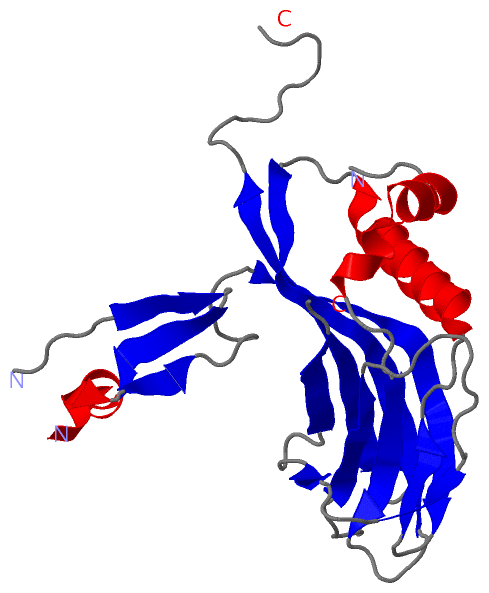
Chains, Units
Summary Information (see also Sequences/Alignments below) |
Ligands, Modified Residues, Ions (0, 0)| (no "Ligand,Modified Residues,Ions" information available for 2WUY) |
Sites (0, 0)| (no "Site" information available for 2WUY) |
SS Bonds (0, 0)| (no "SS Bond" information available for 2WUY) |
Cis Peptide Bonds (3, 3)
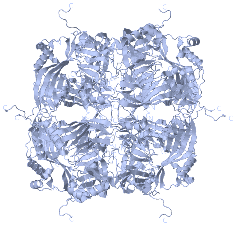
Asymmetric Unit
|
||||||||||||||||
SAPs(SNPs)/Variants (0, 0)| (no "SAP(SNP)/Variant" information available for 2WUY) |
PROSITE Motifs (0, 0)| (no "PROSITE Motif" information available for 2WUY) |
Exons (0, 0)| (no "Exon" information available for 2WUY) |
Sequences/Alignments
Asymmetric UnitChain A from PDB Type:PROTEIN Length:208 aligned with PYHD_NPVAC | P04871 from UniProtKB/Swiss-Prot Length:245 Alignment length:238 17 27 37 47 57 67 77 87 97 107 117 127 137 147 157 167 177 187 197 207 217 227 237 PYHD_NPVAC 8 PTIGRTYVYDNKYYKNLGAVIKNAKRKKHFAEHEIEEATLDPLDNYLVAEDPFLGPGKNQKLTLFKEIRNVKPDTMKLVVGWKGKEFYRETWTRFMEDSFPIVNDQEVMDVFLVVNMRPTRPNRCYKFLAQHALRCDPDYVPHDVIRIVEPSWVGSNNEYRISLAKKGGGCPIMNLHSEYTNSFEQFIDRVIWENFYKPIVYIGTDSAEEEEILLEVSLVFKVKEFAPDAPLFTGPAY 245 SCOP domains ---------------------------------------------------------------------------------------------------------------------------------------------------------------------------------------------------------------------------------------------- SCOP domains CATH domains ---------------------------------------------------------------------------------------------------------------------------------------------------------------------------------------------------------------------------------------------- CATH domains Pfam domains --Polyhedrin-2wuyA01 A:1 0-244 - Pfam domains SAPs(SNPs) ---------------------------------------------------------------------------------------------------------------------------------------------------------------------------------------------------------------------------------------------- SAPs(SNPs) PROSITE ---------------------------------------------------------------------------------------------------------------------------------------------------------------------------------------------------------------------------------------------- PROSITE Transcript ---------------------------------------------------------------------------------------------------------------------------------------------------------------------------------------------------------------------------------------------- Transcript 2wuy A 8 PTIGRTYVYDNKYYKNLGAVIKNA-----------------PLDNYLVAEDPFLGPGKNQKLTLFKEIRNVKPDTMKLVVGWKGKEFYRETWTRFMEDSFPIVNDQEVMDVFLVVNMRPTRPNRCYKFLAQHALRCDPDYVPHDVIRIVEPSWVGSNNEYRISLAK-------------YTNSFEQFIDRVIWENFYKPIVYIGTDSAEEEEILLEVSLVFKVKEFAPDAPLFTGPAY 245 17 27 | - - | 57 67 77 87 97 107 117 127 137 147 157 167 | - 187 197 207 217 227 237 31 49 173 187
|
||||||||||||||||||||
SCOP Domains (0, 0)| (no "SCOP Domain" information available for 2WUY) |
CATH Domains (0, 0)| (no "CATH Domain" information available for 2WUY) |
Pfam Domains (1, 1)
Asymmetric Unit
|
Gene Ontology (3, 3)|
Asymmetric Unit(hide GO term definitions) Chain A (PYHD_NPVAC | P04871)
|
||||||||||||||||||||||||||||||
Interactive Views
|
|||||||||||||||||||||||||||||||||||||||||||||||||||||||||||||||||||||||||||||||||||||||||||||||||||||||||||||||||||||||||||||||||||||||||||||||||||||
Still Images
|
||||||||||||||||
Databases
|
||||||||||||||||||||||||||||||||||||||||||||||||||||||||||||||||||||||||||||||||||||||||||||||||||||||||||||||||||||||||||||||||||||||||||||||||||||||||||||||||
Analysis Tools
|
|||||||||||||||||||||||||||||||||||||||||||||||||||||||||||||
Entries Sharing at Least One Protein Chain (UniProt ID)
Related Entries Specified in the PDB File
|
|