|
|
|
|
 Description
|
|
Compounds
|
||||||||||||||||||||||||||||||||||||||||||||||||
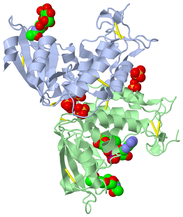
Chains, Units
Summary Information (see also Sequences/Alignments below) |
Ligands, Modified Residues, Ions (3, 10)| Asymmetric/Biological Unit (3, 10) |
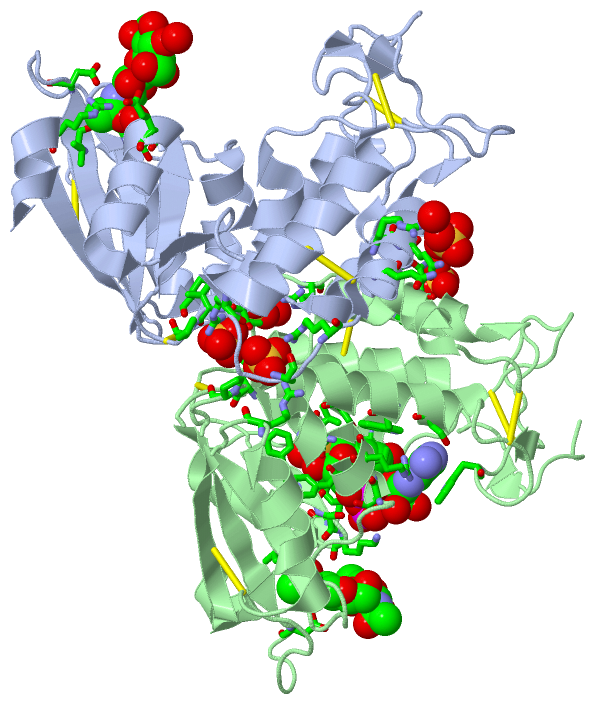
Sites (10, 10)
Asymmetric Unit (10, 10)
|
SS Bonds (10, 10)
Asymmetric/Biological Unit
|
||||||||||||||||||||||||||||||||||||||||||||
Cis Peptide Bonds (4, 4)
Asymmetric/Biological Unit
|
||||||||||||||||||||
SAPs(SNPs)/Variants (0, 0)| (no "SAP(SNP)/Variant" information available for 3GHH) |
PROSITE Motifs (0, 0)| (no "PROSITE Motif" information available for 3GHH) |
Exons (0, 0)| (no "Exon" information available for 3GHH) |
Sequences/Alignments
Asymmetric/Biological UnitChain A from PDB Type:PROTEIN Length:238 aligned with Q9TTF5_BOVIN | Q9TTF5 from UniProtKB/TrEMBL Length:278 Alignment length:238 49 59 69 79 89 99 109 119 129 139 149 159 169 179 189 199 209 219 229 239 249 259 269 Q9TTF5_BOVIN 40 NRWHGAGSTADFQKIIQERCDTYTQTIRPGSRSRNCQAIRQAFMSAFISKDPCKATKEDYNSLINLAPPTVPCGQQVFWSKTKELAHEYAKRRRLMTLEDTLLGYLADGLRWCGEPGSSDLNIWSCPDWRKDCRTNYLSVFWEVLSERFAESACNTVRVVLNGSLENAFDSMSIFGRVEAPNLRPQVELEAWLVHDTGKPPSDSCSGSSIRKLKSILDGRNVKFRCMDNLSRDQFLQR 277 SCOP domains d3ghha_ A: ADP ribosyl cyclase SCOP domains CATH domains ---------------------------------------------------------------------------------------------------------------------------------------------------------------------------------------------------------------------------------------------- CATH domains Pfam domains ---------------------------------------------------------------------------------------------------------------------------------------------------------------------------------------------------------------------------------------------- Pfam domains SAPs(SNPs) ---------------------------------------------------------------------------------------------------------------------------------------------------------------------------------------------------------------------------------------------- SAPs(SNPs) PROSITE ---------------------------------------------------------------------------------------------------------------------------------------------------------------------------------------------------------------------------------------------- PROSITE Transcript ---------------------------------------------------------------------------------------------------------------------------------------------------------------------------------------------------------------------------------------------- Transcript 3ghh A 40 NRWHGAGSTADFQKIIQERCDTYTQTIRPGSRSRNCQAIRQAFMSAFISKDPCKATKEDYNSLINLAPPTVPCGQQVFWSKTKELAHEYAKRRRLMTLEDTLLGYLADGLRWCGEPGSSDLNIWSCPDWRKDCRTNYLSVFWEVLSERFAESACNTVRVVLNGSLENAFDSMSIFGRVQAPNLRPQVELEAWLVHDTGKPPSDSCSGSSIRKLKSILDGRNVKFRCMDNLSRDQFLQR 277 49 59 69 79 89 99 109 119 129 139 149 159 169 179 189 199 209 219 229 239 249 259 269 Chain B from PDB Type:PROTEIN Length:237 aligned with Q9TTF5_BOVIN | Q9TTF5 from UniProtKB/TrEMBL Length:278 Alignment length:237 48 58 68 78 88 98 108 118 128 138 148 158 168 178 188 198 208 218 228 238 248 258 268 Q9TTF5_BOVIN 39 LNRWHGAGSTADFQKIIQERCDTYTQTIRPGSRSRNCQAIRQAFMSAFISKDPCKATKEDYNSLINLAPPTVPCGQQVFWSKTKELAHEYAKRRRLMTLEDTLLGYLADGLRWCGEPGSSDLNIWSCPDWRKDCRTNYLSVFWEVLSERFAESACNTVRVVLNGSLENAFDSMSIFGRVEAPNLRPQVELEAWLVHDTGKPPSDSCSGSSIRKLKSILDGRNVKFRCMDNLSRDQFL 275 SCOP domains d3ghhb_ B: ADP ribosyl cyclase SCOP domains CATH domains --------------------------------------------------------------------------------------------------------------------------------------------------------------------------------------------------------------------------------------------- CATH domains Pfam domains --------------------------------------------------------------------------------------------------------------------------------------------------------------------------------------------------------------------------------------------- Pfam domains SAPs(SNPs) --------------------------------------------------------------------------------------------------------------------------------------------------------------------------------------------------------------------------------------------- SAPs(SNPs) PROSITE --------------------------------------------------------------------------------------------------------------------------------------------------------------------------------------------------------------------------------------------- PROSITE Transcript --------------------------------------------------------------------------------------------------------------------------------------------------------------------------------------------------------------------------------------------- Transcript 3ghh B 39 LNRWHGAGSTADFQKIIQERCDTYTQTIRPGSRSRNCQAIRQAFMSAFISKDPCKATKEDYNSLINLAPPTVPCGQQVFWSKTKELAHEYAKRRRLMTLEDTLLGYLADGLRWCGEPGSSDLNIWSCPDWRKDCRTNYLSVFWEVLSERFAESACNTVRVVLNGSLENAFDSMSIFGRVQAPNLRPQVELEAWLVHDTGKPPSDSCSGSSIRKLKSILDGRNVKFRCMDNLSRDQFL 275 48 58 68 78 88 98 108 118 128 138 148 158 168 178 188 198 208 218 228 238 248 258 268
|
||||||||||||||||||||
SCOP Domains (1, 2)
Asymmetric/Biological Unit
 |
CATH Domains (0, 0)| (no "CATH Domain" information available for 3GHH) |
Pfam Domains (0, 0)| (no "Pfam Domain" information available for 3GHH) |
Gene Ontology (15, 15)|
Asymmetric/Biological Unit(hide GO term definitions) Chain A,B (Q9TTF5_BOVIN | Q9TTF5)
|
||||||||||||||||||||||||||||||||||||||||||||||||||||||||||||||||||||||||||||||||||||||||||||||||||||||||||||
Interactive Views
|
|||||||||||||||||||||||||||||||||||||||||||||||||||||||||||||||||||||||||||||||||||||||||||||||||||||||||||||||||||||||||||||||||||||||||||||||||||||||||||||||||||||||||||||||||||||||||||||||||||||||||||||||||||||||||
Still Images
|
||||||||||||||||
Databases
|
||||||||||||||||||||||||||||||||||||||||||||||||||||||||||||||||||||||||||||||||||||||||||||||||||||||||||||||||||||||||||||||||||||||||||||||||||||||||||||||||
Analysis Tools
|
|||||||||||||||||||||||||||||||||||||||||||||||||||||||||||||
Entries Sharing at Least One Protein Chain (UniProt ID)
Related Entries Specified in the PDB File
|
|