Show PDB file:   
         Plain Text   HTML   (compressed file size)
QuickSearch:   
by PDB,NDB,UniProt,PROSITE Code or Search Term(s)  
(-)Asymmetric Unit
(-)Asym. Unit - sites
(-)Biological Unit 1
(-)Biol. Unit 1 - sites
(-)Biological Unit 2
collapse expand < >
Image Asymmetric Unit
Asymmetric Unit  (Jmol Viewer)
Image Asym. Unit - sites
Asym. Unit - sites  (Jmol Viewer)
Image Biological Unit 1
Biological Unit 1  (Jmol Viewer)
Image Biol. Unit 1 - sites
Biol. Unit 1 - sites  (Jmol Viewer)
Image Biological Unit 2
Biological Unit 2  (Jmol Viewer)

(-) Description

Title :  STRUCTURAL INSIGHTS INTO THE CATALYTIC MECHANISM OF CD38: EVIDENCE FOR A CONFORMATIONALLY FLEXIBLE COVALENT ENZYME-SUBSTRATE COMPLEX.
 
Authors :  P. F. Egea, H. Muller-Steffner, R. M. Stroud, N. Oppenheimer, E. Kellen F. Schuber
Date :  21 Feb 09  (Deposition) - 02 Mar 10  (Release) - 23 Jan 13  (Revision)
Method :  X-RAY DIFFRACTION
Resolution :  1.51
Chains :  Asym. Unit :  A,B
Biol. Unit 1:  A (1x),B (1x)
Biol. Unit 2:  A,B  (1x)
Keywords :  Cd38, Cyclic Adp Ribose, Ecto-Adp-Ribosyl Cyclase, Glycosidase, Hydrolase (Keyword Search: [Gene Ontology, PubMed, Web (Google))
 
Reference :  P. F. Egea, H. Muller-Steffner, I. Kuhn, C. Cakir-Kiefer, N. J. Oppenheimer, R. M. Stroud, E. Kellenberger, F. Schuber
Insights Into The Mechanism Of Bovine Cd38/Nad+Glycohydrolase From The X-Ray Structures Of Its Michaelis Complex And Covalently-Trapped Intermediates.
Plos One V. 7 34918 2012
PubMed-ID: 22529956  |  Reference-DOI: 10.1371/JOURNAL.PONE.0034918

(-) Compounds

Molecule 1 - ECTO-NAD+ GLYCOHYDROLASE (CD38 MOLECULE)
    ChainsA, B
    EC Number3.2.2.5
    EngineeredYES
    Expression SystemPICHIA PASTORIS
    Expression System Taxid4922
    GeneCD38
    MutationYES
    Organism CommonBOVINE,COW,DOMESTIC CATTLE,DOMESTIC COW
    Organism ScientificBOS TAURUS
    Organism Taxid9913

 Structural Features

(-) Chains, Units

  12
Asymmetric Unit AB
Biological Unit 1 (1x)A (1x)B (1x)
Biological Unit 2 (1x)AB

Summary Information (see also Sequences/Alignments below)
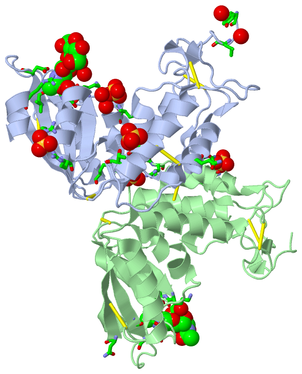
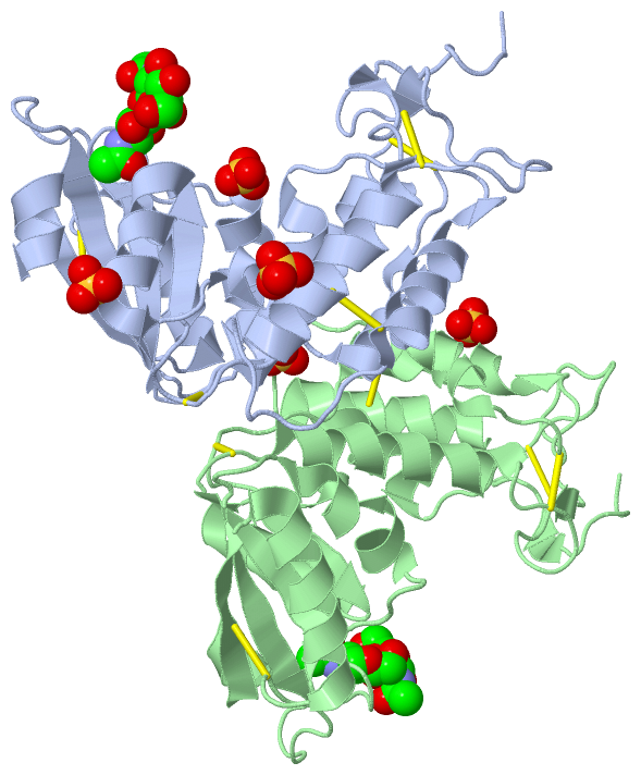
(-) Ligands, Modified Residues, Ions  (2, 9)

Asymmetric Unit (2, 9)
No.NameCountTypeFull Name
1NAG4Ligand/IonN-ACETYL-D-GLUCOSAMINE
2SO45Ligand/IonSULFATE ION
Biological Unit 1 (2, 5)
No.NameCountTypeFull Name
1NAG2Ligand/IonN-ACETYL-D-GLUCOSAMINE
2SO43Ligand/IonSULFATE ION
Biological Unit 2 (2, 9)
No.NameCountTypeFull Name
1NAG4Ligand/IonN-ACETYL-D-GLUCOSAMINE
2SO45Ligand/IonSULFATE ION

(-) Sites  (9, 9)

Asymmetric Unit (9, 9)
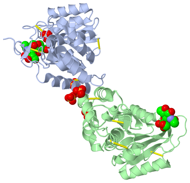
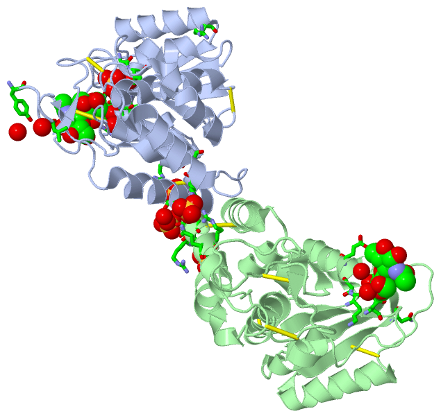
No.NameEvidenceResiduesDescription
1AC1SOFTWARETHR A:61 , THR A:65 , ARG B:71 , SER B:72 , HOH B:332BINDING SITE FOR RESIDUE SO4 A 279
2AC2SOFTWARETYR A:36 , LEU A:39 , ARG A:131 , ARG A:133 , HOH A:486 , HOH A:579 , ASN B:268BINDING SITE FOR RESIDUE SO4 A 280
3AC3SOFTWARELYS A:120 , SER A:212 , ILE A:213 , HOH A:311 , HOH A:511BINDING SITE FOR RESIDUE SO4 A 281
4AC4SOFTWARELYS A:120 , THR A:121 , ASN A:201 , VAL A:233 , ASP A:235 , ARG A:271 , HOH A:323 , NAG A:502 , HOH A:554BINDING SITE FOR RESIDUE NAG A 501
5AC5SOFTWARENAG A:501BINDING SITE FOR RESIDUE NAG A 502
6AC6SOFTWAREARG A:259 , GLY B:69 , SER B:70 , ARG B:71 , ARG B:73BINDING SITE FOR RESIDUE SO4 B 279
7AC7SOFTWAREGLN A:76 , GLN B:76 , ARG B:79 , GLN B:80BINDING SITE FOR RESIDUE SO4 B 280
8AC8SOFTWARELYS B:120 , THR B:121 , ASN B:201 , VAL B:233 , ASP B:235 , HOH B:357 , HOH B:373 , NAG B:502BINDING SITE FOR RESIDUE NAG B 501
9AC9SOFTWAREGLU B:123 , NAG B:501BINDING SITE FOR RESIDUE NAG B 502

(-) SS Bonds  (10, 10)

Asymmetric Unit
No.Residues
1A:59 -A:75
2A:92 -A:172
3A:112 -A:193
4A:152 -A:165
5A:244 -A:265
6B:59 -B:75
7B:92 -B:172
8B:112 -B:193
9B:152 -B:165
10B:244 -B:265

(-) Cis Peptide Bonds  (0, 0)

(no "Cis Peptide Bond" information available for 3GC6)

 Sequence-Structure Mapping

(-) SAPs(SNPs)/Variants  (0, 0)

(no "SAP(SNP)/Variant" information available for 3GC6)

(-) PROSITE Motifs  (0, 0)

(no "PROSITE Motif" information available for 3GC6)

(-) Exons   (0, 0)

(no "Exon" information available for 3GC6)

(-) Sequences/Alignments

Asymmetric Unit
   Reformat: Number of residues per line =  ('0' or empty: single-line sequence representation)
  Number of residues per labelling interval =   
  UniProt sequence: complete  aligned part    
   Show mapping: SCOP domains CATH domains Pfam domains Secondary structure (by author)
SAPs(SNPs) PROSITE motifs Exons
(details for a mapped element are shown in a popup box when the mouse pointer rests over it)
Chain A from PDB  Type:PROTEIN  Length:242
 aligned with Q9TTF5_BOVIN | Q9TTF5 from UniProtKB/TrEMBL  Length:278

    Alignment length:242
                                    45        55        65        75        85        95       105       115       125       135       145       155       165       175       185       195       205       215       225       235       245       255       265       275  
         Q9TTF5_BOVIN    36 YSGLNRWHGAGSTADFQKIIQERCDTYTQTIRPGSRSRNCQAIRQAFMSAFISKDPCKATKEDYNSLINLAPPTVPCGQQVFWSKTKELAHEYAKRRRLMTLEDTLLGYLADGLRWCGEPGSSDLNIWSCPDWRKDCRTNYLSVFWEVLSERFAESACNTVRVVLNGSLENAFDSMSIFGRVEAPNLRPQVELEAWLVHDTGKPPSDSCSGSSIRKLKSILDGRNVKFRCMDNLSRDQFLQR 277
               SCOP domains d3gc6a_ A: ADP ribosyl cyclase                                                                                                                                                                                                                     SCOP domains
               CATH domains -------------------------------------------------------------------------------------------------------------------------------------------------------------------------------------------------------------------------------------------------- CATH domains
               Pfam domains -------------------------------------------------------------------------------------------------------------------------------------------------------------------------------------------------------------------------------------------------- Pfam domains
         Sec.struct. author ........ee....hhhhhhhhhhhhhhhh........hhhhhhhhhhhhhh.......hhhhhhhhhhhh.........eeee.hhhhhhhhhhhhh..hhhhhhhhhhhh................ee.........hhhhhhhhhhhhhhhhh...eeeeeee..........hhhhhhhhhhh....eeeeeeee...........hhhhhhhhhhhhhh..eeeeeeeeehhhhhh. Sec.struct. author
                 SAPs(SNPs) -------------------------------------------------------------------------------------------------------------------------------------------------------------------------------------------------------------------------------------------------- SAPs(SNPs)
                    PROSITE -------------------------------------------------------------------------------------------------------------------------------------------------------------------------------------------------------------------------------------------------- PROSITE
                 Transcript -------------------------------------------------------------------------------------------------------------------------------------------------------------------------------------------------------------------------------------------------- Transcript
                 3gc6 A  36 YSGLNRWHGAGSTADFQKIIQERCDTYTQTIRPGSRSRNCQAIRQAFMSAFISKDPCKATKEDYNSLINLAPPTVPCGQQVFWSKTKELAHEYAKRRRLMTLEDTLLGYLADGLRWCGEPGSSDLNIWSCPDWRKDCRTNYLSVFWEVLSERFAESACNTVRVVLNGSLENAFDSMSIFGRVQAPNLRPQVELEAWLVHDTGKPPSDSCSGSSIRKLKSILDGRNVKFRCMDNLSRDQFLQR 277
                                    45        55        65        75        85        95       105       115       125       135       145       155       165       175       185       195       205       215       225       235       245       255       265       275  

Chain B from PDB  Type:PROTEIN  Length:237
 aligned with Q9TTF5_BOVIN | Q9TTF5 from UniProtKB/TrEMBL  Length:278

    Alignment length:237
                                    48        58        68        78        88        98       108       118       128       138       148       158       168       178       188       198       208       218       228       238       248       258       268       
         Q9TTF5_BOVIN    39 LNRWHGAGSTADFQKIIQERCDTYTQTIRPGSRSRNCQAIRQAFMSAFISKDPCKATKEDYNSLINLAPPTVPCGQQVFWSKTKELAHEYAKRRRLMTLEDTLLGYLADGLRWCGEPGSSDLNIWSCPDWRKDCRTNYLSVFWEVLSERFAESACNTVRVVLNGSLENAFDSMSIFGRVEAPNLRPQVELEAWLVHDTGKPPSDSCSGSSIRKLKSILDGRNVKFRCMDNLSRDQFL 275
               SCOP domains d3gc6b_ B: ADP ribosyl cyclase                                                                                                                                                                                                                SCOP domains
               CATH domains --------------------------------------------------------------------------------------------------------------------------------------------------------------------------------------------------------------------------------------------- CATH domains
               Pfam domains --------------------------------------------------------------------------------------------------------------------------------------------------------------------------------------------------------------------------------------------- Pfam domains
         Sec.struct. author .....ee....hhhhhhhhhhhhhhhh........hhhhhhhhhhhhhh.......hhhhhhhhhhhh.........eeee.hhhhhhhhhhhhh..hhhhhhhhhhhh................ee.........hhhhhhhhhhhhhhhhh...eeeeeee..........hhhhhhhhhhh....eeeeeeee...........hhhhhhhhhhhhhhh.eeeeeeeeehhhhh Sec.struct. author
                 SAPs(SNPs) --------------------------------------------------------------------------------------------------------------------------------------------------------------------------------------------------------------------------------------------- SAPs(SNPs)
                    PROSITE --------------------------------------------------------------------------------------------------------------------------------------------------------------------------------------------------------------------------------------------- PROSITE
                 Transcript --------------------------------------------------------------------------------------------------------------------------------------------------------------------------------------------------------------------------------------------- Transcript
                 3gc6 B  39 LNRWHGAGSTADFQKIIQERCDTYTQTIRPGSRSRNCQAIRQAFMSAFISKDPCKATKEDYNSLINLAPPTVPCGQQVFWSKTKELAHEYAKRRRLMTLEDTLLGYLADGLRWCGEPGSSDLNIWSCPDWRKDCRTNYLSVFWEVLSERFAESACNTVRVVLNGSLENAFDSMSIFGRVQAPNLRPQVELEAWLVHDTGKPPSDSCSGSSIRKLKSILDGRNVKFRCMDNLSRDQFL 275
                                    48        58        68        78        88        98       108       118       128       138       148       158       168       178       188       198       208       218       228       238       248       258       268       

   Legend:   → Mismatch (orange background)
  - → Gap (green background, '-', border residues have a numbering label)
    → Modified Residue (blue background, lower-case, 'x' indicates undefined single-letter code, labelled with number + name)
  x → Chemical Group (purple background, 'x', labelled with number + name, e.g. ACE or NH2)
  extra numbering lines below/above indicate numbering irregularities and modified residue names etc., number ends below/above '|'

 Classification and Annotation

(-) SCOP Domains  (1, 2)

Asymmetric Unit

(-) CATH Domains  (0, 0)

(no "CATH Domain" information available for 3GC6)

(-) Pfam Domains  (0, 0)

(no "Pfam Domain" information available for 3GC6)

(-) Gene Ontology  (15, 15)

Asymmetric Unit(hide GO term definitions)
Chain A,B   (Q9TTF5_BOVIN | Q9TTF5)
molecular function
    GO:0003953    NAD+ nucleosidase activity    Catalysis of the reaction: NAD+ + H2O = nicotinamide + ADP-ribose.
    GO:0016787    hydrolase activity    Catalysis of the hydrolysis of various bonds, e.g. C-O, C-N, C-C, phosphoric anhydride bonds, etc. Hydrolase is the systematic name for any enzyme of EC class 3.
    GO:0016798    hydrolase activity, acting on glycosyl bonds    Catalysis of the hydrolysis of any glycosyl bond.
    GO:0016849    phosphorus-oxygen lyase activity    Catalysis of the cleavage of a phosphorus-oxygen bond by other means than by hydrolysis or oxidation, or conversely adding a group to a double bond.
biological process
    GO:0050853    B cell receptor signaling pathway    A series of molecular signals initiated by the cross-linking of an antigen receptor on a B cell.
    GO:0008152    metabolic process    The chemical reactions and pathways, including anabolism and catabolism, by which living organisms transform chemical substances. Metabolic processes typically transform small molecules, but also include macromolecular processes such as DNA repair and replication, and protein synthesis and degradation.
    GO:0043066    negative regulation of apoptotic process    Any process that stops, prevents, or reduces the frequency, rate or extent of cell death by apoptotic process.
    GO:0045892    negative regulation of transcription, DNA-templated    Any process that stops, prevents, or reduces the frequency, rate or extent of cellular DNA-templated transcription.
    GO:0030890    positive regulation of B cell proliferation    Any process that activates or increases the rate or extent of B cell proliferation.
    GO:0045893    positive regulation of transcription, DNA-templated    Any process that activates or increases the frequency, rate or extent of cellular DNA-templated transcription.
    GO:0042493    response to drug    Any process that results in a change in state or activity of a cell or an organism (in terms of movement, secretion, enzyme production, gene expression, etc.) as a result of a drug stimulus. A drug is a substance used in the diagnosis, treatment or prevention of a disease.
cellular component
    GO:0009986    cell surface    The external part of the cell wall and/or plasma membrane.
    GO:0070062    extracellular exosome    A vesicle that is released into the extracellular region by fusion of the limiting endosomal membrane of a multivesicular body with the plasma membrane. Extracellular exosomes, also simply called exosomes, have a diameter of about 40-100 nm.
    GO:0016020    membrane    A lipid bilayer along with all the proteins and protein complexes embedded in it an attached to it.
    GO:0005886    plasma membrane    The membrane surrounding a cell that separates the cell from its external environment. It consists of a phospholipid bilayer and associated proteins.

 Visualization

(-) Interactive Views

Asymmetric Unit
  Complete Structure
    Jena3D(integrated viewing of ligand, site, SAP, PROSITE, SCOP information)
    WebMol | AstexViewer[tm]@PDBe
(Java Applets, require no local installation except for Java; loading may be slow)
    STRAP
(Java WebStart application, automatic local installation, requires Java; full application with system access!)
    RasMol
(require local installation)
    Molscript (VRML)
(requires installation of a VRML viewer; select preferred view via VRML and generate a mono or stereo PDF format file)
 
  Ligands, Modified Residues, Ions
    NAG  [ RasMol | Jena3D ]  +environment [ RasMol | Jena3D ]
    SO4  [ RasMol | Jena3D ]  +environment [ RasMol | Jena3D ]
 
  Sites
    AC1  [ RasMol ]  +environment [ RasMol ]
    AC2  [ RasMol ]  +environment [ RasMol ]
    AC3  [ RasMol ]  +environment [ RasMol ]
    AC4  [ RasMol ]  +environment [ RasMol ]
    AC5  [ RasMol ]  +environment [ RasMol ]
    AC6  [ RasMol ]  +environment [ RasMol ]
    AC7  [ RasMol ]  +environment [ RasMol ]
    AC8  [ RasMol ]  +environment [ RasMol ]
    AC9  [ RasMol ]  +environment [ RasMol ]
 
  Cis Peptide Bonds
(no "Cis Peptide Bonds" information available for 3gc6)
 
Biological Units
  Complete Structure
    Biological Unit 1  [ Jena3D ]
    Biological Unit 2  [ Jena3D ]

(-) Still Images

Jmol
  protein: cartoon or spacefill or dots and stick; nucleic acid: cartoon and stick; ligands: spacefill; active site: stick
Molscript
  protein, nucleic acid: cartoon; ligands: spacefill; active site: ball and stick

 Databases and Analysis Tools

(-) Databases

Access by PDB/NDB ID
  3gc6
    Family and Domain InformationProDom | SYSTERS
    General Structural InformationGlycoscienceDB | MMDB | NDB | OCA | PDB | PDBe | PDBj | PDBsum | PDBWiki | PQS | PROTEOPEDIA
    Orientation in MembranesOPM
    Protein SurfaceSURFACE
    Secondary StructureDSSP (structure derived) | HSSP (homology derived)
    Structural GenomicsGeneCensus
    Structural NeighboursCE | VAST
    Structure ClassificationCATH | Dali | SCOP
    Validation and Original DataBMRB Data View | BMRB Restraints Grid | EDS | PROCHECK | RECOORD | WHAT_CHECK
 
Access by UniProt ID/Accession number
  Q9TTF5_BOVIN | Q9TTF5
    Comparative Protein Structure ModelsModBase
    Genomic InformationEnsembl
    Protein-protein InteractionDIP
    Sequence, Family and Domain InformationInterPro | Pfam | SMART | UniProtKB/TrEMBL
 
Access by Enzyme Classificator   (EC Number)
  3.2.2.5
    General Enzyme InformationBRENDA | EC-PDB | Enzyme | IntEnz
    PathwayKEGG | MetaCyc
 
Access by Disease Identifier   (MIM ID)
  (no 'MIM ID' available)
    Disease InformationOMIM
 
Access by GenAge ID
  (no 'GenAge ID' available)
    Age Related InformationGenAge

(-) Analysis Tools

Access by PDB/NDB ID
    Domain InformationXDom
    Interatomic Contacts of Structural UnitsCSU
    Ligand-protein ContactsLPC
    Protein CavitiescastP
    Sequence and Secondary StructurePDBCartoon
    Structure AlignmentSTRAP(Java WebStart application, automatic local installation, requires Java; full application with system access!)
    Structure and Sequence BrowserSTING
 
Access by UniProt ID/Accession number
  Q9TTF5_BOVIN | Q9TTF5
    Protein Disorder PredictionDisEMBL | FoldIndex | GLOBPLOT (for more information see DisProt)

 Related Entries

(-) Entries Sharing at Least One Protein Chain (UniProt ID)

UniProtKB/TrEMBL
        Q9TTF5_BOVIN | Q9TTF53gh3 3ghh 3kou 3p5s

(-) Related Entries Specified in the PDB File

(no "Related Entries Specified in the PDB File" available for 3GC6)