Show PDB file:   
         Plain Text   HTML   (compressed file size)
QuickSearch:   
by PDB,NDB,UniProt,PROSITE Code or Search Term(s)  
(-)Asym./Biol. Unit
(-)Asym./Biol. Unit - sites
collapse expand < >
Image Asym./Biol. Unit
Asym./Biol. Unit  (Jmol Viewer)
Image Asym./Biol. Unit - sites
Asym./Biol. Unit - sites  (Jmol Viewer)

(-) Description

Title :  CRYSTAL STRUCTURE OF HIGH-POTENTIAL IRON-SULFUR PROTEIN FROM THERMOCHROMATIUM TEPIDUM AT 0.72 ANGSTROM RESOLUTION
 
Authors :  K. Takeda, K. Kusumoto, Y. Hirano, K. Miki
Date :  11 Jun 09  (Deposition) - 27 Oct 09  (Release) - 05 Mar 14  (Revision)
Method :  X-RAY DIFFRACTION
Resolution :  0.72
Chains :  Asym./Biol. Unit :  A
Keywords :  Iron-Sulfur Cluster, Electron Transport (Keyword Search: [Gene Ontology, PubMed, Web (Google))
 
Reference :  K. Takeda, K. Kusumoto, Y. Hirano, K. Miki
Detailed Assessment Of X-Ray Induced Structural Perturbatio In A Crystalline State Protein.
J. Struct. Biol. V. 169 135 2010
PubMed-ID: 19782139  |  Reference-DOI: 10.1016/J.JSB.2009.09.012

(-) Compounds

Molecule 1 - HIGH-POTENTIAL IRON-SULFUR PROTEIN
    ChainsA
    Organism CommonCHROMATIUM TEPIDUM
    Organism ScientificTHERMOCHROMATIUM TEPIDUM
    Organism Taxid1050
    SynonymHIPIP

 Structural Features

(-) Chains, Units

  1
Asymmetric/Biological Unit A

Summary Information (see also Sequences/Alignments below)

(-) Ligands, Modified Residues, Ions  (3, 8)

Asymmetric/Biological Unit (3, 8)
No.NameCountTypeFull Name
1GOL3Ligand/IonGLYCEROL
2SF41Ligand/IonIRON/SULFUR CLUSTER
3SO44Ligand/IonSULFATE ION

(-) Sites  (8, 8)

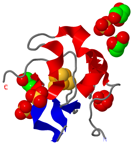
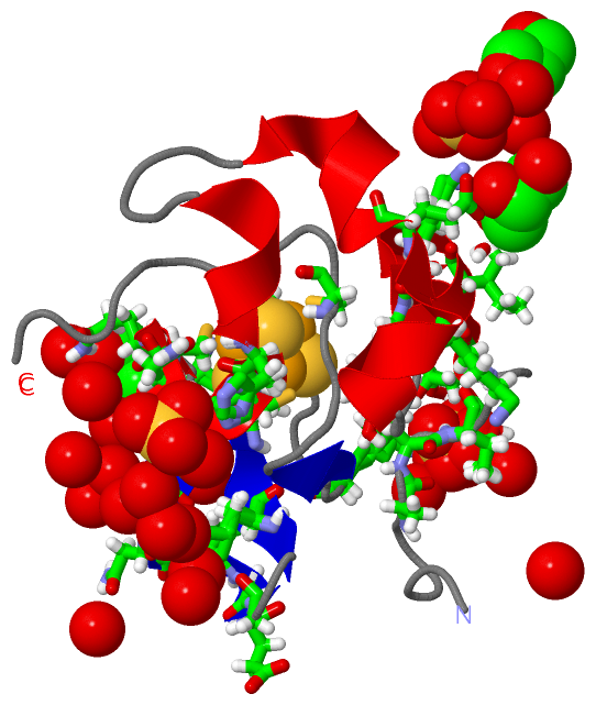
Asymmetric Unit (8, 8)
No.NameEvidenceResiduesDescription
1AC1SOFTWARETYR A:19 , CYS A:43 , CYS A:46 , MET A:49 , CYS A:61 , LEU A:63 , CYS A:75BINDING SITE FOR RESIDUE SF4 A 84
2AC2SOFTWARELYS A:18 , GLY A:54 , GLU A:55 , GLY A:56 , LYS A:59 , GOL A:202 , HOH A:363 , HOH A:364 , HOH A:430BINDING SITE FOR RESIDUE SO4 A 101
3AC3SOFTWAREALA A:6 , VAL A:7 , THR A:8 , ASP A:11 , LYS A:25 , LYS A:67 , HOH A:309 , HOH A:405 , HOH A:425 , HOH A:442 , HOH A:443BINDING SITE FOR RESIDUE SO4 A 102
4AC4SOFTWAREHIS A:42 , GLN A:50 , GLN A:62 , GOL A:201 , HOH A:328 , HOH A:341 , HOH A:364 , HOH A:370 , HOH A:397 , HOH A:408 , HOH A:456BINDING SITE FOR RESIDUE SO4 A 103
5AC5SOFTWAREGLN A:50 , ALA A:51 , ASN A:52 , GOL A:202 , HOH A:380 , HOH A:387 , HOH A:415 , HOH A:431 , HOH A:450 , HOH A:454 , HOH A:459BINDING SITE FOR RESIDUE SO4 A 104
6AC6SOFTWARELYS A:18 , GLU A:27 , ASN A:45 , GLN A:62 , SO4 A:103 , GOL A:203 , HOH A:321 , HOH A:352BINDING SITE FOR RESIDUE GOL A 201
7AC7SOFTWAREGLN A:50 , ALA A:51 , VAL A:53 , LYS A:59 , SO4 A:101 , SO4 A:104 , GOL A:203 , HOH A:350 , HOH A:364 , HOH A:370 , HOH A:459BINDING SITE FOR RESIDUE GOL A 202
8AC8SOFTWAREILE A:15 , GLN A:47 , GLN A:50 , GLN A:62 , GOL A:201 , GOL A:202 , HOH A:364 , HOH A:376 , HOH A:412 , HOH A:454BINDING SITE FOR RESIDUE GOL A 203

(-) SS Bonds  (0, 0)

(no "SS Bond" information available for 3A39)

(-) Cis Peptide Bonds  (0, 0)

(no "Cis Peptide Bond" information available for 3A39)

 Sequence-Structure Mapping

(-) SAPs(SNPs)/Variants  (0, 0)

(no "SAP(SNP)/Variant" information available for 3A39)

(-) PROSITE Motifs  (1, 1)

Asymmetric/Biological Unit (1, 1)
 PROSITEUniProtKBPDB
No.IDACDescriptionIDLocationCountLocation
1HIPIPPS51373 High potential iron-sulfur proteins family profile.HIP_THETI1-83  1A:1-83

(-) Exons   (0, 0)

(no "Exon" information available for 3A39)

(-) Sequences/Alignments

Asymmetric/Biological Unit
   Reformat: Number of residues per line =  ('0' or empty: single-line sequence representation)
  Number of residues per labelling interval =   
  UniProt sequence: complete  aligned part    
   Show mapping: SCOP domains CATH domains Pfam domains Secondary structure (by author)
SAPs(SNPs) PROSITE motifs Exons
(details for a mapped element are shown in a popup box when the mouse pointer rests over it)
Chain A from PDB  Type:PROTEIN  Length:83
 aligned with HIP_THETI | P80176 from UniProtKB/Swiss-Prot  Length:83

    Alignment length:83
                                    10        20        30        40        50        60        70        80   
             HIP_THETI    1 AAPANAVTADDPTAIALKYNQDATKSERVAAARPGLPPEEQHCANCQFMQANVGEGDWKGCQLFPGKLINVNGWCASWTLKAG 83
               SCOP domains d3a39a_ A: HIPIP (high potential iron protein)                                      SCOP domains
               CATH domains 3a39A00 A:1-83 High-Potential Iron-Sulfur Protein, subunit A                        CATH domains
               Pfam domains ----------------------------------------------------------------------------------- Pfam domains
         Sec.struct. author ..........hhhhhhhh...hhhhhhhhhhh....hhhhhhhhhh..eeeeeee..eeee.......ee............. Sec.struct. author
                 SAPs(SNPs) ----------------------------------------------------------------------------------- SAPs(SNPs)
                    PROSITE HIPIP  PDB: A:1-83 UniProt: 1-83                                                    PROSITE
                 Transcript ----------------------------------------------------------------------------------- Transcript
                  3a39 A  1 AAPANAVTADDPTAIALKYNQDATKSERVAAARPGLPPEEQHCANCQFMQANVGEGDWKGCQLFPGKLINVNGWCASWTLKAG 83
                                    10        20        30        40        50        60        70        80   

   Legend:   → Mismatch (orange background)
  - → Gap (green background, '-', border residues have a numbering label)
    → Modified Residue (blue background, lower-case, 'x' indicates undefined single-letter code, labelled with number + name)
  x → Chemical Group (purple background, 'x', labelled with number + name, e.g. ACE or NH2)
  extra numbering lines below/above indicate numbering irregularities and modified residue names etc., number ends below/above '|'

 Classification and Annotation

(-) SCOP Domains  (1, 1)

Asymmetric/Biological Unit

(-) CATH Domains  (1, 1)

Asymmetric/Biological Unit

(-) Pfam Domains  (0, 0)

(no "Pfam Domain" information available for 3A39)

(-) Gene Ontology  (6, 6)

Asymmetric/Biological Unit(hide GO term definitions)
Chain A   (HIP_THETI | P80176)
molecular function
    GO:0051539    4 iron, 4 sulfur cluster binding    Interacting selectively and non-covalently with a 4 iron, 4 sulfur (4Fe-4S) cluster; this cluster consists of four iron atoms, with the inorganic sulfur atoms found between the irons and acting as bridging ligands.
    GO:0009055    electron carrier activity    Any molecular entity that serves as an electron acceptor and electron donor in an electron transport chain. An electron transport chain is a process in which a series of electron carriers operate together to transfer electrons from donors to any of several different terminal electron acceptors to generate a transmembrane electrochemical gradient.
    GO:0051536    iron-sulfur cluster binding    Interacting selectively and non-covalently with an iron-sulfur cluster, a combination of iron and sulfur atoms.
    GO:0046872    metal ion binding    Interacting selectively and non-covalently with any metal ion.
biological process
    GO:0019646    aerobic electron transport chain    A process in which a series of electron carriers operate together to transfer electrons from donors such as NADH and FADH2 to oxygen to generate a transmembrane electrochemical gradient.
    GO:0055114    oxidation-reduction process    A metabolic process that results in the removal or addition of one or more electrons to or from a substance, with or without the concomitant removal or addition of a proton or protons.

 Visualization

(-) Interactive Views

Asymmetric/Biological Unit
  Complete Structure
    Jena3D(integrated viewing of ligand, site, SAP, PROSITE, SCOP information)
    WebMol | AstexViewer[tm]@PDBe
(Java Applets, require no local installation except for Java; loading may be slow)
    STRAP
(Java WebStart application, automatic local installation, requires Java; full application with system access!)
    RasMol
(require local installation)
    Molscript (VRML)
(requires installation of a VRML viewer; select preferred view via VRML and generate a mono or stereo PDF format file)
 
  Ligands, Modified Residues, Ions
    GOL  [ RasMol | Jena3D ]  +environment [ RasMol | Jena3D ]
    SF4  [ RasMol | Jena3D ]  +environment [ RasMol | Jena3D ]
    SO4  [ RasMol | Jena3D ]  +environment [ RasMol | Jena3D ]
 
  Sites
    AC1  [ RasMol ]  +environment [ RasMol ]
    AC2  [ RasMol ]  +environment [ RasMol ]
    AC3  [ RasMol ]  +environment [ RasMol ]
    AC4  [ RasMol ]  +environment [ RasMol ]
    AC5  [ RasMol ]  +environment [ RasMol ]
    AC6  [ RasMol ]  +environment [ RasMol ]
    AC7  [ RasMol ]  +environment [ RasMol ]
    AC8  [ RasMol ]  +environment [ RasMol ]
 
  Cis Peptide Bonds
(no "Cis Peptide Bonds" information available for 3a39)
 

(-) Still Images

Jmol
  protein: cartoon or spacefill or dots and stick; nucleic acid: cartoon and stick; ligands: spacefill; active site: stick
Molscript
  protein, nucleic acid: cartoon; ligands: spacefill; active site: ball and stick

 Databases and Analysis Tools

(-) Databases

Access by PDB/NDB ID
  3a39
    Family and Domain InformationProDom | SYSTERS
    General Structural InformationGlycoscienceDB | MMDB | NDB | OCA | PDB | PDBe | PDBj | PDBsum | PDBWiki | PQS | PROTEOPEDIA
    Orientation in MembranesOPM
    Protein SurfaceSURFACE
    Secondary StructureDSSP (structure derived) | HSSP (homology derived)
    Structural GenomicsGeneCensus
    Structural NeighboursCE | VAST
    Structure ClassificationCATH | Dali | SCOP
    Validation and Original DataBMRB Data View | BMRB Restraints Grid | EDS | PROCHECK | RECOORD | WHAT_CHECK
 
Access by UniProt ID/Accession number
  HIP_THETI | P80176
    Comparative Protein Structure ModelsModBase
    Genomic InformationEnsembl
    Protein-protein InteractionDIP
    Sequence, Family and Domain InformationInterPro | Pfam | SMART | UniProtKB/SwissProt
 
Access by Enzyme Classificator   (EC Number)
  (no 'Enzyme Classificator' available)
    General Enzyme InformationBRENDA | EC-PDB | Enzyme | IntEnz
    PathwayKEGG | MetaCyc
 
Access by Disease Identifier   (MIM ID)
  (no 'MIM ID' available)
    Disease InformationOMIM
 
Access by GenAge ID
  (no 'GenAge ID' available)
    Age Related InformationGenAge

(-) Analysis Tools

Access by PDB/NDB ID
    Domain InformationXDom
    Interatomic Contacts of Structural UnitsCSU
    Ligand-protein ContactsLPC
    Protein CavitiescastP
    Sequence and Secondary StructurePDBCartoon
    Structure AlignmentSTRAP(Java WebStart application, automatic local installation, requires Java; full application with system access!)
    Structure and Sequence BrowserSTING
 
Access by UniProt ID/Accession number
  HIP_THETI | P80176
    Protein Disorder PredictionDisEMBL | FoldIndex | GLOBPLOT (for more information see DisProt)

 Related Entries

(-) Entries Sharing at Least One Protein Chain (UniProt ID)

UniProtKB/Swiss-Prot
        HIP_THETI | P801761eyt 1iua 2ams 2fla 3a38 5d8v 5wqq 5wqr

(-) Related Entries Specified in the PDB File

1eyt CRYSTAL STRUCTURE OF THE SAME PROTEIN AT 1.5 ANGSTROM RESOLUTION
1iua CRYSTAL STRUCTURE OF THE SAME PROTEIN AT 0.8 ANGSTROM RESOLUTION
3a38 CRYSTAL STRUCTURE OF THE SAME PROTEIN AT 0.7 ANGSTROM RESOLUTION