Asymmetric Unit (9, 9)
| No. | Name | Evidence | Residues | Description |
|---|
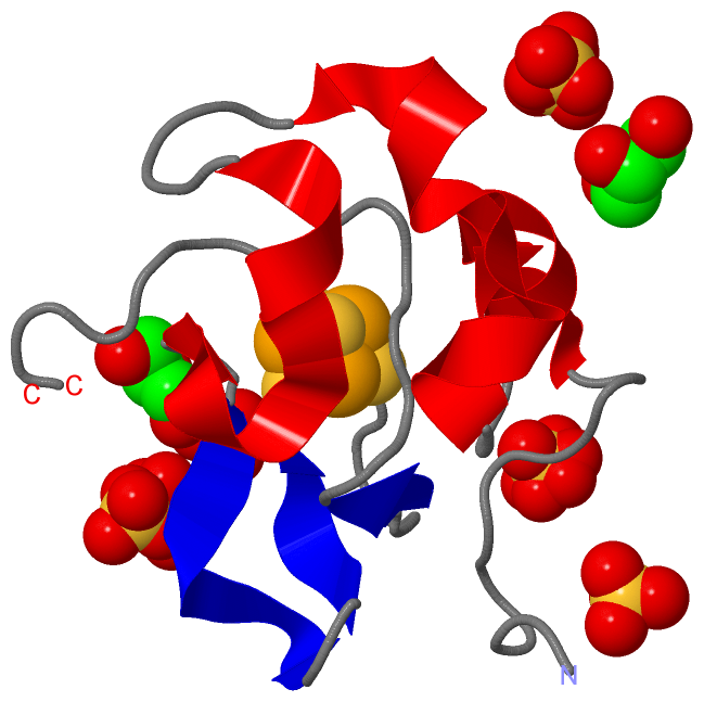
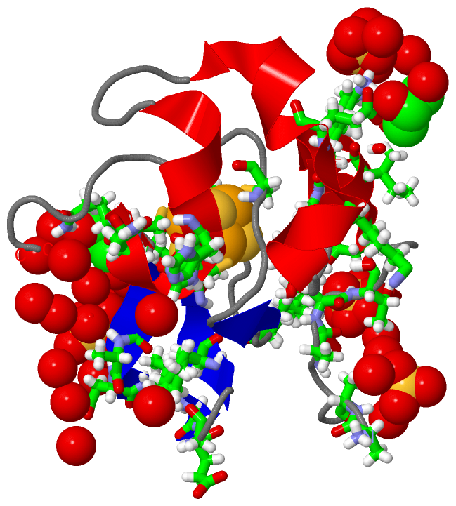
| 1 | AC1 | SOFTWARE | TYR A:19 , CYS A:43 , CYS A:46 , CYS A:61 , LEU A:63 , CYS A:75 | binding site for residue SF4 A 101 |
| 2 | AC2 | SOFTWARE | LYS A:18 , GLY A:54 , GLU A:55 , GLY A:56 , LYS A:59 , GOL A:108 , GOL A:109 , HOH A:206 , HOH A:261 , HOH A:275 , HOH A:321 | binding site for residue SO4 A 102 |
| 3 | AC3 | SOFTWARE | GLN A:50 , ALA A:51 , ASN A:52 , GOL A:109 , HOH A:204 , HOH A:210 , HOH A:234 , HOH A:273 , HOH A:285 | binding site for residue SO4 A 103 |
| 4 | AC4 | SOFTWARE | HIS A:42 , GLN A:50 , GLN A:62 , GOL A:107 , HOH A:203 , HOH A:215 , HOH A:219 , HOH A:241 , HOH A:261 , HOH A:269 , HOH A:274 | binding site for residue SO4 A 104 |
| 5 | AC5 | SOFTWARE | ALA A:6 , VAL A:7 , THR A:8 , ASP A:11 , LYS A:25 , LYS A:67 , HOH A:208 , HOH A:209 , HOH A:221 , HOH A:262 , HOH A:283 , HOH A:289 | binding site for residue SO4 A 105 |
| 6 | AC6 | SOFTWARE | ALA A:1 , ALA A:2 , HOH A:205 , HOH A:209 , HOH A:221 , HOH A:227 , HOH A:236 , HOH A:283 , HOH A:299 , HOH A:311 | binding site for residue SO4 A 106 |
| 7 | AC7 | SOFTWARE | LYS A:18 , GLU A:27 , ASN A:45 , GLN A:62 , SO4 A:104 , GOL A:108 , HOH A:203 , HOH A:263 , HOH A:338 | binding site for residue GOL A 107 |
| 8 | AC8 | SOFTWARE | ILE A:15 , GLN A:47 , GLN A:50 , GLN A:62 , SO4 A:102 , GOL A:107 , GOL A:109 , HOH A:218 , HOH A:228 , HOH A:234 , HOH A:261 | binding site for residue GOL A 108 |
| 9 | AC9 | SOFTWARE | GLN A:50 , ALA A:51 , VAL A:53 , SO4 A:102 , SO4 A:103 , GOL A:108 , HOH A:210 , HOH A:223 , HOH A:241 , HOH A:261 | binding site for residue GOL A 109 |
|
 Description