|
|
|
|
 Description
|
|
Compounds
|
||||||||||||||||||||||||||||||||||||||||||||||||
Chains, Units
Summary Information (see also Sequences/Alignments below) |
Ligands, Modified Residues, Ions (2, 2)| Asymmetric/Biological Unit (2, 2) |
Sites (2, 2)
Asymmetric Unit (2, 2)
|
SS Bonds (2, 2)
Asymmetric/Biological Unit
|
||||||||||||
Cis Peptide Bonds (2, 2)
Asymmetric/Biological Unit
|
||||||||||||
SAPs(SNPs)/Variants (1, 1)
Asymmetric/Biological Unit (1, 1)
|
|||||||||||||||||||||||||||||||||||||||||||||||||||||
PROSITE Motifs (1, 1)
Asymmetric/Biological Unit (1, 1)
|
||||||||||||||||||||||||
Exons (4, 4)
Asymmetric/Biological Unit (4, 4)
|
||||||||||||||||||||||||||||||||||||||||||||||||||||||||||||||||||||||||||||||||||||||||||||||||
Sequences/Alignments
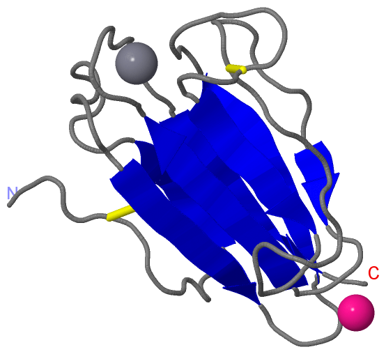
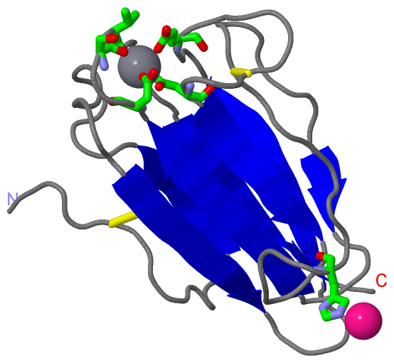
Asymmetric/Biological UnitChain A from PDB Type:PROTEIN Length:119 aligned with TSG6_HUMAN | P98066 from UniProtKB/Swiss-Prot Length:277 Alignment length:119 140 150 160 170 180 190 200 210 220 230 240 TSG6_HUMAN 131 HAKECGGVFTDPKQIFKSPGFPNEYEDNQICYWHIRLKYGQRIHLSFLDFDLEDDPGCLADYVEIYDSYDDVHGFVGRYCGDELPDDIISTGNVMTLKFLSDASVTAGGFQIKYVAMDP 249 SCOP domains ----------------------------------------------------------------------------------------------------------------------- SCOP domains CATH domains ----------------------------------------------------------------------------------------------------------------------- CATH domains Pfam domains ----CUB-2wnoA01 A:135-244 ----- Pfam domains SAPs(SNPs) -------------R--------------------------------------------------------------------------------------------------------- SAPs(SNPs) PROSITE ----CUB PDB: A:135-247 UniProt: 135-247 -- PROSITE Transcript 1 (1) 1.---------------------------------------------------------------------------Exon 1.5a --------------------------- Transcript 1 (1) Transcript 1 (2) -Exon 1.4 PDB: A:132-208 UniProt: 132-208 -------------Exon 1.6 PDB: A:222-249 Transcript 1 (2) 2wno A 131 HAKECGGVFTDPKQIFKSPGFPNEYEDNQICYWHIRLKYGQRIHLSFLDFDLEDDPGCLADYVEIYDSYDDVHGFVGRYCGDELPDDIISTGNVMTLKFLSDASVTAGGFQIKYVAMDP 249 140 150 160 170 180 190 200 210 220 230 240
|
||||||||||||||||||||
SCOP Domains (0, 0)| (no "SCOP Domain" information available for 2WNO) |
CATH Domains (0, 0)| (no "CATH Domain" information available for 2WNO) |
Pfam Domains (1, 1)
Asymmetric/Biological Unit
|
Gene Ontology (6, 6)|
Asymmetric/Biological Unit(hide GO term definitions) Chain A (TSG6_HUMAN | P98066)
|
||||||||||||||||||||||||||||||||||||||||||||||||
Interactive Views
|
||||||||||||||||||||||||||||||||||||||||||||||||||||||||||||||||||||||||||||||||||||||||||||||||||||||||||||||||||||||||||||||||||||||||||||
Still Images
|
||||||||||||||||
Databases
|
||||||||||||||||||||||||||||||||||||||||||||||||||||||||||||||||||||||||||||||||||||||||||||||||||||||||||||||||||||||||||||||||||||||||||||||||||||||||||||||||
Analysis Tools
|
|||||||||||||||||||||||||||||||||||||||||||||||||||||||||||||
Entries Sharing at Least One Protein Chain (UniProt ID)
Related Entries Specified in the PDB File
|
|