|
|
|
|
 Description
|
|
Compounds
|
||||||||||||||||||||||||||||||||||||||||||||||||||||||||
Chains, Units
Summary Information (see also Sequences/Alignments below) |
Ligands, Modified Residues, Ions (0, 0)| (no "Ligand,Modified Residues,Ions" information available for 1O7B) |
Sites (0, 0)| (no "Site" information available for 1O7B) |
SS Bonds (2, 2)
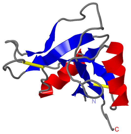
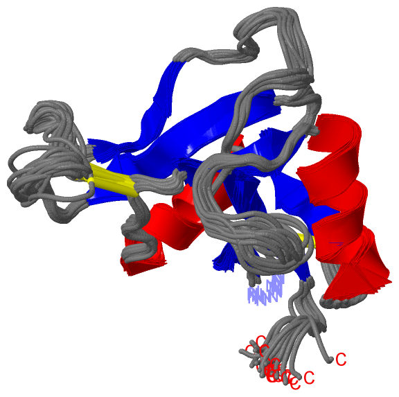
NMR Structure
|
||||||||||||
Cis Peptide Bonds (0, 0)| (no "Cis Peptide Bond" information available for 1O7B) |
SAPs(SNPs)/Variants (0, 0)| (no "SAP(SNP)/Variant" information available for 1O7B) |
PROSITE Motifs (2, 2)
NMR Structure (2, 2)
|
||||||||||||||||||||||||||||||||||||||||||||||||||||||||||||||||
Exons (3, 3)
NMR Structure (3, 3)
|
||||||||||||||||||||||||||||||||||||||||||||||||||||||||||||||||||||||||||||||||||||||||||||||||
Sequences/Alignments
NMR StructureChain T from PDB Type:PROTEIN Length:98 aligned with TSG6_HUMAN | P98066 from UniProtKB/Swiss-Prot Length:277 Alignment length:98 45 55 65 75 85 95 105 115 125 TSG6_HUMAN 36 GVYHREARSGKYKLTYAEAKAVCEFEGGHLATYKQLEAARKIGFHVCAAGWMAKGRVGYPIVKPGPNCGFGKTGIIDYGIRLNRSERWDAYCYNPHAK 133 SCOP domains d1o7bt_ T: TSG-6, Link module SCOP domains CATH domains 1o7bT00 T:1-98 Mannose-Binding Protein A, subunit A CATH domains Pfam domains Xlink-1o7bT01 T:1-93 ----- Pfam domains SAPs(SNPs) -------------------------------------------------------------------------------------------------- SAPs(SNPs) PROSITE (1) LINK_2 PDB: T:1-94 UniProt: 36-129 ---- PROSITE (1) PROSITE (2) ----------------------LINK_1 PDB: T:23-68 UniProt: 58-103 ------------------------------ PROSITE (2) Transcript 1 (1) Exon 1.2 PDB: T:1-43 UniProt: 32-78 -----------------------------------------------------1. Transcript 1 (1) Transcript 1 (2) ------------------------------------------Exon 1.3a PDB: T:43-97 UniProt: 78-132 - Transcript 1 (2) 1o7b T 1 GVYHREARSGKYKLTYAEAKAVCEFEGGHLATYKQLEAARKIGFHVCAAGWMAKGRVGYPIVKPGPNCGFGKTGIIDYGIRLNRSERWDAYCYNPHAK 98 10 20 30 40 50 60 70 80 90
|
||||||||||||||||||||
SCOP Domains (1, 1)
NMR Structure
 |
CATH Domains (1, 1)
NMR Structure
|
Pfam Domains (1, 1)
NMR Structure
|
Gene Ontology (6, 6)|
NMR Structure(hide GO term definitions) Chain T (TSG6_HUMAN | P98066)
|
||||||||||||||||||||||||||||||||||||||||||||||||
Interactive Views
|
||||||||||||||||||||||||||||||||||||||||||||||||||||||||||||||||||||||||||||||||||||||||||||||||||||||||||||||||||||
Still Images
|
||||||||||||||||
Databases
|
||||||||||||||||||||||||||||||||||||||||||||||||||||||||||||||||||||||||||||||||||||||||||||||||||||||||||||||||||||||||||||||||||||||||||||||||||||||||||||||||
Analysis Tools
|
|||||||||||||||||||||||||||||||||||||||||||||||||||||||||||||
Entries Sharing at Least One Protein Chain (UniProt ID)
Related Entries Specified in the PDB File
|
|